Case Reports
. 2019 Feb;120(4):435-443.
doi: 10.1038/s41416-018-0359-4. Epub 2019 Feb 12.

Drug sensitivity testing on patient-derived sarcoma cells predicts patient response to treatment and identifies c-Sarc inhibitors as active drugs for translocation sarcomas

Affiliations
Case Reports

Drug sensitivity testing on patient-derived sarcoma cells predicts patient response to treatment and identifies c-Sarc inhibitors as active drugs for translocation sarcomas

Bertha A Brodin et al. Br J Cancer. 2019 Feb.

Abstract

Background: Heterogeneity and low incidence comprise the biggest challenge in sarcoma diagnosis and treatment. Chemotherapy, although efficient for some sarcoma subtypes, generally results in poor clinical responses and is mostly recommended for advanced disease. Specific genomic aberrations have been identified in some sarcoma subtypes but few of them can be targeted with approved drugs.

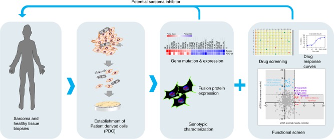
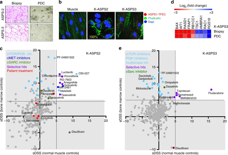
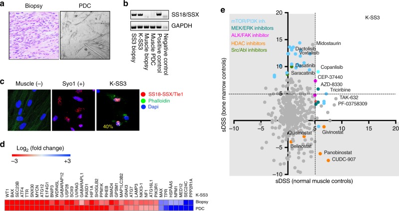
Methods: We cultured and characterised patient-derived sarcoma cells and evaluated their sensitivity to 525 anti-cancer agents including both approved and non-approved drugs. In total, 14 sarcomas and 5 healthy mesenchymal primary cell cultures were studied. The sarcoma biopsies and derived cells were characterised by gene panel sequencing, cancer driver gene expression and by detecting specific fusion oncoproteins in situ in sarcomas with translocations.

Results: Soft tissue sarcoma cultures were established from patient biopsies with a success rate of 58%. The genomic profile and drug sensitivity testing on these samples helped to identify targeted inhibitors active on sarcomas. The cSrc inhibitor Dasatinib was identified as an active drug in sarcomas carrying chromosomal translocations. The drug sensitivity of the patient sarcoma cells ex vivo correlated with the response to the former treatment of the patient.

Conclusions: Our results show that patient-derived sarcoma cells cultured in vitro are relevant and practical models for genotypic and phenotypic screens aiming to identify efficient drugs to treat sarcoma patients with poor treatment options.


Conflict of interest statement

The authors declare no competing interests.

Figures

Fig. 1
Fig. 1
Model of precision medicine for sarcoma patients. Illustration of our precision medicine model showing the process for the establishment of patient-derived cells (PDC) from patient biopsies, the characterisation of the PDC by gene panel sequencing, cancer driver gene expression and fusion oncoprotein expression in situ; and the drug sensitivity testing where active target inhibitors are identified for the specific PDC. The results of the drug screens are reported back to the referring physicians in order to nominate a potential treatment for refractory patients
Fig. 2
Fig. 2
Alveolar rhabdomyosarcoma patient-derived cells (K-RMS1). a Giemsa staining of the fine needle aspiration biopsy (FNA) showing high content of rhabdomyosarcoma cells and a light microscopy picture (×10) of the derived PDC. b RT-PCR showing the expression of PAX3-FOXO1A in the PDC (K-RMS-1) after 2 and 8 weeks of in vitro culturing. RH30 is an alveolar rhabdomyosarcoma cell line used as a positive control. Primary muscle cells were used as negative control. c Heatmap illustrating cancer driver genes expressed in K-RMS-1 at the time of drug screening. Relative expression (normalised to muscle cells) is expressed as log2 fold change. Values were calculated using the Livak method. d Plot showing the selective drug sensitivity scores (sDSS) of K-RMS1 in relation to normal bone marrow mononuclear cells (Y-axis), and healthy mesenchymal cell controls (X-axis). The patient treatment at the time of biopsy is highlighted in red
Fig. 3
Fig. 3
Alveolar soft part sarcoma patient-derived cells (K-ASPS2 and K-ASPS3). a Haematoxylin and eosin staining of formalin-fixed ASPS biopsies and the derived PDC (visualised by light microscopy). b Proximity ligation assay showing nuclear expression of the ASPS1-TFE3 fusion protein in the K-ASPS2 cells and cytoplasmic signals in K-ASPS3. Muscle cells as negative control. c Expression of cancer driver genes determined by Q-RT-PCRT in the K-ASPS3 relative to normal muscle cells. d Plot showing the drug activity in K-ASPS2 and e K-ASPS3 respectively. Patient treatment at the time of biopsy is highlighted in red
Fig. 4
Fig. 4
Synovial sarcoma patient-derived cells (K-SS3). a Haematoxylin and eosin staining showing spindle-like cells, characteristic of synovial sarcoma. The derived PDC (K-SS3) is visualised by light microscopy. b RT-PCR showing the expression of SS18-SSX in the biopsy, PDC and in control Syo1 cells. c Proximity ligation assay showing the expression of the SS18-SSX/Tle fusion protein complex in 40% (yellow) of K-SS3 cells and in the synovial sarcoma cell line Syo-1. d Heatmap comparing cancer driver gene expression in the synovial sarcoma biopsy and derived PDC. e Plot showing the selective drug sensitivity scores (sDSS) in relation to normal bone marrow mononuclear cells (Y-axis) and healthy mesenchymal cell controls (X-axis)
Fig. 5
Fig. 5
Ewing’s sarcoma patient-derived cells K-ES1 and K-ES2. a Giemsa-stained fine needle aspiration (FNA) biopsy and PDC (K-ES1) visualised by light microscopy. b Proximity ligation assay showing the expression of the EWS1-FLI1 fusion protein in K-ES1 and K-ES2 and in the Ewing’s sarcoma cell line SK-ES. c Cancer driver gene expression in K-ES1. d Drug activity in K-ES1 and f K-ES2 expressed as selective drug sensitivity scores (sDSS). e CT scans showing a rib metastasis of ES1 patient donor (yellow circles) before and after taxane treatment

References

    1. Fletcher, C. D. M., Bridge, J. A., Hogendoorn, P. & Mertens, F. WHO Classification of Tumours of Soft Tissue and Bone 4th edn (IARC Press, Lyon, 2013).
    1. Bramwell, V. H., Anderson, D., Charette, M. L., Sarcoma Disease Site G. Doxorubicin-based chemotherapy for the palliative treatment of adult patients with locally advanced or metastatic soft tissue sarcoma. Cochrane Database Syst. Rev. CD003293 3, (2003). - PMC - PubMed
    1. Le Cesne A, et al. Randomized phase III study comparing conventional-dose doxorubicin plus ifosfamide versus high-dose doxorubicin plus ifosfamide plus recombinant human granulocyte-macrophage colony-stimulating factor in advanced soft tissue sarcomas: a trial of the European Organization for Research and Treatment of Cancer/Soft Tissue and Bone Sarcoma Group. J. Clin. Oncol. 2000;18:2676–2684. doi: 10.1200/JCO.2000.18.14.2676. - DOI - PubMed
    1. Verma S, et al. Meta-analysis of ifosfamide-based combination chemotherapy in advanced soft tissue sarcoma. Cancer Treat. Rev. 2008;34:339–347. doi: 10.1016/j.ctrv.2008.01.005. - DOI - PubMed
    1. Helman LJ, Meltzer P. Mechanisms of sarcoma development. Nat. Rev. Cancer. 2003;3:685–694. doi: 10.1038/nrc1168. - DOI - PubMed

Publication types