. 2019 Jan;6(1):014002.
doi: 10.1117/1.JMI.6.1.014002. Epub 2019 Jan 29.

Evaluation of segmentation algorithms for optical coherence tomography images of ovarian tissue

Affiliations

Evaluation of segmentation algorithms for optical coherence tomography images of ovarian tissue

Travis W Sawyer et al. J Med Imaging (Bellingham). 2019 Jan.

Abstract

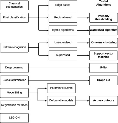
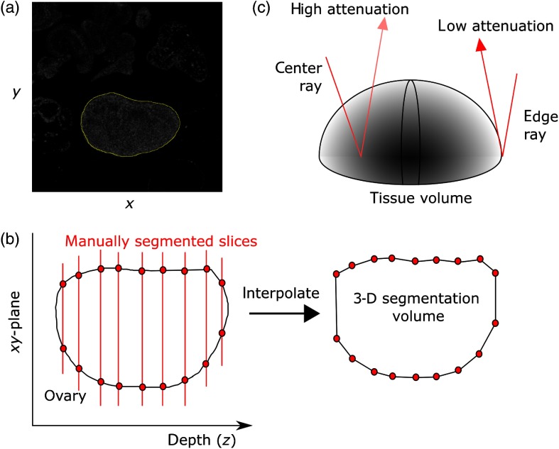
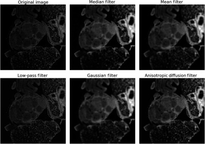
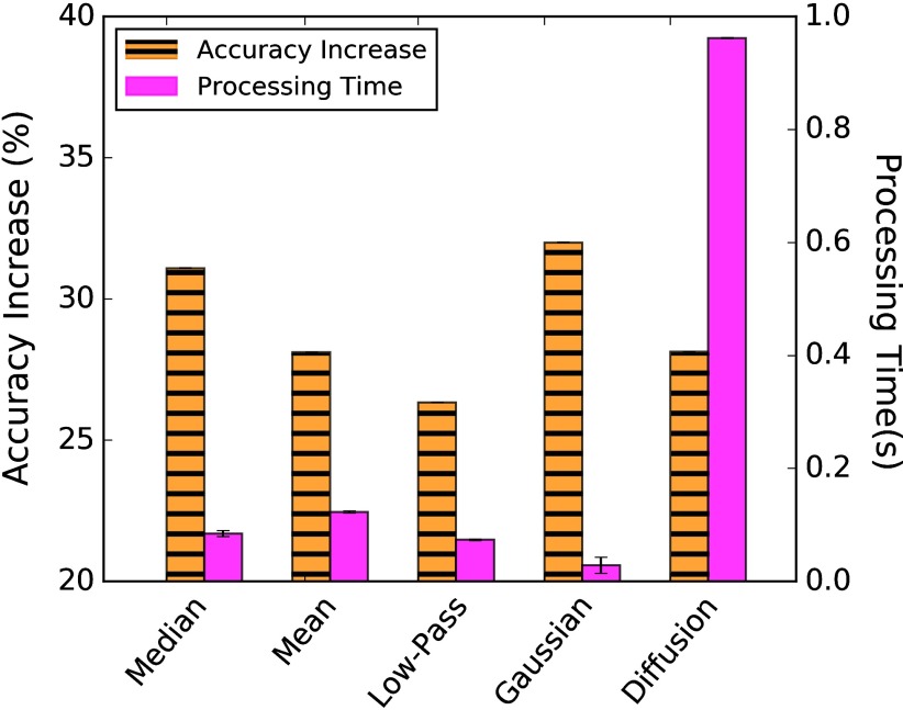
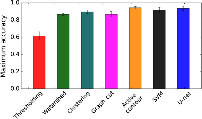
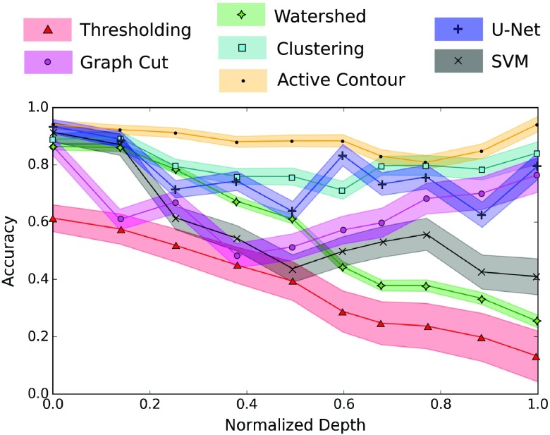
Ovarian cancer has the lowest survival rate among all gynecologic cancers predominantly due to late diagnosis. Early detection of ovarian cancer can increase 5-year survival rates from 40% up to 92%, yet no reliable early detection techniques exist. Optical coherence tomography (OCT) is an emerging technique that provides depth-resolved, high-resolution images of biological tissue in real-time and demonstrates great potential for imaging of ovarian tissue. Mouse models are crucial to quantitatively assess the diagnostic potential of OCT for ovarian cancer imaging; however, due to small organ size, the ovaries must first be separated from the image background using the process of segmentation. Manual segmentation is time-intensive, as OCT yields three-dimensional data. Furthermore, speckle noise complicates OCT images, frustrating many processing techniques. While much work has investigated noise-reduction and automated segmentation for retinal OCT imaging, little has considered the application to the ovaries, which exhibit higher variance and inhomogeneity than the retina. To address these challenges, we evaluate a set of algorithms to segment OCT images of mouse ovaries. We examine five preprocessing techniques and seven segmentation algorithms. While all preprocessing methods improve segmentation, Gaussian filtering is most effective, showing an improvement of 32 % ± 1.2 % . Of the segmentation algorithms, active contours performs best, segmenting with an accuracy of 94.8 % ± 1.2 % compared with manual segmentation. Even so, further optimization could lead to maximizing the performance for segmenting OCT images of the ovaries.

Keywords: image processing; image segmentation; optical coherence tomography; ovarian cancer.


Figures

Fig. 1
Fig. 1
Image segmentation algorithms can be decomposed into different classes, depending on the mathematics involved. We test seven of the most commonly found techniques for medical image segmentation, each of which belongs to a different class of algorithm.
Fig. 2
Fig. 2
(a) Individual slices of the OCT image stack were manually segmented using ImageJ. (b) This was repeated throughout the image depth and interpolated to yield the final segmented volume. (c) Due to the absorbing nature of tissue, as the imaging depth increases, the signal within the tissue is expected to decrease, while the signal at the edges will remain high.
Fig. 3
Fig. 3
(a) Five preprocessing techniques were investigated to suppress speckle noise in OCT images of the ovaries. (b) We tested mean, (c) median, (d) low-pass, (e) Gaussian filters, (f) in addition to anisotropic diffusion filtering. Each image here is a single en face slice.
Fig. 4
Fig. 4
Average increase in segmentation accuracy (orange) and processing time (magenta) of different filtering techniques. The Gaussian filter performs best in both categories, while the median filter also exhibits high accuracy and rapid processing time.
Fig. 5
Fig. 5
Highest accuracy obtained by each algorithm for a single 2-D slice throughout the image depth. Active contour modeling performs the best, followed by K-means clustering and the SVM.
Fig. 6
Fig. 6
Segmentation accuracy as a function of image depth. We see that an active contour model maintains high accuracy throughout the depth while thresholding; the watershed algorithm and the SVM degrade rapidly throughout the depth. Clustering and graph cutting perform reasonably well, but have a larger variation than active contours.
Fig. 7
Fig. 7
Propagating active contour snake throughout the tissue depth maintains high accuracy for delineating the segmentation boundary throughout the tissue depth, from (a) the most superficial slice to (b) shallow, (c) midrange, and (d) deep slices. The overall signal is attenuated as the depth increases, which introduces challenges with other segmentation techniques.
Fig. 8
Fig. 8
Increasing the sampling depth for propagating the active contour through (a) the image leads to a linear decrease in accuracy, (b) as well as an added interpolation error. The processing time for an image stack decreases with an increased sampling depth, suggesting that an optimal sampling depth can yield rapid processing time while maintaining high accuracy.

References

    1. Barnholtz-Sloan J. S., et al. , “Ovarian cancer: changes in patterns at diagnosis and relative survival over the last three decades,” Am. J. Obstet. Gynecol. 189(4), 1120–1127 (2003).AJOGAH10.1067/S0002-9378(03)00579-9 - DOI - PubMed
    1. Maringe C., et al. , “Stage at diagnosis and ovarian cancer survival: evidence from the International Cancer Benchmarking Partnership,” Gynecol. Oncol. 127(1), 75–82 (2012).GYNOA310.1016/j.ygyno.2012.06.033 - DOI - PubMed
    1. Carlson K. J., Skates S. J., Singer D. E., “Screening for ovarian cancer,” Ann. Intern. Med. 121, 124–132 (1994).10.7326/0003-4819-121-2-199407150-00009 - DOI - PubMed
    1. Moyer V. A., “Screening for ovarian cancer: U.S. preventive services task force reaffirmation recommendation statement,” Ann. Intern. Med. 157(12), 900–904 (2012).10.7326/0003-4819-157-11-201212040-00539 - DOI - PubMed
    1. Huang D., et al. , “Optical coherence tomography,” Science 254(5035), 1178–1181 (1991).SCIEAS10.1126/science.1957169 - DOI - PMC - PubMed