. 2019 Feb 1;5(2):eaav0282.
doi: 10.1126/sciadv.aav0282. eCollection 2019 Feb.

X-ray Fourier ptychography

Affiliations

X-ray Fourier ptychography

Klaus Wakonig et al. Sci Adv. .

Abstract

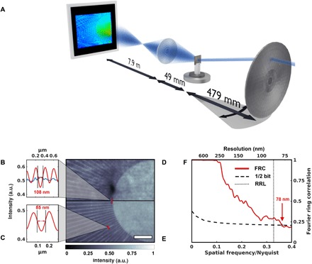
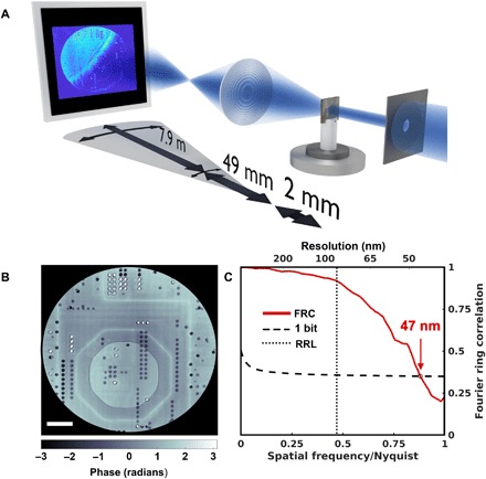
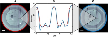
To a large extent, the performance of imaging systems is determined by their objectives, which affect properties as varied as collection efficiency, resolving power, and image distortions. Such limitations can be addressed by so-called aperture synthesis, a technique used, for instance, in radar, astronomy, and, increasingly, microscopy. Here, we apply such techniques to x-ray imaging and demonstrate how Fourier ptychography can be used at transmission x-ray microscopes to increase resolution, provide quantitative absorption and phase contrast, and allow for corrections of lens aberrations. We anticipate that such methods will find common and frequent applications, alleviating a number of limitations imposed by x-ray optical elements, offering an alternative approach to phase contrast imaging, and providing novel opportunities to mitigate radiation damage.


Figures

Fig. 1
Fig. 1. X-ray Fourier ptychography by scanning the illumination direction.
(A) A sketch of the experimental setup. By inserting a movable pinhole (not shown) close to the fixed condenser, a standard TXM setup can be modified for Fourier ptychography as the change in illumination direction can be achieved by selecting individual subfields of the condenser. (B to E) A comparison of the flat-field corrected full-field image (D) and the magnitude of an x-ray Fourier ptychographic reconstruction (E) highlights the improvement in image quality and resolution. Scale bar, 2 μm. The test pattern’s last line cut indicates a linewidth of 150 nm, which is resolved with both techniques. Yet, the innermost lines, while being washed out in the full-field image, are resolved in the Fourier ptychographic reconstruction (B) (blue and red, respectively). Even smaller features with a width of 85 nm are visible in the Fourier ptychographic reconstruction (C). (F) A Fourier ring correlation (FRC) between two subsets of the Fourier ptychographic scan confirms the improvement in resolution. The dotted line marks the Rayleigh resolution limit (RRL).
Fig. 2
Fig. 2. X-ray Fourier ptychography by scanning the objective lens.
(A) A sketch of the experimental setup. Both objective lens and detector are scanned perpendicular to the optical axis. (B) The reconstructed phase image of an ASIC. Scale bar, 5 μm. (C) The resolution for a reconstruction using an FZP with an outermost zone width of 70 nm, a diameter of 100 μm, and a scan range of 80 μm was estimated using the Fourier ring correlation (FRC) between two independent scans to 47 nm, i.e., significantly below the Rayleigh resolution limit (RRL) of 85 nm for a conventional TXM, marked by the dotted line.
Fig. 3
Fig. 3. Confirmation of quantitativeness.
(A and C) A reconstruction of the same ASIC, measured with Fourier ptychography (A) and conventional ptychography (C) (16). Scale bar, 2 μm. Resolution estimate for (A) is 47 nm and that for (C) is 41 nm (16). (B) The phase of the x-ray Fourier ptychographic reconstruction was compared to its conventional ptychographic counterpart. (C) The line cuts of both reconstructed phases reveal that the phase profiles match and thus a quantitative reconstruction can be provided (red for the Fourier ptychographic reconstruction and blue for “conventional” ptychography).

References

    1. Matsuyama S., Yasuda S., Yamada J., Okada H., Kohmura Y., Yabashi M., Ishikawa T., Yamauchi K., 50-nm-resolution full-field X-ray microscope without chromatic aberration using total-reflection imaging mirrors. Sci. Rep. 7, 46358 (2017). - PMC - PubMed
    1. Seiboth F., Schropp A., Scholz M., Wittwer F., Rödel C., Wünsche M., Ullsperger T., Nolte S., Rahomäki J., Parfeniukas K., Giakoumidis S., Vogt U., Wagner U., Rau C., Boesenberg U., Garrevoet J., Falkenberg G., Galtier E. C., Ja Lee H., Nagler B., Schroer C. G., Perfect X-ray focusing via fitting corrective glasses to aberrated optics. Nat. Commun. 8, 14623 (2017). - PMC - PubMed
    1. Chapman H. N., Nugent K. A., Coherent lensless X-ray imaging. Nat. Photonics 4, 833–839 (2010).
    1. Pfeiffer F., X-ray ptychography. Nat. Photonics 12, 9–17 (2018).
    1. Thibault P., Dierolf M., Bunk O., Menzel A., Pfeiffer F., Probe retrieval in ptychographic coherent diffractive imaging. Ultramicroscopy 109, 338–343 (2009). - PubMed

LinkOut - more resources