. 2019 Apr;161(4):645-656.
doi: 10.1007/s00701-019-03835-2. Epub 2019 Feb 12.

Protective distal side-to-side neurorrhaphy in proximal nerve injury-an experimental study with rats

Affiliations

Protective distal side-to-side neurorrhaphy in proximal nerve injury-an experimental study with rats

Henrikki Rönkkö et al. Acta Neurochir (Wien). 2019 Apr.

Abstract

Background: Side-to-side neurorrhaphy may protect the denervated end organ and preserve the initial connection with proximal stump. We examined the effect of protective side-to-side anastomosis on nerve and end organ regeneration in proximal nerve injury model.

Methods: The left common peroneal nerve of 24 Sprague Dawley rats was proximally transected. In groups B and C, side-to-side neurorrhaphy was performed distally between the peroneal and tibial nerves without (group B) and with (group C) partial donor nerve axotomy inside the epineural window. Group A served as an unprotected control. After 26 weeks, the proximal transection was repaired with end-to-end neurorrhaphy on all animals. Regeneration was followed during 12 weeks with the walk track analysis. Morphometric studies and wet muscle mass calculations were conducted at the end of the follow-up period.

Results: The results of the walk track analysis were significantly better in groups B and C compared to group A. Groups B and C showed significantly higher wet mass ratios of the tibialis anterior and extensor digitorum longus muscle compared to group A. Group C showed significantly higher morphometric values compared to group A. Group B reached higher values of the fibre count, fibre density, and percentage of the fibre area compared to group A.

Conclusions: Protective distal side-to-side neurorrhaphy reduced muscle atrophy and had an improving effect on the morphometric studies and walk track analysis. Distal side-to-side neurorrhaphy does not prevent the regenerating axons to grow from the proximal stump to achieve distal nerve stump.

Keywords: Chronic denervation; Nerve injury; Nerve regeneration; Nerve repair; Peripheral nerves; Side-to-side repair.


Conflict of interest statement

Conflict of interest

The authors declare that they have no conflict of interest.

Ethical approval

All procedures performed in studies involving animals were in accordance with the ethical standards of the institution or practice at which the studies were conducted. This article does not contain any studies with human participants performed by any of the authors.

Figures
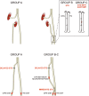
Fig. 1
Fig. 1
Schematic representation of interventions. In the first operation (upper panel), in all groups, the common peroneal nerve (CPN) is proximally transected and left unrepaired. In group B, distal side-to-side (STS) neurorrhaphy is performed without intentional axonal injury to the nerves. In group C, donor axonal injury to the extent of half of the nerve is done to the donor nerve inside the epineural windows and side-to-side anastomosis is performed similarly to the previous group. After 26 weeks, the second operation (lower panel) is performed similarly in all groups; proximal injury is repaired in an end-to-end fashion. The red marks show the nerve biopsy sites
Fig. 2
Fig. 2
Results of the walk track analysis. Groups B and C show significantly higher peroneal function index (PFI) values compared to group A from 8 weeks onward. There are no significant differences between groups B and C. The data are analysed with the analysis of covariance with Tukey–Kramer adjustment for multiple comparisons. *p < 0.05, **p < 0.01, ***p < 0.001 comparison of group A to C, +p < 0.05, ++p < 0.01, +++p < 0.001 comparison of group A to B. Error bar, ± 1 SD
Fig. 3
Fig. 3
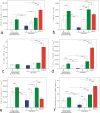
Morphometric results of the common peroneal nerve. Group C shows statistically significantly higher values of all morphometric outcomes except mean fibre area compared to group A. Group B reaches higher values of fibre count (b), fibre density (e), and percentage of the fibre area (f) compared to group A. When comparing groups B and C, group C shows higher values of fibre count (b), total fibre area (d), fibre density (e), and percentage of the fibre area (f) both at the site between neurorrhaphies and distal to common peroneal nerve. *p < 0.05, **p < 0.01, ***p < 0.001. Bars express the mean values, error bar ± 1 SD
Fig. 4
Fig. 4
Nerve sections of the distal common peroneal nerve. Morphometric analyses are done with whole-nerve cross-sections. The nerve structure is better preserved in groups B (b) and C (c) compared to group A (a). In groups B and C, the fibre density is high, but the fibre area is smaller compared to intact nerve (d). Neurofilament staining
Fig. 5
Fig. 5
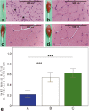
HE-stained biopsies and macroscopic figures of the tibialis anterior muscle of the intervention groups (a–c) and control sample from the contralateral side (d). In group A (a), the muscle fibres are small and angular-shaped (asterisk). In groups B and C, focal signs of atrophy can be seen. The wet mass ratio (e) is significantly higher in groups B and C compared to group A. In group A, the wet muscle mass value of tibialis anterior (TA) muscle is 22 (6.2)% of the contralateral side value. In groups B and C, the values are 54 (9.6)% and 62 (8.5)%, respectively. *p < 0.05, **p < 0.01, ***p < 0.001. Bars express the mean values, error bar ± 1 SD
Fig. 6
Fig. 6
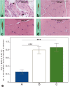
HE-stained biopsies and macroscopic figures of the extensor digitorum longus muscle of the intervention groups (a–c) and control sample from the contralateral side (d). Clear signs of atrophy and replacement of adipose tissue (asterisk) are seen in group A. In groups B and C, the muscle cells are large, but there are focal signs of atrophy. The wet mass ratio (e) is significantly higher in groups B and C compared to group A. In group A, the wet muscle mass value of extensor digitorum longus (EDL) muscle is 26 (5.2)%, in group B 80 (9.3)%, and in group C 86 (10.1)% of the contralateral side values. *p < 0.05, **p < 0.01, ***p < 0.001. Bars express the mean values, error bar ± 1 SD
Fig. 7
Fig. 7
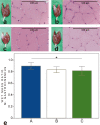
HE-stained biopsies and macroscopic figures of the gastrocnemius muscle of the intervention groups (a–c) and control sample from the contralateral side (d). Muscle architecture was well preserved in all groups. However, mild focal signs of atrophy were detected in groups B and C. In wet mass ratio calculations (e), group A (89 (6.0) %) got higher values compared to group C (82 (7.8) %). Groups B (83 (4.9) %) and C did not differ. *p < 0.05, **p < 0.01, ***p < 0.001. Bars express the mean values, error bar ± 1 SD

Similar articles

Cited by

References

    1. Bain JR, Mackinnon SE, Hunter DA. Functional evaluation of complete sciatic, peroneal, and posterior tibial nerve lesions in the rat. Plast Reconstr Surg. 1989;83:129–138. doi: 10.1097/00006534-198901000-00024. - DOI - PubMed
    1. Bain JR, Veltri KL, Chamberlain D, Fahnestock M. Improved functional recovery of denervated skeletal muscle after temporary sensory nerve innervation. Neuroscience. 2001;103:503–510. doi: 10.1016/S0306-4522(00)00577-7. - DOI - PubMed
    1. Baltzer HMD, Woo AMD, Oh CMD, Moran SLMD. Comparison of ulnar intrinsic function following supercharge end-to-side anterior interosseous-to-ulnar motor nerve transfer: a matched cohort study of proximal ulnar nerve injury patients. Plast Reconstr Surg. 2016;138:1264–1272. doi: 10.1097/PRS.0000000000002747. - DOI - PubMed
    1. Barbour J, Yee A, Kahn LC, Mackinnon SE. Supercharged end-to-side anterior interosseous to ulnar motor nerve transfer for intrinsic musculature reinnervation. J Hand Surg Am. 2012;37:2150–2159. doi: 10.1016/j.jhsa.2012.07.022. - DOI - PubMed
    1. Brown CJ, Mackinnon SE, Evans PJ, Bain JR, Makino AP, Hunter DA, Hare GM. Self-evaluation of walking-track measurement using a sciatic function index. Microsurgery. 1989;10:226–235. doi: 10.1002/micr.1920100317. - DOI - PubMed

Publication types