

Ulcer-associated cell lineage expresses genes involved in regeneration and is hallmarked by high neutrophil gelatinase-associated lipocalin (NGAL) levels
- PMID: 30746716
- PMCID: PMC6618036
- DOI: 10.1002/path.5258
Ulcer-associated cell lineage expresses genes involved in regeneration and is hallmarked by high neutrophil gelatinase-associated lipocalin (NGAL) levels
Abstract
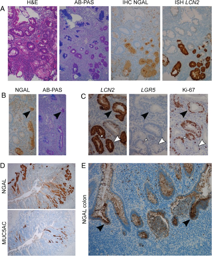
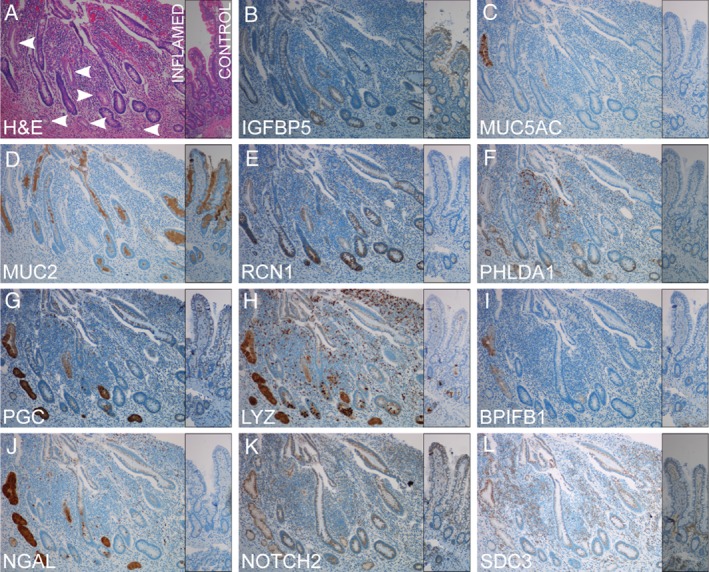
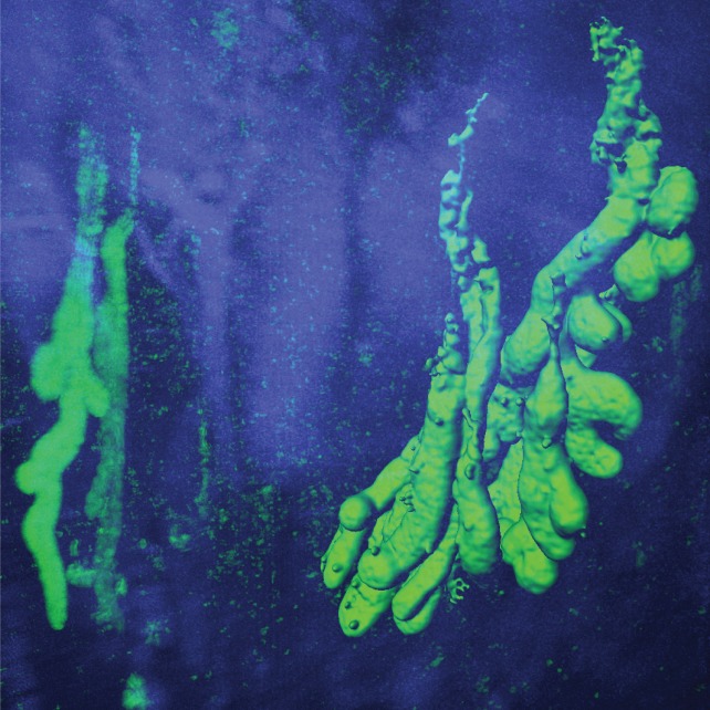
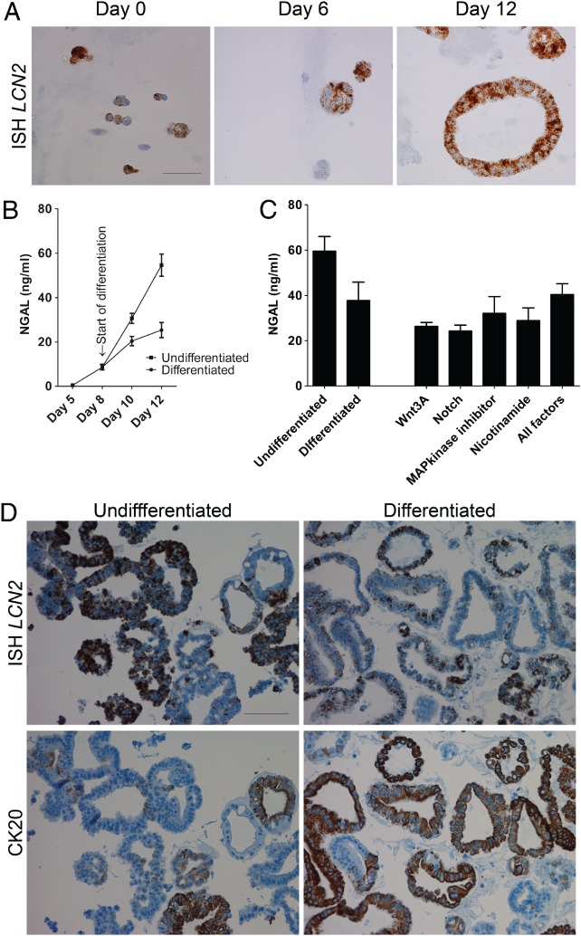
Neutrophil gelatinase-associated lipocalin (NGAL), also known as Lipocalin 2, is an antimicrobial protein, encoded by the gene LCN2, strongly upregulated in inflammatory bowel disease (IBD) and a promising biomarker for IBD. Here we demonstrate that NGAL is highly expressed in all parts of pyloric metaplasia, also known as the ulcer-associated cell lineage (UACL), a metaplastic cell lineage suggested to play a role in wound healing in Crohn's disease (CD). We further show NGAL expression in regenerative intestinal crypts and in undifferentiated patient-derived colonoids. This indicates that NGAL is important in the tissue regeneration process. The remarkable overexpression of NGAL in UACL led us to explore the pathobiology of these cells by transcriptome-wide RNA sequencing. This study is, to our knowledge, the first to characterize the UACL at this level. Biopsies with UACL and inflamed non-UACL epithelium from the terminal ileum of CD patients and epithelium from healthy controls were laser capture microdissected for RNA sequencing. Among the 180 genes differentially expressed between UACL and control epithelium, the ten most-upregulated genes specific for UACL were MUC5AC, PGC, MUC6, MUC5B, LCN2, POU2AF1, MUC1, SDC3, IGFBP5, and SLC7A5. PDX1 was among the most upregulated in both UACL and inflamed non-UACL epithelium. Immunohistochemistry and iDisco 3D visualization was used to characterize UACL histo-morphologically, and to validate protein expression of 11 selected differentially expressed genes. Among these genes, LCN2, NOTCH2, PHLDA1, IGFBP5, SDC3, BPIFB1, and RCN1 have previously not been linked to UACL. Gene expression results were analyzed for functional implications using MetaCore, showing that differentially expressed genes are enriched for genes involved in cell migration and motility, and for biomarkers of gastrointestinal neoplasia. These results support a role for UACL as part of the reepithelialization process during and after destructive intestinal inflammation. © 2019 The Authors. The Journal of Pathology published by John Wiley & Sons Ltd on behalf of Pathological Society of Great Britain and Ireland.
Keywords: CD; IBD; NGAL; UACL; pyloric metaplasia; regeneration; wound healing.
© 2019 The Authors. The Journal of Pathology published by John Wiley & Sons Ltd on behalf of Pathological Society of Great Britain and Ireland.
Figures
Similar articles
-
Expression of neutrophil gelatinase-associated lipocalin (NGAL) in the gut in Crohn's disease.Cell Tissue Res. 2018 Nov;374(2):339-348. doi: 10.1007/s00441-018-2860-8. Epub 2018 Jun 5. Cell Tissue Res. 2018. PMID: 29869714 Free PMC article.
-
Altered expression of CDX-2, PDX-1 and mucin core proteins in "Ulcer-associated cell lineage (UACL)" in Crohn's disease.J Mol Histol. 2008 Apr;39(2):161-8. doi: 10.1007/s10735-007-9149-7. Epub 2007 Oct 24. J Mol Histol. 2008. PMID: 17957487
-
Ulcer-associated cell lineage ('pyloric metaplasia') in Crohn's disease: a lectin histochemical study.J Pathol. 1993 Sep;171(1):13-9. doi: 10.1002/path.1711710105. J Pathol. 1993. PMID: 8229451
-
Role of oncogene 24p3 neutrophil gelatinase-associated lipocalin (NGAL) in digestive system cancers.Postepy Hig Med Dosw (Online). 2016 Jan 4;70(0):1026-1031. doi: 10.5604/17322693.1220397. Postepy Hig Med Dosw (Online). 2016. PMID: 27708207 Review.
-
Neutrophil Gelatinase-Associated Lipocalin as a Biomarker of Allograft Function After Renal Transplantation: Evaluation of the Current Status and Future Insights.Artif Organs. 2018 Jan;42(1):8-14. doi: 10.1111/aor.13039. Epub 2017 Dec 20. Artif Organs. 2018. PMID: 29266311 Free PMC article. Review.
Cited by
-
Physiological hypoxia improves growth and functional differentiation of human intestinal epithelial organoids.Front Immunol. 2023 Jan 27;14:1095812. doi: 10.3389/fimmu.2023.1095812. eCollection 2023. Front Immunol. 2023. PMID: 36793710 Free PMC article.
-
Cellular Plasticity, Reprogramming, and Regeneration: Metaplasia in the Stomach and Beyond.Gastroenterology. 2022 Feb;162(2):415-430. doi: 10.1053/j.gastro.2021.10.036. Epub 2021 Oct 30. Gastroenterology. 2022. PMID: 34728185 Free PMC article. Review.
-
The Lipocalin2 Gene is Regulated in Mammary Epithelial Cells by NFκB and C/EBP In Response to Mycoplasma.Sci Rep. 2020 May 6;10(1):7641. doi: 10.1038/s41598-020-63393-x. Sci Rep. 2020. PMID: 32376831 Free PMC article.
-
Single-cell integration reveals metaplasia in inflammatory gut diseases.Nature. 2024 Nov;635(8039):699-707. doi: 10.1038/s41586-024-07571-1. Epub 2024 Nov 20. Nature. 2024. PMID: 39567783 Free PMC article.
-
Weighted Gene Coexpression Network Analysis Identifies Key Genes and Pathways Associated with Idiopathic Pulmonary Fibrosis.Med Sci Monit. 2019 Jun 9;25:4285-4304. doi: 10.12659/MSM.916828. Med Sci Monit. 2019. PMID: 31177264 Free PMC article.
References
-
- Thorsvik S, Damas JK, Granlund AV, et al. Fecal neutrophil gelatinase‐associated lipocalin as a biomarker for inflammatory bowel disease. J Gastroenterol Hepatol 2017; 32: 128–135. - PubMed
-
- Wright NA, Pike C, Elia G. Induction of a novel epidermal growth factor‐secreting cell lineage by mucosal ulceration in human gastrointestinal stem cells. Nature 1990; 343: 82–85. - PubMed
Publication types
MeSH terms
Substances
LinkOut - more resources
Full Text Sources
Medical
Molecular Biology Databases
Research Materials
Miscellaneous
