Review
. 2019 Jul;25(7):825-836.
doi: 10.1111/cns.13105. Epub 2019 Feb 11.

Mitochondrial integrity in neurodegeneration

Affiliations
Review

Mitochondrial integrity in neurodegeneration

Katrina Cowan et al. CNS Neurosci Ther. 2019 Jul.

Abstract

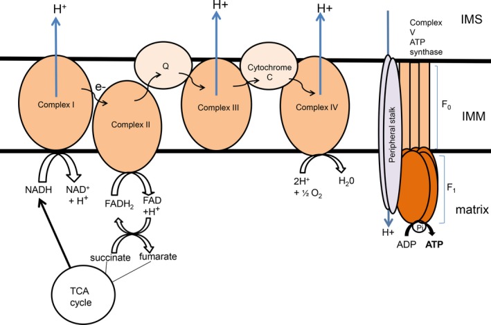
The mitochondrion is a unique organelle with a diverse range of functions. Mitochondrial dysfunction is a key pathological process in several neurodegenerative diseases. Mitochondria are mostly important for energy production; however, they also have roles in Ca2+ homeostasis, ROS production, and apoptosis. There are two major systems in place, which regulate mitochondrial integrity, mitochondrial dynamics, and mitophagy. These two processes remove damaged mitochondria from cells and protect the functional mitochondrial population. These quality control systems often become dysfunctional during neurodegenerative diseases, such as Parkinson's and Alzheimer's disease, causing mitochondrial dysfunction and severe neurological symptoms.

Keywords: cytotoxicity; mitochondrion; mitophagy; neurodegeneration.


Conflict of interest statement

The authors declare no conflict of interest.

Figures

Figure 1
Figure 1
The Tim and Tom complexes. The key pore proteins are Tom40 and Tim23, for the OMM and the IMM, respectively. In the Tom complex, smaller subunits Tom5, Tom6, and Tom7 are within the OMM. Larger subunits Tom20, Tom22, Tom37, Tom70, and Tom71 are receptor proteins, which bind to proteins and guide them into the IMS. The main Tim complex is comprised of additional subunits Tim11, Tim17, and Tim44. Proteins enter to the matrix via the Tom and Tim complexes. Proteins such as alpha‐synuclein bind to the receptor Tom20, which then guides them into the Tom40 pore (blue line)
Figure 2
Figure 2
Structure of the ETC The ETC is comprised of five main complexes which span the IMM. In complex I, NADH, which is produced in the TCA cycle, is reduced to NAD+ and H+ ions. The H+ ions are pumped out into the IMS and the electrons (e−) are passed onto complex II, which catalyzes the reduction in FADH2 to FAD. Complex II is also a part of the TCA cycle, and FAD can also be oxidized to form FADH2, which in turn reduces succinate to form fumarate in the TCA cycle. Electrons are passed through the coenzyme Q, reducing ubiquinone into ubiquinol, and then into complex III. Cytochrome C allows the flow of electrons from complex III to complex IV. At complex IV, H+ ions in the matrix react with O2 molecules to form H2O. In addition H+ ions are also pumped out into the IMS. At complex V, also known as ATP synthase, H+ ion, which have accumulated in the IMS, then pass down the concentration gradient back into the matrix. This H+ ion flow causes the F0F1 ATPase to rotate, which catalyzes the phosphorylation of ADP, generating ATP molecules into the matrix
Figure 3
Figure 3
Mitochondrion‐dependent apoptosis. When the mitochondrion is damaged, BH3‐only proteins activate Bak and/or Bax, causing permeabilization of the OMM (red line representing the permeabilization of the OMM). Bcl‐2 proteins including Bcl‐2, Bcl‐x, and MCL inhibit Bak/Bax activity. Once the OMM is permeabilized, several IMS‐localized proteins, such as cytochrome C, transport out of the mitochondrial in the cytosol (dotted line represents the movement of cytochrome C). Cytochrome C activates Apaf‐1, which in turn binds to caspase‐9, to form a complex, the apoptosome. This complex then activates caspase‐3 and caspase‐7, and leads to apoptosis. In addition to cytochrome C, the IMS‐localized protein Smac/Diablo enters the cytosol and inhibits XIAP, which is a caspase‐3 and caspase‐7 inhibitor. This leads to higher levels of caspase‐3 and caspase‐7 in the cytosol
Figure 4
Figure 4
Pink1 and Parkin in mitophagy in healthy and dysfunction mitochondria. A, In normal healthy mitochondria, Pink1 is predominantly expressed in the IMM. Pink1 is then translocated to the cytosol, where Pink1 is digested by the proteasome. Pink1 does not ubiquitinate OMM proteins and the cytosolic protein Parkin, and so Parkin is not activated. B, When mitochondria are extremely damaged, the Pink1‐Parkin pathway is activated. Pink1, instead of being degraded by the proteasome, is translocated to the OMM, which leads to its accumulation on the OMM. Pink1 phosphorylates both ubiquitin on the OMM and Parkin. The E3 ligase activity of Parkin is initiated, Parkin then ubiquitinates protein substrates, such as Miro, on the OMM, and also recruits the autophagosome to the mitochondrion for mitochondrial degradation
Figure 5
Figure 5
An overview of mitochondrial dysfunction in PD (asyn), AD (Aβ), and HD (mHtt). Protein aggregates formed in these three neurodegenerative diseases negatively impact mitochondrial function. Although the exact mechanisms are not entirely understood, protein aggregates can lead to a disruption in the mitochondrial membrane potential, resulting in decreased ATP production. This disruption in membrane potential can lead to the permeabilization of the OMM, by opening the mitochondrial permeability transition pore (mPTP), leading to the release of pro‐apoptotic molecules such as cytochrome C. The impairment of the mitochondrial membrane potential can also prevent the import of mitochondrial structural proteins, such as the subunit cytochrome b2, which are normally produced in the nucleus and are translocated into the mitochondria through the Tom and Tim complexes. Protein aggregates can also disrupt mitochondrial dynamics (Drp1 and Opa1), leading to abnormal mitochondrial number and fragmentation. When the mitochondrion is damaged beyond repair, the mitophagy process is activated, which involves the recruitment of Pink1 to the OMM, which in turn recruits Parkin from the cytosol and activates it. The activation of Parkin then leads to the formation of the autophagosome, which degrades the damaged mitochondrion, removing it from the general mitochondrial population

References

    1. Veltri KL, Espiritu M, Singh G. Distinct genomic copy number in mitochondria of different mammalian organs. J Cell Physiol. 1990;143(1):160‐164. - PubMed
    1. Bertram R, Gram Pedersen M, Luciani DS, Sherman A. A simplified model for mitochondrial ATP production. J Theor Biol. 2006;243(4):575‐586. - PubMed
    1. Gunter TE, Buntinas L, Sparagna GC, Gunter KK. The Ca2+ transport mechanisms of mitochondria and Ca2+ uptake from physiological‐type Ca2+ transients. Biochim Biophys Acta. 1998;1366(1‐2):5‐15. - PubMed
    1. Scarpa A, Graziotti P. Mechanisms for intracellular calcium regulation in heart. I. Stopped‐flow measurements of Ca++ uptake by cardiac mitochondria. J Gen Physiol. 1973;62(6):756‐772. - PMC - PubMed
    1. Maharjan S, Oku M, Tsuda M, Hoseki J, Sakai Y. Mitochondrial impairment triggers cytosolic oxidative stress and cell death following proteasome inhibition. Sci Rep. 2014;4:5896. - PMC - PubMed

Publication types