. 2019 Mar;19(3):2087-2096.
doi: 10.3892/mmr.2019.9887. Epub 2019 Jan 22.

Anti‑inflammatory effects of Dendropanax morbifera in lipopolysaccharide‑stimulated RAW264.7 macrophages and in an animal model of atopic dermatitis

Affiliations

Anti‑inflammatory effects of Dendropanax morbifera in lipopolysaccharide‑stimulated RAW264.7 macrophages and in an animal model of atopic dermatitis

Gang-Sik Choo et al. Mol Med Rep. 2019 Mar.

Abstract

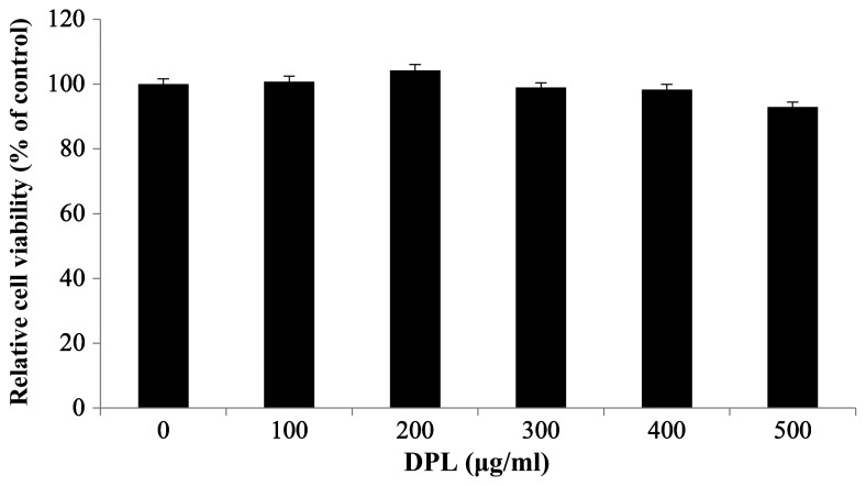
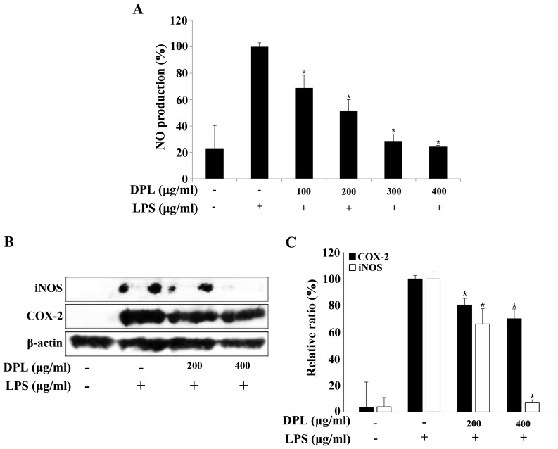
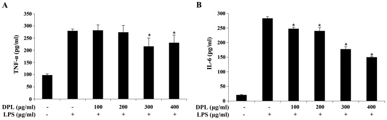
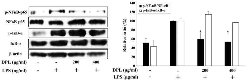
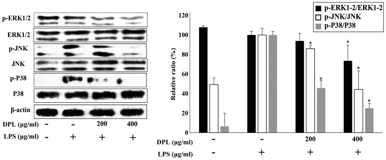
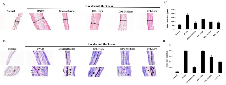
Dendropanax morbifera (D. morbifera), known as Dendro, means 'omnipotent drug' (Panax), and has been called the panacea tree. Various studies on D. morbifera are currently ongoing, aiming to determine its medicinal uses. The present study investigated the anti‑inflammatory effects and underlying mechanism of a natural extract of D. morbifera leaves (DPL) in lipopolysaccharide (LPS)‑stimulated RAW264.7 macrophages. In the present study, the following assays and models were used: MTT assay, nitric oxide (NO) assay, western blotting, ELISA and mouse models of atopic dermatitis. DPL extract markedly reduced the production of NO, inducible NO synthase and interleukin‑6, as well as the nuclear translocation of nuclear factor‑κB (NF‑κB). Additionally, the LPS‑induced activation of extracellular signal‑regulated kinase 1/2 (ERK1/2), P38 and c‑Jun N‑terminal kinase (JNK) was suppressed by DPL extract. Taken together, these results indicate that NF‑κB, ERK1/2, P38 and JNK may be potential molecular targets of DPL extract in the LPS‑induced inflammatory response. Subsequently, the present study investigated the effects of DPL extract in a 2,4‑dinitrochlorobenzene‑induced atopic dermatitis mouse model. Ear thickness, serum immunoglobulin E levels and histological analysis revealed that the DPL extract was effective in attenuating the inflammatory response. These results indicate that DPL extract has anti‑inflammatory potential and may be developed as a botanical drug to treat atopic dermatitis.


Figures

Figure 1.
Figure 1.
Effects of DPL extract on the viability of RAW264.7 cells. RAW264.7 cells (1×10⁵ cells/ml) were treated with 100, 200, 300, 400 and 500 µg/ml DPL extract for 24 h, and cell viability was determined by an MTT assay. Each bar represents the mean ± standard deviation calculated from three independent experiments. DPL, Dendropanax morbifera leaf.
Figure 2.
Figure 2.
Effects of DPL extract on LPS-induced iNOS and COX-2 protein levels and NO production in RAW264.7 cells. (A) RAW264.7 cells were pre-incubated with 100, 200, 300, and 400 µg/ml DPL extract for 2 h, and then treated with 1 µg/ml LPS for an additional 24 h. NO was measured using the Griess reaction. (B) The cells were sampled and lysed following 24 h treatment, and iNOS and COX-2 protein levels were determined by western blotting. β-actin was used as a loading control. (C) Data analysis was performed using ImageJ software by measuring the integrated band densities following background subtraction. Each bar represents the mean ± standard deviation calculated from three independent experiments. *P<0.05 vs. LPS only treatment. DPL, Dendropanax morbifera leaf; LPS, lipopolysaccharide; iNOS, inducible nitric oxide synthase; NO, nitric oxide; COX2, cyclooxygenase 2.
Figure 3.
Figure 3.
Effects of DPL extract on LPS-induced pro-inflammatory cytokine production in RAW264.7 cells. RAW264.7 cells were treated with LPS (1 µg/ml) in the presence or absence of DPL extract at the indicated concentrations for 24 h. (A) TNF-α and (B) IL-6 in the culture supernatant were measured by ELISA. Each bar represents the mean ± standard deviation calculated from three independent experiments. *P<0.05 vs. LPS only treatment. DPL, Dendropanax morbifera leaf; LPS, lipopolysaccharide; TNF-α, tumor necrosis factor-α; IL, interleukin.
Figure 4.
Figure 4.
Effects of DPL extract on the degradation or phosphorylation of NF-κB/p65 and IκB-α protein in LPS-induced RAW264.7 cells. RAW264.7 cells were treated with LPS (1 µg/ml) in the presence or absence of DPL extract at the indicated concentrations for 24 h. NF-κB phosphorylation and IκB-α degradation were assessed by western blotting. Data analysis was performed using ImageJ software by measuring the integrated band densities following background subtraction. Each bar represents the mean ± standard deviation calculated from three independent experiments. *P<0.05 vs. LPS only treatment. DPL, Dendropanax morbifera leaf; LPS, lipopolysaccharide; NF-κB, nuclear factor-κB; IκB-α, NF-κB inhibitor-α; p-, phosphorylated.
Figure 5.
Figure 5.
Effects of DPL extract on the MAPK signaling pathway in LPS-induced RAW264.7 cells. RAW264.7 cells were treated with LPS (1 µg/ml) in the presence or absence of DPL extract at the indicated concentrations for 24 h. MAPKs were assessed by western blot analysis of whole cell lysates. Each bar represents the mean ± standard deviation calculated from three independent experiments. *P<0.05 vs. LPS only treatment. DPL, Dendropanax morbifera leaf; LPS, lipopolysaccharide; MAPK, mitogen-activated protein kinase; ERK1/2, extracellular signal-regulated kinase 1/2; JNK, c-Jun N-terminal kinase; p-, phosphorylated.
Figure 6.
Figure 6.
Effects of DPL extract on DNCB-induced AD in BALB/c mice. (A) Schematic diagram presenting the timeline employed for the induction of AD and treatment with DPL extract. (B) Ear thickness during the course of AD. (C) The lymph nodes were photographed to record morphological changes. (D) Levels of serum IgE were measured by ELISA. Blood samples were collected on day 10 post-induction. Data are expressed as the mean ± standard error of the mean. *P<0.05 vs. DNCB. DPL, Dendropanax morbifera leaf; LPS, lipopolysaccharide; DNCB, 2,4-dinitrochlorobenzene; AD, atopic dermatitis; IgE, immunoglobulin E.
Figure 7.
Figure 7.
Effects of DPL extract on tissue inflammation and the infiltration of immune cells in atopic dermatitis mice. (A) Hematoxylin and eosin (red) and (B) toluidine blue (blue) staining was performed, and the cells were examined under a light microscope (magnification, ×200). The arrows indicate mast cells. (C) Skin thickness and (D) the numbers of mast cells were measured. Data are expressed as the mean ± standard error of the mean. *P<0.05 vs. DNCB. DPL, Dendropanax morbifera leaf; LPS, lipopolysaccharide; DNCB, 2,4-dinitrochlorobenzene.

References

    1. Urabe K, Aroca P, Tsukamoto K, Mascagna D, Paulumbo A, Prota G, Hearing VJ. The inherent cytotoxicity of melanin precursors: A revision. Biochim Biophys Acta 1221. 1994:272–278. - PubMed
    1. Voss GT, Oliveira RL, de Souza JF, Duarte LFB, Fajardo AR, Alves D, Luchese C, Wilhelm EA. Therapeutic and technological potential of 7-chloro-4-phenylselanyl quinoline for the treatment of atopic dermatitis-like skin lesions in mice. Mater Sci Eng C Mater Biol Appl. 2018;84:90–98. doi: 10.1016/j.msec.2017.11.026. - DOI - PubMed
    1. Hwang SW, Kang JH, Seol JE, Seo JK, Lee DBR, Sung HS. The correlation between SCORAD index and instrumental assessment in evaluation of atopic dermatitis severity. Korean J Dermatol. 2010;48:266–271. (In Korean)
    1. Leung DY. Pathogenesis of atopic dermatitis. J Allergy Clin Immun. 1999;104:S99–S108. doi: 10.1016/S0091-6749(99)70051-5. - DOI - PubMed
    1. Leung V, Hartwell R, Yang H, Ghahary A, Ko F. Bioactive nanofibres for wound healing applications. Journal of Fiber Bioengineering and Informatics. 2011;4:1–14. doi: 10.3993/jfbi04201101. - DOI

MeSH terms