. 2019 Apr 1;129(4):1713-1726.
doi: 10.1172/JCI123386. Epub 2019 Mar 18.

Nonimmune cell-derived ICOS ligand functions as a renoprotective αvβ3 integrin-selective antagonist

Affiliations

Nonimmune cell-derived ICOS ligand functions as a renoprotective αvβ3 integrin-selective antagonist

Kwi Hye Koh et al. J Clin Invest. .

Abstract

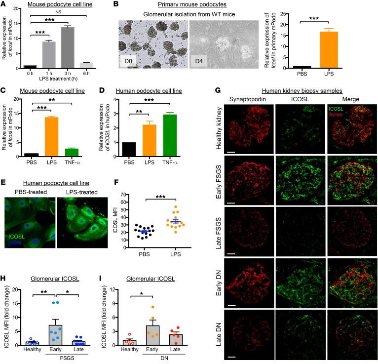
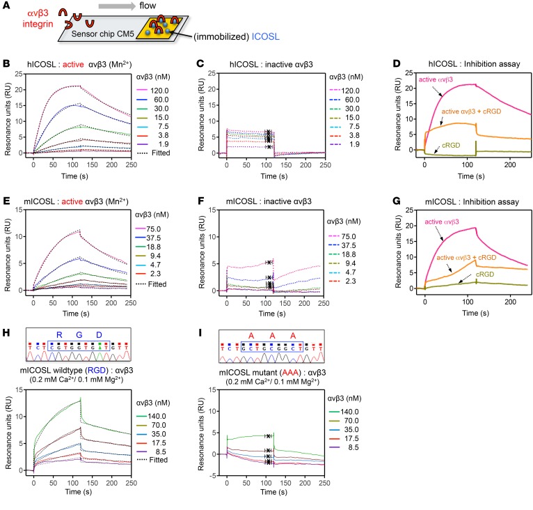
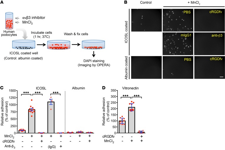
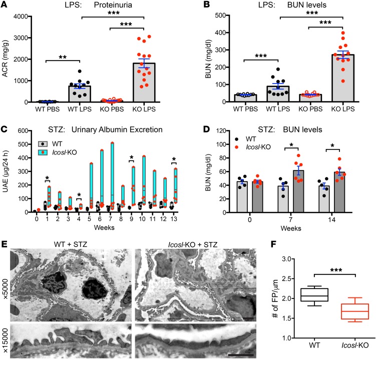
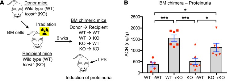
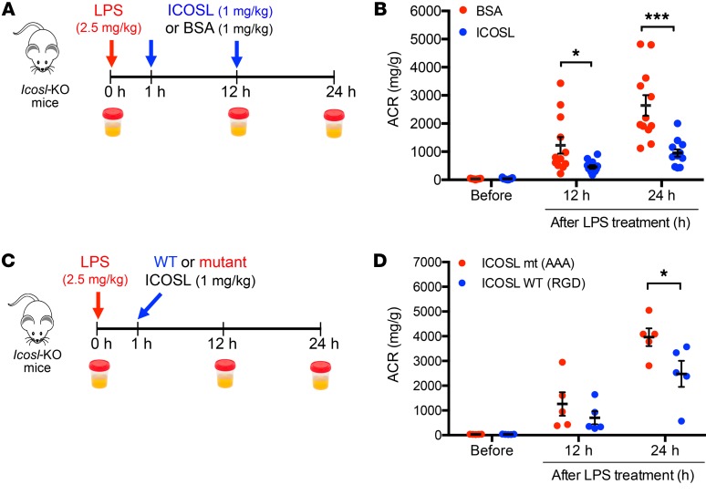
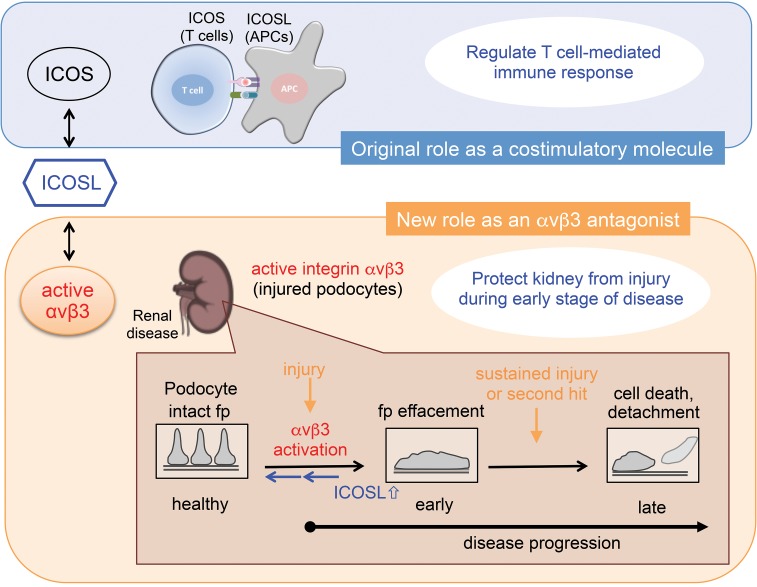
Soluble urokinase receptor (suPAR) is a circulatory molecule that activates αvβ3 integrin on podocytes, causes foot process effacement, and contributes to proteinuric kidney disease. While active integrin can be targeted by antibodies and small molecules, endogenous inhibitors haven't been discovered yet. Here we report what we believe is a novel renoprotective role for the inducible costimulator ligand (ICOSL) in early kidney disease through its selective binding to podocyte αvβ3 integrin. Contrary to ICOSL's immune-regulatory role, ICOSL in nonhematopoietic cells limited the activation of αvβ3 integrin. Specifically, ICOSL contains the arginine-glycine-aspartate (RGD) motif, which allowed for a high-affinity and selective binding to αvβ3 and modulation of podocyte adhesion. This binding was largely inhibited either by a synthetic RGD peptide or by a disrupted RGD sequence in ICOSL. ICOSL binding favored the active αvβ3 rather than the inactive form and showed little affinity for other integrins. Consistent with the rapid induction of podocyte ICOSL by inflammatory stimuli, glomerular ICOSL expression was increased in biopsies of early-stage human proteinuric kidney diseases. Icosl deficiency in mice resulted in an increased susceptibility to proteinuria that was rescued by recombinant ICOSL. Our work identified a potentially novel role for ICOSL, which serves as an endogenous αvβ3-selective antagonist to maintain glomerular filtration.

Keywords: Chronic kidney disease; Integrins; Nephrology.


Conflict of interest statement

Conflict of interest: EH, YC, and JR are inventors on a patent application (62676070) of inducible costimulator ligand for use as a renal therapeutic. JR is cofounder and stock holder of TRISAQ, a biotech company with multiple products for kidney disease.

Figures

Figure 1
Figure 1. Increased ICOSL expression is an early cellular response to renal injury.
Relative mRNA expression values measured by quantitative PCR targeting ICOSL in both mouse (AC; mPodo) and human (D; hPodo) podocyte cell lines. (A) qPCR analysis of Icosl mRNA in mouse podocyte cell lines 1, 3, or 6 hours after 50 μg/ml LPS treatment, normalized with the expression level of Gapdh and presented relative to the expression of Icosl in untreated control cells. (B) Primary podocyte isolation from BALB/c mice by Dynabead perfusion followed by 50 μg/ml LPS treatment for 3 hours. The cells were cultured and harvested, and relative expression levels of Icosl were measured by qPCR. (C) Relative mRNA expression levels of Icosl in mouse podocyte cell lines treated with 50 μg/ml LPS or 100 ng/ml TNF-α for 3 hours. (D) Relative expression levels of ICOSL in human podocyte cell lines following the same treatments as in C. Representative images (E) and quantification (F) of immunofluorescence staining of ICOSL protein in human podocytes treated with 50 μg/ml LPS (orange dots in F) or PBS (black dots in F) as control. For quantification, cells were individually defined by tracing cell borders, and the levels of ICOSL protein expression were measured by mean fluorescence intensity (MFI) using ImageJ software (n = 15 cells/group). (GI) Human kidney biopsy samples were double-stained to detect ICOSL (green) and synaptopodin (Synpo; red) by immunofluorescence staining. As depicted, analysis groups include healthy control and kidney tissues from patients with FSGS or DN at early/late stages (n = 5/group). The confocal micrographs were analyzed for glomerular expression of ICOSL by manually selecting glomeruli, defined by synaptopodin, as the region of interest. Representative confocal microscopic images are shown in G. Scale bars, 50 μm. ICOSL MFI in FSGS (H) or DN (I) groups was normalized to that of healthy controls, and data are presented as fold changes. Data are shown as mean ± SEM; *P < 0.05, **P < 0.01, ***P < 0.001; 1-way ANOVA with Dunnett’s multiple comparison test (A, C, and D) or with Tukey’s multiple comparison test (H and I) and Student’s 2-tailed, unpaired t test (B and F).
Figure 2
Figure 2. ICOSL binds to active αvβ3 integrin through its RGD motif.
(A) Schematic of a gold surface with ICOSL protein on a sensor chip CM5 and associated protein (αvβ3 integrin) over which buffer is flown in SPR assay. (BI) SPR sensorgrams depicting interaction of immobilized human ICOSL (hICOSL, BD) or mouse ICOSL (mICOSL, EI) with αvβ3 integrin. These bindings were tested in the presence (B and E, active form of αvβ3) or absence (C and F, inactive form of one with EDTA in the binding buffer) of Mn2+. (D and G) SPR used in an inhibition experiment with cRGDfv. Injection of αvβ3 integrin only (D, 120 nM αvβ3 or G, 150 nM αvβ3) resulted in a binding signal for immobilized hICOSL or mICOSL alone (pink line). Preincubation with cRGDfv (3 μM or 15 μM) significantly reduced the binding for ICOSL, indicating that the RGD peptide competes with ICOSL for binding to αvβ3 (orange line). cRGDfv alone was used as a control (green line). (H and I) SPR sensorgrams showing the binding between WT (H) or mutant (I) mICOSL protein and αvβ3 integrin in the presence of physiologically relevant divalent ions, Ca2+ (0.2 mM) and Mg2+ (0.1 mM). The average KD values were determined from at least 3 independent experiments. Rate constants (ka and kd) were determined by kinetic fitting (black dotted line) of the sensorgrams using 1-to-1 Langmuir binding equation, and KD values for B, E, and H were calculated by kd/ka (B, KD = 16.2 ± 4.0 nM for hICOSL/αvβ3 with Mn2+; E, KD = 24.2 ± 6.5 nM for mICOSL/αvβ3 with Mn2+; H, KD = 21.3 ± 1.2 nM for WT mICOSL/αvβ3 with Ca2+/Mg2+). KD values for C, F, and I were calculated from steady-state affinity fittings (C, KD = 411.8 ± 164.1 nM for hICOSL/αvβ3; F, KD ≥ 2 mM for mICOSL/αvβ3; I, KD = 0.83 ± 0.8 mM for mutant mICOSL/αvβ3 with Ca2+/Mg2+).
Figure 3
Figure 3. ICOSL regulates αvβ3 integrin–dependent adhesion in human podocytes.
(A) Schematic representation of the protocol to measure relative cell adhesion levels in cultured human podocytes. Image analysis and quantification by high-content screening technology were described in Methods. (B) Phase-contrast microscopy images show that cultured human podocytes confer enhanced adhesion to ICOSL mediated by β3 integrin treated with Mn2+, but do not adhere on albumin (protein control). Increased adhesion levels were completely prevented by incubation with the integrin inhibitors, including cRGD peptide and anti-β3 integrin antibody. Scale bar 100 μm. (C) Quantification of the cell adhesion using the images in B. ICOSL induced cell adhesion to RGD-dependent β3 integrin on cultured podocytes. (D) Cell adhesion analysis of cultured podocytes plated on vitronectin. Data are shown as mean ± SD; ***P < 0.001; 1-way ANOVA with Tukey’s multiple comparison test (C and D).
Figure 4
Figure 4. ICOSL plays a protective role during kidney injury.
(A and B), BALB/c WT and Icosl-KO mice were injected with either PBS or LPS (2.5 mg/kg body weight), then urine and blood were collected 24 hours later (n = 10 for WT PBS, n = 10 for WT LPS, n = 14 for KO PBS, n = 14 for KO LPS). (A) Urinary albumin and creatinine were measured using a mouse albumin ELISA kit and a creatinine assay kit, respectively. ACR ratio (mg/g) was calculated and used as a parameter to determine proteinuria. (B) Renal function was evaluated by measuring BUN levels as described in Methods. (C and D) Both BALB/c WT (black dots) and Icosl-KO (red dots) mice developed hyperglycemia after STZ injection (n = 5–6 per group). (C) The floating bar graph indicates urinary albumin excretion levels. (D) BUN levels. (E) Transmission electron microscope (TEM) analysis of PFA-fixed kidney glomeruli from STZ-induced WT and Icosl-KO mice (14 weeks after STZ injection). Top, TEM images displaying capillary loops at ×5000 magnification. Bottom, high magnification of podocyte foot processes (×15,000) highlighting mild effacement in the WT group and more severe effacement in the Icosl-KO group. Scale bars, 2 μm. (F) Quantification of foot process (FP) effacement using the TEM images (E). Boxes and line represent mean ± SEM and whiskers showing minimum and maximum points (n = 10 biological samples per group). Data are mean ± SEM; *P < 0.05, **P < 0.01, ***P < 0.001; 1-way ANOVA with Tukey’s multiple comparison test (A and B) or multiple unpaired t test with the Holm-Sidak comparisons test (C and D) or Student’s 2-tailed, unpaired t test (F).
Figure 5
Figure 5. Loss of nonhematopoietic ICOSL aggravates LPS-induced kidney injury.
(A) Schematic experimental design for BM chimeric mice generation. In brief, BM cells were isolated from donor mice (WT and Icosl-KO) and then transferred into irradiated recipient mice (WT and Icosl-KO) on day 0. The 4 types of BM chimera mice were produced: WT donor cells into WT recipients, WT donor cells into KO recipients, KO donor cells into WT recipients, and KO donor cells into KO recipients. Six weeks after engraftment, the BM chimeric mice were injected with LPS (2 mg/kg body weight). (B) Urine was harvested from each group of mice 24 hours after LPS administration (n = 5–7 per group from 2 independent experiments). Data are mean ± SEM; *P < 0.05, ***P < 0.001; 1-way ANOVA with Tukey’s multiple comparison test.
Figure 6
Figure 6. Administration of ICOSL reverses proteinuria in LPS-injected Icosl-KO mice.
Schematic representation of the experimental design (A). In brief, Icosl-KO mice were challenged with 2.5 mg/kg LPS, then injected intravenously (i.v.) with mouse ICOSL protein (1 mg/kg) or BSA (1 mg/kg) as a protein control 1 and 12 hours after LPS injection. Urine samples were collected at time points, 0, 12, and 24 hours after LPS administration for ACR measurement. (B) ACR levels in Icosl-KO mice treated with either ICOSL protein (blue dots) or BSA (red dots) (n = 12 per group from 2 independent experiments). Schematic representation of the experimental design (C). Icosl-KO mice were challenged with 2.5 mg/kg LPS, then injected i.v. with either WT (RGD) or mutant (AAA) mouse ICOSL protein (1 mg/kg) 1 hour after LPS injection. (D) ACR levels in Icosl-KO mice treated with either WT (blue dots) or mutant (red dots) ICOSL (n = 5 per group from 1 experiment). Data are mean ± SEM; *P < 0.05, ***P < 0.001; multiple unpaired t test with the Holm-Sidak comparisons test (B and D).
Figure 7
Figure 7. Schematic model of ICOSL’s functions.
In this study, it is shown that ICOSL binds podocyte αvβ3 integrin through its RGD motif. Kidney injury results in a rapid increase of ICOSL expression, leading to podocyte protection by blocking active αvβ3 integrin. ICOSL acts as a regulatory brake to modulate active αvβ3 integrin–mediated signaling. Fp, foot process.

References

    1. Takada Y, Ye X, Simon S. The integrins. Genome Biol. 2007;8(5):215. doi: 10.1186/gb-2007-8-5-215. - DOI - PMC - PubMed
    1. Pozzi A, Zent R. Integrins in kidney disease. J Am Soc Nephrol. 2013;24(7):1034–1039. doi: 10.1681/ASN.2013010012. - DOI - PMC - PubMed
    1. Kreidberg JA, Symons JM. Integrins in kidney development, function, and disease. Am J Physiol Renal Physiol. 2000;279(2):F233–F242. doi: 10.1152/ajprenal.2000.279.2.F233. - DOI - PubMed
    1. Fogo AB. Mechanisms of progression of chronic kidney disease. Pediatr Nephrol. 2007;22(12):2011–2022. doi: 10.1007/s00467-007-0524-0. - DOI - PMC - PubMed
    1. Reiser J, Altintas MM. Podocytes. F1000Res. 2016;5(F1000 Faculty Rev):114. - PubMed

Publication types

MeSH terms

Substances