Review
. 2019 May;8(5):466-477.
doi: 10.1002/sctm.18-0282. Epub 2019 Feb 12.

Concise Review: Update on Retinal Pigment Epithelium Transplantation for Age-Related Macular Degeneration

Affiliations
Review

Concise Review: Update on Retinal Pigment Epithelium Transplantation for Age-Related Macular Degeneration

Marco Zarbin et al. Stem Cells Transl Med. 2019 May.

Abstract

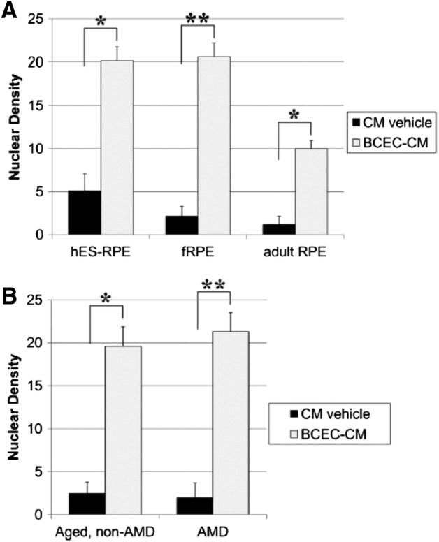
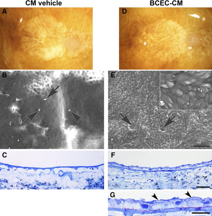
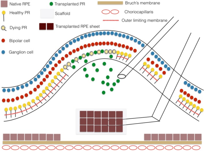
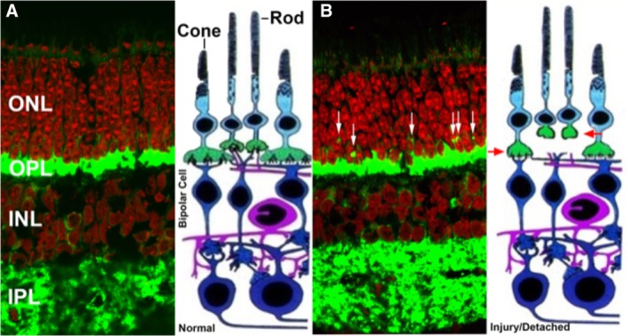
Retinal cell therapy can have the objectives of rescue (i.e., modulation of metabolic abnormalities primarily for sight preservation) as well as replacement (i.e., replace cells lost due to injury or disease for sight restoration as well as preservation). The first clinical trials of retinal pigment epithelium (RPE) transplantation for vision-threatening complications of age-related macular degeneration (AMD) have begun with some preliminary signs of success (e.g., improvement in vision in some patients, anatomic evidence of transplant-host integration with some evidence of host photoreceptor recovery, long-term survival of autologous induced pluripotent stem cell-derived RPE transplants without immune suppression) as well as limitations (e.g., limited RPE suspension survival in the AMD eye, limited tolerance for long-term systemic immune suppression in elderly patients, suggestion of uncontrolled cell proliferation in the vitreous cavity). RPE survival on aged and AMD Bruch's membrane can be improved with chemical treatment, which may enhance the efficacy of RPE suspension transplants in AMD patients. Retinal detachment, currently used to deliver transplanted RPE cells to the subretinal space, induces disjunction of the first synapse in the visual pathway: the photoreceptor-bipolar synapse. This synaptic change occurs even in areas of attached retina near the locus of detachment. Synaptic disjunction and photoreceptor apoptosis associated with retinal detachment can be reduced with Rho kinase inhibitors. Addition of Rho kinase inhibitors may improve retinal function and photoreceptor survival after subretinal delivery of cells either in suspension or on scaffolds.

Keywords: Autologous stem cell transplantation; Cell transplantation; Clinical trials; Embryonic stem cells; Experimental models; Induced pluripotent stem cells; Retina.


Conflict of interest statement

Dr. Zarbin is a paid consultant for Cell Cure, Chengdu Kanghong Biotechnology Co., Coherus Biosciences, Inc., Daiichi Sankyo, Frequency Therapeutics, Genentech/Roche, Healios KK, Inc., Iridex, Isarna Therapeutics, Makindus, Novartis Pharma AG, Ophthotech Corp., and Percept Corp. Work on improving RPE survival on AMD Bruch's membrane is covered in U.S. Patent no. 9598672, issued March 21, 2017 (“Production of extracellular matrix, conditioned media and uses thereof.” Inventors: Sugino I (Madison, NJ), Zarbin, M. (Chatham, NJ), Sun Q. (West Orange, NJ), Birge R. (New York, NY). The other authors indicated no potential conflicts of interest.

Figures

Figure 1
Figure 1
Nuclear densities of cells seeded on aged submacular Bruch's membrane explants after 21‐day culture in conditioned media (CM) vehicle or bovine corneal endothelial cell (BCEC)‐CM (paired explants from the same donor). (A): Nuclear density comparison of retinal pigment epithelium (RPE) cells derived from hESC‐RPE (n = 6), cultured human fetal RPE (fRPE, n = 22), and cultured human adult RPE (donor ages 58, 71, and 78 years; n = 7). Within each group, significant differences were observed between cells cultured in CM vehicle and cells cultured in BCEC‐CM. The nuclear density of cells cultured in CM vehicle was not statistically different between groups. The nuclear densities of hES‐RPE and fRPE were not significantly different from each other but were significantly higher than the nuclear density of adult RPE cells after culture in BCEC‐CM. (B): Comparison of nuclear densities of fRPE on age‐matched, non‐AMD versus AMD Bruch's membrane at day 21. Explants seeded with fRPE on aged Bruch's membrane (n = 9) were compared with explants seeded on age‐related macular degeneration (AMD) submacular Bruch's membrane (n = 13). No significant differences were observed in the nuclear densities of fRPE on non‐AMD versus AMD explants for a given medium, although the nuclear density was significantly higher in the presence of BCEC‐CM versus CM vehicle. Nuclear density values are counts of nuclei of cells directly in contact with Bruch's membrane, expressed as mean nuclear density per millimeter Bruch's membrane. Bars indicate mean ± SE nuclear density. *, p < .05; **, p < .001. Reproduced with permission from Sugino et al. 55.
Figure 2
Figure 2
Paired explants from an 82‐year‐old woman with geographic atrophy, seeded with fetal retinal pigment epithelium (RPE) cells. The patient's clinical history noted age‐related macular degeneration (AMD) for 20 years. (A, D): Post‐mortem clinical photographs showing subfoveal geographic atrophy before RPE cell seeding. In conditioned media (CM) vehicle, (B) only a few dead cells (arrows) and cellular debris are present on the explant surface. (C): No cells are present on Bruch's membrane surface. In bovine corneal endothelial cell (BCEC)‐CM, (E) RPE cells fully resurface Bruch's membrane in the area of geographic atrophy with a few very small defects (arrows). Localized areas of multilayering are present. Cell surfaces show abundant apical processes (inset). (F): In this field, cells resurfacing the BCEC‐CM explant are predominantly bilayered. Cells directly on Bruch's membrane are small and tightly packed; flat cells appear to overlie the cells in contact with Bruch's membrane. (G): Flattened cell processes overlying cells on top of Bruch's membrane are indicated by arrowheads. The cell processes contain vesicles. CM vehicle nuclear density (ND), 0; BCEC‐CM ND, 19.61 ± 0.43. Scale bars: 100 μm (E); 20 μm (E, inset); 50 μm (F); 20 μm (G). Toluidine blue staining. Reproduced with permission from Sugino et al. 58.
Figure 3
Figure 3
Schematic drawing illustrating subretinal injection of a suspension of rod photoreceptor precursor cells as might be done for a patient with photoreceptor degeneration due to a retinal dystrophy. The cells integrate into the retina preferentially in areas of external limiting membrane breakdown. Also shown is subretinal delivery of a retinal pigment epithelium (RPE) sheet on a scaffold to replace a localized RPE defect on Bruch's membrane as could occur in patients with geographic atrophy. Cell delivery to the subretinal space requires creating a localized retinal detachment. Reproduced with permission from Zarbin 15.
Figure 4
Figure 4
Injury‐induced synaptic disjunction. (A): Normal retina labeled for synaptic protein (SV2, green) and nuclei (red). (B): After detachment, rod terminals retract from the outer plexiform layer into the outer nuclear layer (white arrows) and cone terminals either round up or flatten due to reduction in invaginations in the cone terminals (red arrows, injured cone and rod terminals). Cone terminals are not readily evident in the photomicrograph (visualized as a reduction in invaginations in the cone terminals) due to the paucity of cones in this region but are illustrated in the adjacent schematic drawing. Abbreviations: INL, inner nuclear layer; IPL inner plexiform layer.
Figure 5
Figure 5
Effect of the ROCK inhibitor, Y27632. On axonal retraction by photoreceptors after retinal detachment in vivo. (A, C): Representative confocal images of control detached retina (BD) and detached retina treated with 10 mM Y27632 (YD) labeled for SV2 (green) and nuclei (red). SV2 labels rod presynaptic terminals. SV2‐labeled spots (white arrows) in the outer nuclear layer (ONL) indicate axonal retraction. (B, D): Binary images created from (A) and (C). SV2‐labeled spots are indicated with red arrows. The number of labeled pixels in the ONL delimited by the red borders was determined and divided by the length of examined ONL. (E): Normal retina without detachment is shown for comparison. (F, G): Treatment with 1 mM and 10 mM Y27632. Comparison of SV2‐labeled pixels per unit retinal length in different retinal areas (BC and BD, attached and detached areas, respectively, in the eye using balanced salt solution [BSS] for detachment; YC and YD, attached and detached areas, respectively, in the eye using Y27632 for detachment). In both treatment groups, there are no significant differences between BC and BD in SV2‐labeled pixels. For 1 mM Y27632, labeled pixels in YD were 34.5% less than labeled pixels in BD (*, p = .02; n = 48 retinal sections; 16 samples; four pigs). There was also a reduction in pixel labeling to 38.7% in YD compared with that in YC (p = .06; n = 48 retinal sections; 16 samples; four pigs). For 10 mM Y27632, labeled pixels in YD and YC were 43.7% (*, p = .02) and 29.9% (*, p = .04), respectively, less than labeled pixels in BD. Thus, only with 10 mM Y27632 was there a reduction in pixel labeling in the YC area compared with that in BD. Finally, there was also a significant reduction in labeled pixels in YD compared with that in YC by 24.4% (**, p = .009; n = 48 retinal sections; 16 samples; four pigs). Reproduced with permission from Wang et al. 71.

References

    1. Wong WL, Su X, Li X et al. Global prevalence of age‐related macular degeneration and disease burden projection for 2020 and 2040: A systematic review and meta‐analysis. Lancet Glob Health 2014;2:e106–e116. - PubMed
    1. Zarbin MA. Current concepts in the pathogenesis of age‐related macular degeneration. Arch Ophthalmol 2004;122:598–614. - PubMed
    1. Gehrs KM, Jackson JR, Brown EN et al. Complement, age‐related macular degeneration and a vision of the future. Arch Ophthalmol 2010;128:349–358. - PMC - PubMed
    1. Green WR, Enger C. Age‐related macular degeneration histopathologic studies. The 1992 Lorenz E. Zimmerman Lecture. Ophthalmology 1993;100:1519–1535. - PubMed
    1. Ambati J, Atkinson JP, Gelfand BD. Immunology of age‐related macular degeneration. Nat Rev Immunol 2013;13:438–451. - PMC - PubMed

Publication types