. 2019 Mar 21;4(6):e125348.
doi: 10.1172/jci.insight.125348.

A unique androgen excess signature in idiopathic intracranial hypertension is linked to cerebrospinal fluid dynamics

Affiliations

A unique androgen excess signature in idiopathic intracranial hypertension is linked to cerebrospinal fluid dynamics

Michael W O'Reilly et al. JCI Insight. .

Abstract

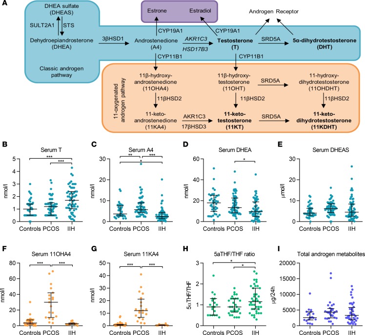
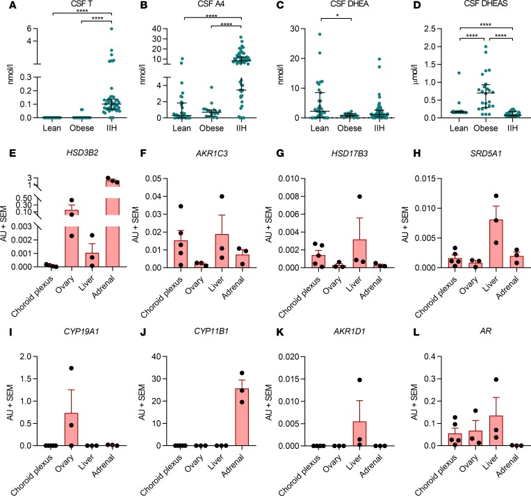
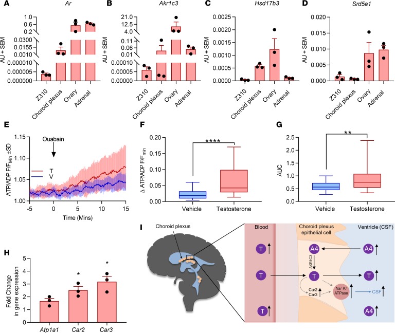
Idiopathic intracranial hypertension (IIH) is a condition of unknown etiology, characterized by elevated intracranial pressure frequently manifesting with chronic headaches and visual loss. Similar to polycystic ovary syndrome (PCOS), IIH predominantly affects obese women of reproductive age. In this study, we comprehensively examined the systemic and cerebrospinal fluid (CSF) androgen metabolome in women with IIH in comparison with sex-, BMI-, and age-matched control groups with either simple obesity or PCOS (i.e., obesity and androgen excess). Women with IIH showed a pattern of androgen excess distinct to that observed in PCOS and simple obesity, with increased serum testosterone and increased CSF testosterone and androstenedione. Human choroid plexus expressed the androgen receptor, alongside the androgen-activating enzyme aldoketoreductase type 1C3. We show that in a rat choroid plexus cell line, testosterone significantly enhanced the activity of Na+/K+-ATPase, a surrogate of CSF secretion. We demonstrate that IIH patients have a unique signature of androgen excess and provide evidence that androgens can modulate CSF secretion via the choroid plexus. These findings implicate androgen excess as a potential causal driver and therapeutic target in IIH.

Keywords: Endocrinology; Neurological disorders; Neuroscience.


Conflict of interest statement

Conflict of interest: The authors have declared that no conflict of interest exists.

Figures

Figure 1
Figure 1. Serum and urinary androgen metabolism.
(A) Androgen pathways. (BE) Serum androgen concentrations in obese controls (n = 40), PCOS patients (n = 60), and IIH patients (n = 70). (F and G) Serum 11-oxygenated androgens in obese controls (n = 35), PCOS patients (n = 20), and IIH patients (n = 13). (H and I) Urinary steroid excretion in obese controls (n = 15), PCOS patients (n = 30), and IIH patients (n = 40). 5α-Reductase activity is indicated by the ratio of 5α-tetrahydrocortisol/tetrahydrocortisol (5α-THF/THF), and total androgen metabolite excretion was calculated as the sum of androsterone plus etiocholanolone. Data presented as median and interquartile range. *P < 0.05, **P < 0.01, ***P < 0.001; Kruskal-Wallis test.
Figure 2
Figure 2. CSF androgen metabolism.
Lean controls (mixed non-IIH neurological diseases) (n = 31), obese controls (healthy volunteers with obesity) (n = 19), and IIH patients (n = 55). (A) Testosterone (T) and (B) androstenedione (A4) were significantly higher in the IIH cohort compared with both control groups (P < 0.0001 for both). (C) CSF DHEA levels were higher in lean compared with obese controls but did not differ between controls and women with IIH. (D) CSF DHEAS levels were significantly higher in control women with obesity than in both lean controls and women with IIH (P < 0.0001). (EL) HSD3B2, AKR1C3, HSD17B3, SRD5A1, CYP19A1, CYP11B1, AKR1D1, and AR mRNA expression in human choroid plexus (n = 5), ovary (n = 3), liver (n = 3), and adrenal (n = 3) tissue. Axes not uniform due to differences in mRNA expression. (AD) Presented as median and interquartile range. *P < 0.05, ****P < 0.0001; Kruskal-Wallis test. (EL) Presented as mean ± SEM.
Figure 3
Figure 3. Functional effect of testosterone on rodent choroid plexus.
(AD) mRNA expression in female rats normalized to ribosomal 18S; n = 3 biological replicates (mean ± SEM). Z310 cells are immortalized choroid plexus epithelial cells. (EG) Z310 cells incubated with 100 nM testosterone for 2 days increased the ΔATP/ADP ratio at 15 minutes and AUC (AU) of the ratio over 15 minutes, indicating increased Na+/K+-ATPase activity, a surrogate measure of CSF production (n = 39 cells from 3 pooled coverslips). V, vehicle; F, intensity for cell in particular frame; Fmin, lowest fluorescence intensity at baseline. Data presented as mean ± SD in E; box and whisker plots in F and G present data as median and interquartile range, showing minimum and maximum values. (H) mRNA expression of carbonic anhydrases II and III (Car2 and Car3) and Na+/K+-ATPase (Atp1a1) at 48 hours in testosterone-treated (versus vehicle) Z310 cells (n = 3 repeats, mean ± SEM). Mann-Whitney U test used for EH; *P < 0.05, **P < 0.01, and ****P < 0.0001. (I) Concept figure linking systemic and CSF androgen activation and impact on CSF secretion at the choroid plexus.

References

    1. Mollan SP, Ali F, Hassan-Smith G, Botfield H, Friedman DI, Sinclair AJ. Evolving evidence in adult idiopathic intracranial hypertension: pathophysiology and management. J Neurol Neurosurg Psychiatry. 2016;87(9):982–992. doi: 10.1136/jnnp-2015-311302. - DOI - PMC - PubMed
    1. Mulla Y, Markey KA, Woolley RL, Patel S, Mollan SP, Sinclair AJ. Headache determines quality of life in idiopathic intracranial hypertension. J Headache Pain. 2015;16:521. - PMC - PubMed
    1. Mollan SP, et al. Idiopathic intracranial hypertension: consensus guidelines on management. J Neurol Neurosurg Psychiatry. 2018;89(10):1088–1100. doi: 10.1136/jnnp-2017-317440. - DOI - PMC - PubMed
    1. Mollan SP, Aguiar M, Evison F, Frew E, Sinclair AJ. The expanding burden of idiopathic intracranial hypertension. Eye (Lond) 2019;33:478–485. - PMC - PubMed
    1. Markey KA, Mollan SP, Jensen RH, Sinclair AJ. Understanding idiopathic intracranial hypertension: mechanisms, management, and future directions. Lancet Neurol. 2016;15(1):78–91. doi: 10.1016/S1474-4422(15)00298-7. - DOI - PubMed

Publication types