. 2019 Dec 17;29(11):4763-4774.
doi: 10.1093/cercor/bhz009.

Motivational Impairment is Accompanied by Corticoaccumbal Dysfunction in the BACHD-Tg5 Rat Model of Huntington's Disease

Affiliations

Motivational Impairment is Accompanied by Corticoaccumbal Dysfunction in the BACHD-Tg5 Rat Model of Huntington's Disease

Natalie E Zlebnik et al. Cereb Cortex. .

Abstract

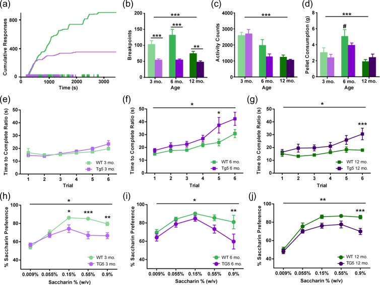
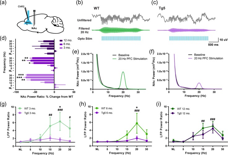
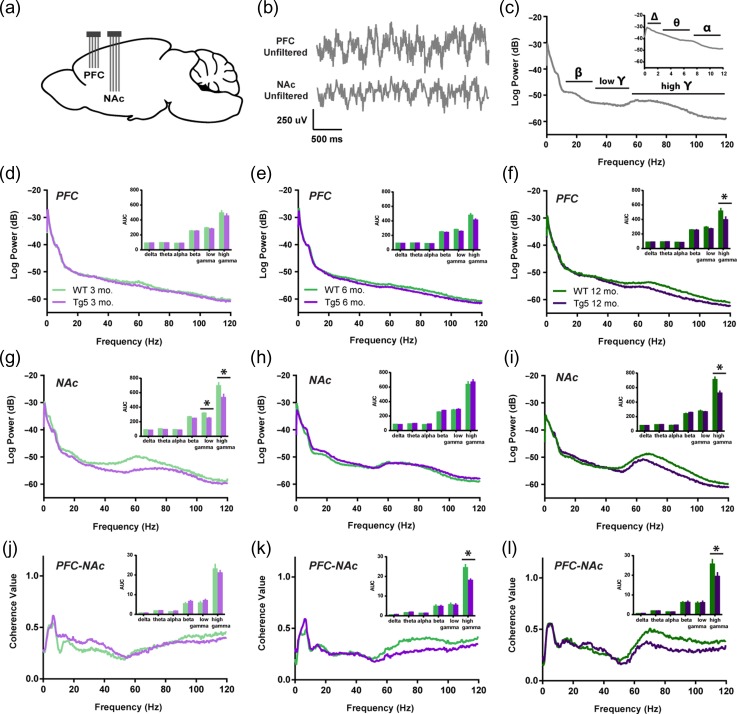
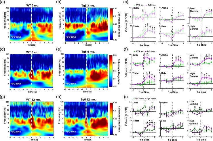
Neuropsychiatric symptoms, such as avolition, apathy, and anhedonia, precede the onset of debilitating motor symptoms in Huntington's disease (HD), and their development may give insight into early disease progression and treatment. However, the neuronal and circuit mechanisms of premanifest HD pathophysiology are not well-understood. Here, using a transgenic rat model expressing the full-length human mutant HD gene, we find early and profound deficits in reward motivation in the absence of gross motor abnormalities. These deficits are accompanied by significant and progressive dysfunction in corticostriatal processing and communication among brain areas critical for reward-driven behavior. Together, our results define early corticostriatal dysfunction as a possible pathogenic contributor to psychiatric disturbances and may help identify potential pharmacotherapeutic targets for the treatment of HD.

Keywords: Huntington’s disease; corticostriatal; motivation; nucleus accumbens; prefrontal cortex.


Figures

Figure 1.
Figure 1.
Early and sustained motivational and hedonic deficits in the absence of gross motor abnormalities in HD. (a) Representative cumulative response records from 3 mo. WT and Tg5 rats performing the PR task. (b) Tg5 rats show reduced breakpoints under a PR schedule across all ages (n = 89). (c) Significant age-related decline in locomotor activity counts in both genotypes (n = 44). (d) Similar consumption of highly-palatable food pellets under free-access conditions across all ages in Tg5 vs. WT rats (n = 50). At 6 mo., WT rats consume significantly more pellets than at 12 mo. (eg) On matched PR trials, Tg5 rats show similar latency as WT rats to complete each response ratio at 3 mo. of age before declining in performance at 6 and 12 mo. of age (n = 73). (hj) Tg5 rats demonstrate lower preference for saccharin solution than WT rats at all ages (n = 50). Unless otherwise stated, data are represented as mean ± SEM; *P < 0.05, **P < 0.01, ***P < 0.001 vs. Tg5; # P < 0.05 vs. other ages.
Figure 2.
Figure 2.
Optogenetic stimulation reveals compromised integrity of corticostriatal communication in HD. (a) ChR2 transduction and optogenetic stimulation site in the PFC and extracellular tetrode recording site in the NAc. (b,c) Upper: Representative unfiltered and filtered NAc LFP recordings during 20 Hz optogenetic stimulation of the PFC in 3 mo. rats. Lower: Representative FFTs of NAc power at baseline and during 20 Hz PFC stimulation. (d) Evoked NAc power across frequency ranges as percent change from WT. Tg5 rats had much smaller changes in NAc power following optogenetic PFC stimulation compared to WT rats at 3 and 6 mo. but not 12 mo. (n = 21, *P < 0.05, **P < 0.01, ***P < 0.001 vs. NL). (eg) Power ratio from NAc LFP recordings across PFC stimulation frequencies. Evoked LFP power is greatly attenuated in Tg5 compared to WT rats at 3 mo. and 6 mo. but not 12 mo. (n = 44, *P < 0.05 vs. Tg5; # P < 0.05, ## P < 0.01, ### P < 0.001 vs. NL). Unless otherwise stated, data are represented as mean ± SEM.
Figure 3.
Figure 3.
Progressive corticostriatal network dysfunction in HD. (a) Multi-electrode extracellular recording sites in the PFC and NAc. (b) Representative simultaneous LFP recordings collected during PR task performance. (c) Representative power spectrum illustrating functional frequency bandwidths [Δ: 0–3 Hz, θ: 3.1–7 Hz, α: 7.1–12 Hz, β: 12.1–30 Hz, low γ: 30.1–55 Hz, high γ: 55.1–120 Hz]. Inset: Magnification of 0–12 Hz range. (df) Intra-PFC power spectra demonstrate attenuated high gamma power at 12 mo. in Tg5 vs. WT rats. Insets: AUC of frequency bandwidth power (n = 47). (gi) Intra-NAc power spectra reveal genotype differences in gamma frequency power across ages. Compared to WT rats, Tg5 rats display lower power in both low and high gamma frequency ranges at 3 mo., normalization of power at 6 mo., and then reemergence of abnormally lower power in the high gamma range at 12 mo. Insets: AUC of frequency bandwidth power (n = 48). (jl) LFP–LFP coherence between the PFC and NAc is diminished in Tg5 rats at 6 and 12 mo., but not 3 mo. Insets: AUC of frequency bandwidth coherence (n = 47). Unless otherwise stated, data are represented as mean or mean ± SEM. *P < 0.05 vs. Tg5.
Figure 4.
Figure 4.
Abnormal reward-evoked changes in corticostriatal network dynamics in HD. Left: Comparison of WT and Tg5 average wavelet spectrograms demonstrates dysregulation of LFP–LFP coherence among the PFC and NAc regions upon reward delivery (0 s, dashed line). Right: Frequency-specific PFC–NAc coherence within the 10-s window centered around reward delivery and normalized to the first second of the window. (a–c) At 3 mo., there were robust elevations in PFC–NAc coherence in response to reward delivery in both WT and Tg5 but few differences in coherence among the genotypes (n = 12). (df) By 6 mo., increases in LFP–LFP coherence at reward delivery were present, and Tg5 vs. WT rats showed prominent potentiation of network coherence upon reward delivery across most frequency bandwidths, including delta, theta, alpha, low gamma, and high gamma (n = 13). (gi) At 12 mo., abnormally elevated PFC–NAc coherence in the theta frequency range persisted in Tg5 compared to WT rats. However, there was overall attenuation of evoked coherence with normalization of coherence magnitude in the delta, alpha, low gamma, and high gamma bandwidths and with significantly lower coherence in the low gamma frequency range pre-reward delivery in the Tg5 vs. WT rats (n = 10). Unless otherwise stated, data are represented as mean or mean ± SEM. *P < 0.05, **P < 0.01, ***P < 0.001, ****P < 0.0001 vs. Tg5; WT: † P < 0.05 vs. Bin 1; Tg5: & P < 0.05 vs. Bin 1.
Figure 5.
Figure 5.
Reward-evoked corticostriatal theta synchrony predicts motivational state in WT but not HD rats. Among all WT animals, higher peak reward-evoked PFC–NAc coherence in the theta frequency range (3.1–7 Hz) was correlated with a greater number of reinforcers earned on the PR task (n = 17; R2 = 0.4188, P < 0.01). Consistent with abnormally elevated reward-evoked theta coherence in the Tg5 animals across all ages, no strong relationship among theta coherence and PR task performance was measured in the Tg5 rats (n = 18; R2 = 0.01913, ns).

Similar articles

Cited by

References

    1. Abada Y-SK, Nguyen HP, Schreiber R, Ellenbroek B. 2013. Assessment of motor function, sensory motor gating and recognition memory in a novel BACHD transgenic rat model for huntington disease. PLoS One. 8:e68584. - PMC - PubMed
    1. Abeles M. 1991. Corticonics: Neural Circuits of the Cerebral Cortex [WWW Document]. Camb Core. URL /core/books/corticonics/7BF149062695412A32FFC1255C98B410
    1. Akwei-Sekyere S. 2015. Powerline noise elimination in biomedical signals via blind source separation and wavelet analysis. PeerJ. 3:e1086. - PMC - PubMed
    1. Alexopoulos GS, Hoptman MJ, Yuen G, Kanellopoulos D, Seirup JK, Lim KO, Gunning FM. 2013. Functional connectivity in apathy of late-life depression: a preliminary study. J Affect Disord. 149:398–405. - PMC - PubMed
    1. André VM, Cepeda C, Fisher YE, Huynh M, Bardakjian N, Singh S, Yang XW, Levine MS. 2011. Differential electrophysiological changes in striatal output neurons in Huntington’s disease. J Neurosci Off J Soc Neurosci. 31:1170–1182. - PMC - PubMed

Publication types