. 2019 Feb 1;77(1):ftz007.
doi: 10.1093/femspd/ftz007.

Accumulation of EBI3 induced by virulent Mycobacterium tuberculosis inhibits apoptosis in murine macrophages

Affiliations

Accumulation of EBI3 induced by virulent Mycobacterium tuberculosis inhibits apoptosis in murine macrophages

Jia-Hui Deng et al. Pathog Dis. .

Abstract

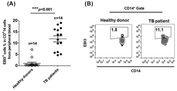
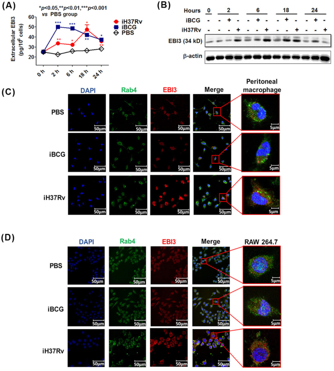
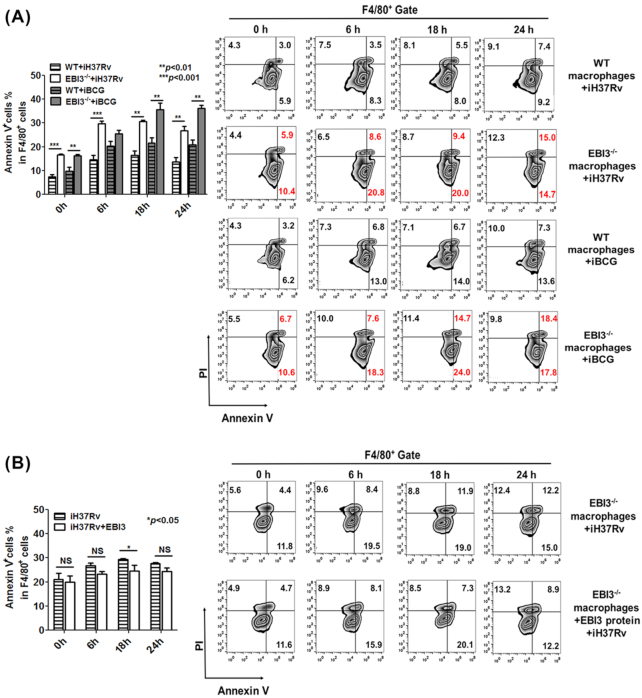
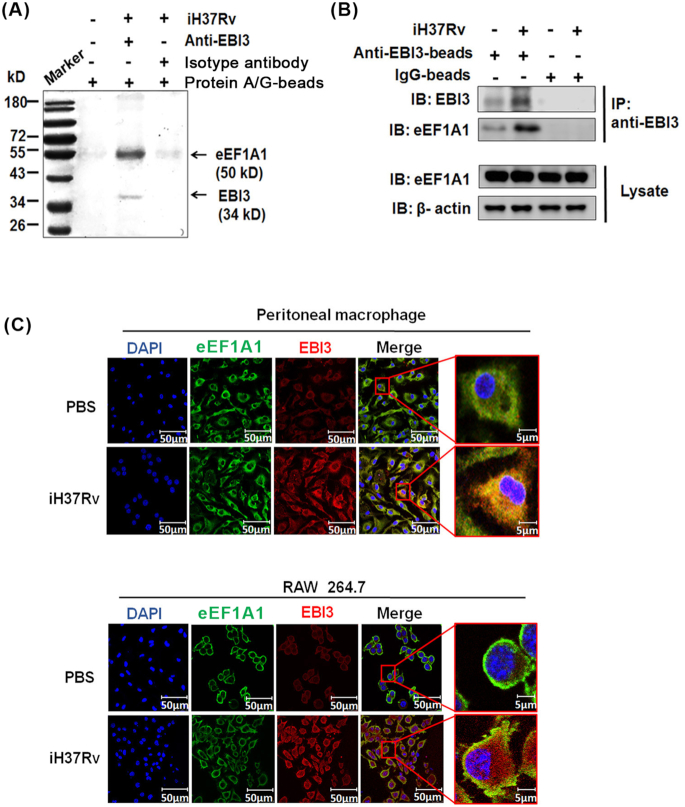
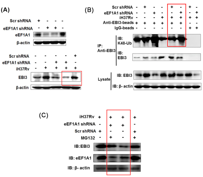
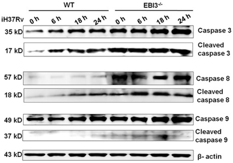
Macrophages are the primary host target cells of Mycobacterium tuberculosis (M. tb). As a subunit of immunoregulatory cytokines IL-27 and IL-35, Epstein-Barr virus-induced gene 3 (EBI3) has typically been explored as the secreted form and assessed in terms of its effects triggered by extracellular EBI3. However, little is known about intracellular EBI3 function. In the current study, we report that EBI3 production by macrophages is elevated in TB patients. We further demonstrate that increased EBI3 accumulates in virulent M. tb-treated murine macrophages. Eukaryotic translation elongation factor 1-alpha 1 (eEF1A1) binds to intracellular EBI3 to reduce Lys48 (K48)-linked ubiquitination of EBI3, leading to EBI3 accumulation. Moreover, the intracellular EBI3 inhibits caspase-3-mediated apoptosis in M. tb-treated macrophages. Herein, we propose a novel mechanism for accumulating intracellular EBI3 and its regulation of macrophage apoptosis in response to virulent M. tb.

Keywords: M. tb; EBI3; apoptosis; eEF1A1; macrophage; ubiquitination.


Figures

Figure 1.
Figure 1.
EBI3 production by CD14+ macrophages is elevated in TB patients. The percentages of EBI3+ cells in CD14+ human monocytes from peripheral blood were determined by FCM. (A) Pooled data and (B) Representative dot plots. The data in (A) are shown as mean ± SD (n = 14).
Figure 2.
Figure 2.
Increased EBI3 accumulates in virulent M. tb-treated murine macrophages. (A) and (B) Murine peritoneal macrophages were stimulated with iH37Rv or iBCG for 2 h, 6 h, 18 h and 24 h. (A) Extracellular EBI3 level in cell culture supernatant was determined by ELISA. The data are shown as mean ± SD (n = 3). (B) Intracellular EBI3 was determined by immunoblot analysis. The iH37Rv/iBCG-treated cell pellets were washed with PBS, and employed to determine the intracellular EBI3. (C) Peritoneal macrophages and (D) RAW 264.7 cells were stimulated with iH37Rv or iBCG for 6 h. Intracellular EBI3 levels were determined by confocal microscopy.
Figure 3.
Figure 3.
Accumulated intracellular EBI3 inhibits apoptosis in iH37Rv-treated macrophages.(A) Apoptosis was increased in iH37Rv/iBCG-treated EBI3−/− macrophages. WT and EBI3−/− peritoneal macrophages were treated with iH37Rv/iBCG (MOI 1:10) for 6 h, 18 h and 24 h. Apoptotic cells were analyzed by FCM. Left panel, pool data. The data are shown as mean ± SD (n = 3). Right panel, representative dot plots. (B) Extracellular EBI3 had a limited effect on apoptosis in iH37Rv-treated EBI3−/− macrophages. The recombinant mouse EBI3 protein (50 pg/106 cells/ml) was added to the EBI3−/− macrophages, and the cells were stimulated with iH37Rv (MOI 1:10). Apoptosis in the EBI3−/− macrophages was determined by FCM.
Figure 4.
Figure 4.
eEF1A1 binds to EBI3 in iH37Rv-treated macrophages. (A) eEF1A1 was identified as EBI3 binding protein. RAW 264.7 cells were stimulated with iH37Rv for 6 h, and the cell lysate was incubated with anti-EBI3 antibody and protein A/G magnetic beads. Precipitated samples were analyzed by SDS-PAGE. The specific band was identified by LC-MS/MS analysis. eEF1A1 peptides were confirmed by peptide mass fingerprinting (PMF). (B) The binding between eEF1A1 and EBI3 was determined by pull-down and immunoblot analysis. Cell lysates were incubated with anti-EBI3 antibody and protein A/G magnetic beads. Precipitated samples were analyzed by anti-EBI3 and anti-eEF1A1 antibodies. (C)Peritoneal macrophages and RAW 264.7 cells were stimulated with iH37Rv for 6 h. Intracellular EBI3 and eEF1A1 were determined by confocal microscopy.
Figure 5.
Figure 5.
eEF1A1 reduces K48-linked EBI3 ubiquitination in iH37Rv-treated macrophages. (A) EBI3 expression was reduced in eEF1A1-silenced macrophages upon iH37Rv stimulation. RAW 264.7 cells were transfected with eEF1A1 shRNA. After 24 h of transfection, cells were stimulated with iH37Rv for 6 h. Upper panel, after 24 h of transfection, eEF1A1 expression was determined by immunoblot analysis. Lower panel, after 6 h of iH37Rv stimulation, EBI3 expression was determined in eEF1A1-slienced cells. (B) eEF1A1 inhibited K48-linked ubiquitination of EBI3 in iH37Rv-treated cells. RAW 264.7 cells were transfected with eEF1A1 shRNA and stimulated with iH37Rv. Supernatant from the cell lysate was incubated with anti-EBI3 antibody followed by incubation with protein A/G magnetic beads. The precipitated samples were subjected to immunoblot analysis with anti-EBI3 antibody and K48-linkage specific polyubiquitin antibody. (C) eEF1A1-silienced RAW 264.7 cells were treated with proteasome inhibitor MG132 (2 nM) prior to iH37Rv treatment. The EBI3 production were measured by immunoblot analysis with anti-EBI3 antibody.
Figure 6.
Figure 6.
Intracellular EBI3 inhibits caspase-3 activation in iH37Rv-treated macrophages. Activation levels of caspase-3, caspase-8 and caspase-9 were increased in iH37Rv-treated EBI3−/− macrophages. WT and EBI3−/− macrophages were treated with iH37Rv for 6 h, 18 h and 24 h. Activation of caspase-3, caspase-8 and caspase-9 was determined by immunoblot analysis.

Similar articles

Cited by

References

    1. Abbas W, Kumar A, Herbein G. The eEF1A proteins: at the crossroads of oncogenesis, apoptosis, and viral infections. Front Oncol. 2015;5:75. - PMC - PubMed
    1. Basaraba RJ, Bielefeldt-Ohmann H, Eschelbach EK et al. .. Increased expression of host iron-binding proteins precedes iron accumulation and calcification of primary lung lesions in experimental tuberculosis in the guinea pig. Tuberculosis (Edinb). 2008;88:69–79. - PMC - PubMed
    1. Behar SM, Divangahi M, Remold HG. Evasion of innate immunity by Mycobacterium tuberculosis: is death an exit strategy? Nat Rev Microbiol. 2010;8:668–74. - PMC - PubMed
    1. Blanch A, Robinson F, Watson IR et al. .. Eukaryotic translation elongation factor 1-alpha 1 inhibits p53 and p73 dependent apoptosis and chemotherapy sensitivity. PLoS One. 2013;8:e66436. - PMC - PubMed
    1. Bohme J, Rossnagel C, Jacobs T et al. .. Epstein-Barr virus-induced gene 3 suppresses T helper type 1, type 17 and type 2 immune responses after Trypanosoma cruzi infection and inhibits parasite replication by interfering with alternative macrophage activation. Immunology. 2016;147: 338–48. - PMC - PubMed

Publication types

MeSH terms