Review
. 2019 Jul 13;31(7):465-475.
doi: 10.1093/intimm/dxz012.

Immunotherapy for skin cancer

Affiliations
Review

Immunotherapy for skin cancer

Kelly G Paulson et al. Int Immunol. .

Abstract

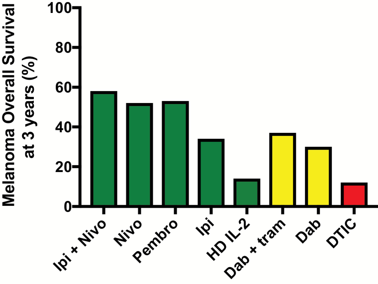
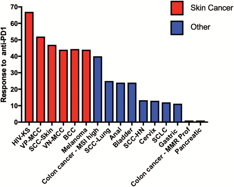
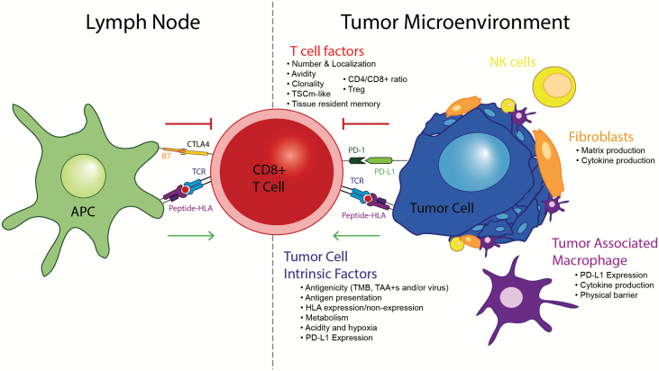
Among all tumor types, skin cancers are profoundly sensitive to immunotherapy. Indeed, the recently reported response rates for anti-PD-1 (anti-programmed-death 1) therapy for cutaneous malignant melanomas (MM), Merkel cell carcinomas, basal cell carcinomas, cutaneous squamous cell carcinomas and Kaposi sarcomas are all above 40%. This unique immunogenicity renders skin cancers as a paradigm for tumor-immune interactions and is driven by high mutational burdens, over-expressed tumor antigens and/or viral antigens. However, despite the clear demonstration of immunologic cure of skin cancer in some patients, most tumors develop either early (primary) or late (adaptive) resistance to immunotherapy. Resistance mechanisms are complex, and include contributions of tumor cell-intrinsic, T cell and microenvironment factors that have been recently further elucidated with the advent of single-cell technologies. This review will focus on the exciting progress with immunotherapy for skin cancers to date, and also our current understanding of the mechanisms of resistance to immunotherapy.

Keywords: Merkel cell; PD-1; cutaneous malignancy; immune checkpoint; melanoma.


Figures

Fig. 1.
Fig. 1.
Overall survival from selected first-line therapies for metastatic melanoma. The graph shows 3-year overall survival rates for selected immunotherapies, targeted therapies and chemotherapies (5, 6, 44, 62, 127, 128). As there are differences between the selected studies in terms of entry criteria, as well as change over time in availability of second-line therapies, this graph is intended to illustrate the marked improvement in overall survival with recently FDA-approved immunotherapies and not intended for treatment decision-making. Ipi, ipilimumab; Nivo, nivolumab; Pembro, pembrolizumab; HD IL-2, high-dose IL-2; Dab, dabrafenib; tram, trametinib; DTIC = dacarbazine. Green indicates immune therapy; yellow indicates targeted therapy; red indicates chemotherapy.
Fig. 2.
Fig. 2.
Response to anti-PD-1 blockade in advanced skin cancers and other solid tumors. Data from representative trials of anti-PD1 agents in skin cancers (6, 20, 27, 80, 82) and non-skin cancers are shown (17, 32, 129–133); for selected gastrointestinal malignancies [DNA mismatch repair (MMR)-proficient colon cancer, pancreatic cancer, gastric cancer] summary data are as reported from Yarchoan et al. (17). Where possible data are from first-line therapy; however, for some cancers only 2L+ data are available. Data include all comers irrespective of PD-L1 tumor status. Only anti-PD-1 monotherapy is included. Reported response rates to anti-PD-1 monotherapy in skin cancers are consistently >30–40% and are substantially higher than reported response rates for most other solid tumors. SCC-HN, squamous cell carcinoma of the head and neck; SCLC, small cell lung cancer.
Fig. 3.
Fig. 3.
Development of vitiligo-like hypopigmentation in a patient being treated with immunotherapy. Top panel: standard photograph. Bottom photograph: Wood’s lamp. Vitiligo/hypopigmentation arose in a patient on immune checkpoint inhibitor immunotherapy.
Fig. 4.
Fig. 4.
Contributors to immunotherapy response/resistance. A number of factors contribute to immunotherapy response/resistance, which broadly fall into three major categories: tumor cell-intrinsic, T cell and microenvironment. Please see text for additional details. TAA+s indicate positivity for tumor-associated antigens.
Fig. 5.
Fig. 5.
Patterns of T-cell recognition of skin tumors. Three major patterns of T-cell recognition are observed across tumor types including melanomas and MCC: T-cell desert, T-cell exclusion and T-cell infiltration (134, 135). These patterns partially predict anti-PD-1 immunotherapy responsiveness (104). Top row: schematic indicating the localization pattern of T cells (red) compared with tumor (blue). Bottom row: three VP-MCC tumors (blue) exhibiting each of the three described patterns; in the bottom row, blue staining indicates CD56 (marks MCC) and red staining indicates anti-CD3 (marks T cells).

References

    1. Siegel R. L., Miller K. D. and Jemal A. 2018. Cancer statistics, 2018. CA Cancer J. Clin. 68:7. - PubMed
    1. Paulson K. G., Park S. Y., Vandeven N. A. et al. . 2018. Merkel cell carcinoma: current US incidence and projected increases based on changing demographics. J. Am. Acad. Dermatol. 78:457. - PMC - PubMed
    1. Palyca P., Koshenkov V. P., and Mehnert J. M. 2014. Developments in the treatment of locally advanced and metastatic squamous cell carcinoma of the skin: a rising unmet need. Am. Soc. Clin. Oncol. Educ. Book 34:e397. - PMC - PubMed
    1. Rogers H. W., Weinstock M. A., Feldman S. R. and Coldiron B. M. 2015. Incidence estimate of nonmelanoma skin cancer (keratinocyte carcinomas) in the U.S. population, 2012. JAMA Dermatol. 151:1081. - PubMed
    1. Robert C., Thomas L., Bondarenko I. et al. . 2011. Ipilimumab plus dacarbazine for previously untreated metastatic melanoma. N. Engl. J. Med. 364:2517. - PubMed

Publication types

Substances