

Comparing vasopressin and oxytocin fiber and receptor density patterns in the social behavior neural network: Implications for cross-system signaling
- PMID: 30753840
- PMCID: PMC7469073
- DOI: 10.1016/j.yfrne.2019.02.001
Comparing vasopressin and oxytocin fiber and receptor density patterns in the social behavior neural network: Implications for cross-system signaling
Abstract
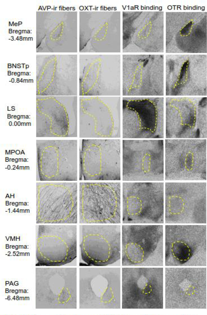
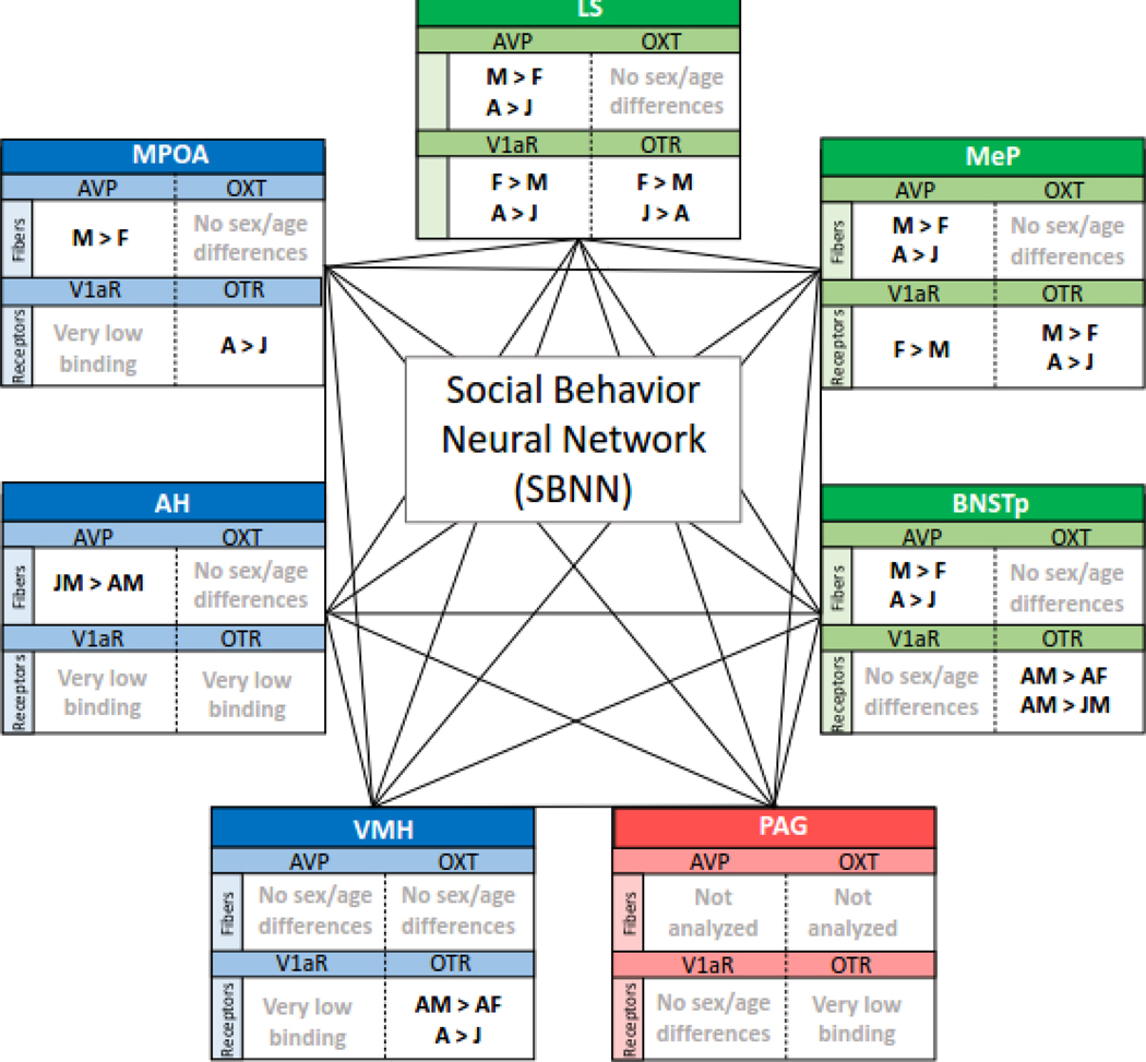
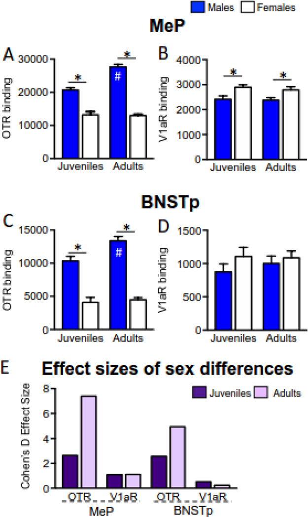
Vasopressin (AVP) and oxytocin (OXT) regulate social behavior by binding to their canonical receptors, the vasopressin V1a receptor (V1aR) and oxytocin receptor (OTR), respectively. Recent studies suggest that these neuropeptides may also signal via each other's receptors. The extent to which such cross-system signaling occurs likely depends on anatomical overlap between AVP/OXT fibers and V1aR/OTR expression. By comparing AVP/OXT fiber densities with V1aR/OTR binding densities throughout the rat social behavior neural network (SBNN), we propose the potential for cross-system signaling in four regions: the medial amygdala (MeA), bed nucleus of the stria terminalis (BNSTp), medial preoptic area, and periaqueductal grey. We also discuss possible implications of corresponding sex (higher in males versus females) and age (higher in adults versus juveniles) differences in AVP fiber and OTR binding densities in the MeA and BNSTp. Overall, this review reveals the need to unravel the consequences of potential cross-system signaling between AVP and OXT systems in the SBNN for the regulation of social behavior.
Keywords: Bed nucleus of the stria terminalis; Cross-talk; Medial amygdala; Medial preoptic area; Oxytocin; Oxytocin receptor; Periaqueductal grey; Social behavior; V1a receptor; Vasopressin.
Copyright © 2019 Elsevier Inc. All rights reserved.
Figures
Similar articles
-
Age and sex differences in oxytocin and vasopressin V1a receptor binding densities in the rat brain: focus on the social decision-making network.Brain Struct Funct. 2017 Mar;222(2):981-1006. doi: 10.1007/s00429-016-1260-7. Epub 2016 Jul 7. Brain Struct Funct. 2017. PMID: 27389643 Free PMC article.
-
Quantitative mapping reveals age and sex differences in vasopressin, but not oxytocin, immunoreactivity in the rat social behavior neural network.J Comp Neurol. 2017 Aug 1;525(11):2549-2570. doi: 10.1002/cne.24216. Epub 2017 May 8. J Comp Neurol. 2017. PMID: 28340511 Free PMC article.
-
Social status in mouse social hierarchies is associated with variation in oxytocin and vasopressin 1a receptor densities.Horm Behav. 2019 Aug;114:104551. doi: 10.1016/j.yhbeh.2019.06.015. Epub 2019 Aug 13. Horm Behav. 2019. PMID: 31279703
-
Cross-talk among oxytocin and arginine-vasopressin receptors: Relevance for basic and clinical studies of the brain and periphery.Front Neuroendocrinol. 2018 Oct;51:14-24. doi: 10.1016/j.yfrne.2017.10.004. Epub 2017 Oct 18. Front Neuroendocrinol. 2018. PMID: 29054552 Free PMC article. Review.
-
Vasopressin and oxytocin receptor systems in the brain: Sex differences and sex-specific regulation of social behavior.Front Neuroendocrinol. 2016 Jan;40:1-23. doi: 10.1016/j.yfrne.2015.04.003. Epub 2015 May 4. Front Neuroendocrinol. 2016. PMID: 25951955 Free PMC article. Review.
Cited by
-
Beyond the binary: Characterizing the relationships between sex and neuropeptide receptor binding density measures in the rat brain.Horm Behav. 2024 Mar;159:105471. doi: 10.1016/j.yhbeh.2023.105471. Epub 2023 Dec 21. Horm Behav. 2024. PMID: 38128247 Free PMC article.
-
Emerging roles for microglia and microbiota in the development of social circuits.Brain Behav Immun Health. 2021 Jul 15;16:100296. doi: 10.1016/j.bbih.2021.100296. eCollection 2021 Oct. Brain Behav Immun Health. 2021. PMID: 34589789 Free PMC article.
-
Oxytocin and vasopressin: Signalling, behavioural modulation and potential therapeutic effects.Br J Pharmacol. 2022 Apr;179(8):1544-1564. doi: 10.1111/bph.15481. Epub 2021 May 21. Br J Pharmacol. 2022. PMID: 33817785 Free PMC article. Review.
-
Hypothalamic Vasopressin Neurons Enable Maternal Thermoregulatory Behaviors.bioRxiv [Preprint]. 2025 Jan 23:2025.01.23.634569. doi: 10.1101/2025.01.23.634569. bioRxiv. 2025. PMID: 40196592 Free PMC article. Preprint.
-
The psychedelic-peptide paradox: a hormetic hypothesis.Compr Psychoneuroendocrinol. 2025 Jun 2;23:100303. doi: 10.1016/j.cpnec.2025.100303. eCollection 2025 Aug. Compr Psychoneuroendocrinol. 2025. PMID: 40584160 Free PMC article. Review.
References
-
- Albers HE, Rowland CM, Ferris CF (1991) Arginine-vasopressin immunoreactivity is not altered by photoperiod or gonadal hormones in the Syrian hamster (Mesocricetus auratus). Brain Research. 539(1): 137–42. - PubMed
-
- Arakawa H, Arakawa K, Deak T (2010) Oxytocin and vasopressin in the medial amygdala differentially modulate approach and avoidance behavior toward illness-related social odor, Neuroscience. 171(4):1141–51. - PubMed
Publication types
MeSH terms
Substances
Grants and funding
LinkOut - more resources
Full Text Sources
Miscellaneous
