. 2019 Apr 1:88:251-265.
doi: 10.1016/j.actbio.2019.02.007. Epub 2019 Feb 10.

Hydrophobic and antimicrobial dentin: A peptide-based 2-tier protective system for dental resin composite restorations

Affiliations

Hydrophobic and antimicrobial dentin: A peptide-based 2-tier protective system for dental resin composite restorations

Dina G Moussa et al. Acta Biomater. .

Abstract

Dental caries, i.e., tooth decay mediated by bacterial activity, is the most widespread chronic disease worldwide. Carious lesions are commonly treated using dental resin composite restorations. However, resin composite restorations are prone to recurrent caries, i.e., reinfection of the surrounding dental hard tissues. Recurrent caries is mainly a consequence of waterborne and/or biofilm-mediated degradation of the tooth-restoration interface through hydrolytic, acidic and/or enzymatic challenges. Here we use amphipathic antimicrobial peptides to directly coat dentin to provide resin composite restorations with a 2-tier protective system, simultaneously exploiting the physicochemical and biological properties of these peptides. Our peptide coatings modulate dentin's hydrophobicity, impermeabilize it, and are active against multispecies biofilms derived from caries-active individuals. Therefore, the coatings hinder water penetration along the otherwise vulnerable dentin/restoration interface, even after in vitro aging, and increase its resistance against degradation by water, acids, and saliva. Moreover, they do not weaken the resin composite restorations mechanically. The peptide-coated highly-hydrophobic dentin is expected to notably improve the service life of resin composite restorations and to enable the development of entirely hydrophobic restorative systems. The peptide coatings were also antimicrobial and thus, they provide a second tier of protection preventing re-infection of tissues in contact with restorations. STATEMENT OF SIGNIFICANCE: We present a technology using designer peptides to treat the most prevalent chronic disease worldwide; dental caries. Specifically, we used antimicrobial amphipathic peptides to coat dentin with the goal of increasing the service life of the restorative materials used to treat dental caries, which is nowadays 5 years on average. Water and waterborne agents (enzymes, acids) degrade restorative materials and enable re-infection at the dentin/restoration interface. Our peptide coatings will hinder degradation of the restoration as they produced highly hydrophobic and antimicrobial dentin/material interfaces. We anticipate a high technological and economic impact of our technology as it can notably reduce the lifelong dental bill of patients worldwide. Our findings can enable the development of restorations with all-hydrophobic and so, more protective components.

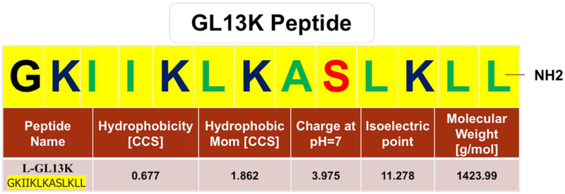
Keywords: Antimicrobial peptide; Dental restoration; Dentin; GL13K; Hydrophobic coating; Recurrent caries.


Conflict of interest statement

Competing financial interests

The authors confirm that there are no known conflicts of interest associated with this publication and there has been no significant financial support for this work that could have influenced its outcome

Figures

Figure 1.
Figure 1.. Amphipathic antimicrobial peptide, GL13K.
Amino-acid sequence of the GL13K peptide showing hydrophobic (green) and hydrophilic (red) residues, including positively charged amino acids (dark blue). Reported physical and chemical properties of GL13K peptide were obtained from the BaAMPs-Biofilm-active AMPs database (http://www.baamps.it/browse?task=peptide.display&ID=106, last access September 04, 2018).
Figure 2.
Figure 2.. Peptide coatings on dentin.
Schematics of the different procedures for dentin conditioning/coating with amphipathic peptides. See text for detailed description of sample preparation. AAMP: amphipathic antimicrobial peptide; EtOH: absolute ethanol solution.
Figure 3.
Figure 3.. Method of preparation of restored dentin discs.
The restored discs were used for assessing fracture resistance using the diametral compression test or permeability at the d/r interface using micro-computed tomography (micro-CT, μCT). See text for the detailed description of this method of sample preparation.
Figure 4.
Figure 4.. Hydrophobic dentin and its impermeability before and after chemical, mechanical and saliva-mediated challenges.
A) Dynamic water contact angles (WCA, average value, n=7) for cut and etched bovine dentin (controls) and dentin treated with amphipathic peptides following different protocols (see materials and methods section for description of the different dentin treatments); B) sessile water drop images and final WCA (average ± standard deviation, n=7) on bovine dentin before (cut and etched) and after peptide treatments; C) dentin impermeability: copper sulfate acidic dye penetration at the superficial and pulp overlaying dentin visualized using transmitted or reflected light illumination; D) sessile water drop images and final WCA (average ± standard deviation, n=5) on buffer treated and peptide coated bovine dentin, before and after ultrasonication and saliva challenges, E) dentin impermeability after saliva-mediated challenges: copper sulfate dye penetration through dentin visualized using transmitted or reflected light illumination.
Figure 5.
Figure 5.. Micro-CT analysis of impermeability at the dentin-composite interface.
(A) Top view and (B) side view of representative restored bovine dentin discs for each tested group showing silver nitrate (AgNO3) dye leakage (converted to yellow for easier visualization); (C) one representative stack of four restored dentin discs for each tested group showing the 3D rendering of the dye that leaked through dentin/composite interfaces and reconstruction of the micro-CT scans for one representative CWBA whole stack (last column). Quantification of the penetrated AgNO3 volume along dentin-composite interface (n=7) before (D) and (E) after aging by water storage and thermal cycling. C: (restored roots using composite with bonding agent; C13: restored roots using composite with bonding agent on GL13K-coated dentin; C13WBA: restored roots using composite without bonding agent on GL13K-coated dentin; CWBA: restored roots using composite without bonding agent. Ends of horizontal bars connect groups with statistically significant different silver nitrate penetration volume (*, p-value < 0.05). Circles denote moderate outliers.
Figure 6.
Figure 6.. Fracture resistance of restored dentin discs.
A) Diametral compression test setting; B) boxplots of fracture loads of the restored dentin discs prepared with different treatments (n=22). C: restored roots using composite with bonding agent; C13: restored roots using composite with bonding agent on GL13K-coated dentin; C13WBA: restored roots using composite without bonding agent on GL13K-coated dentin; CWBA: restored roots using composite without bonding agent. Ends of horizontal bars connect groups with statistically significant different fracture loads (*, p-value < 0.05). Circles denote moderate outliers.
Figure 7.
Figure 7.. Antibiofilm potency of D-GL13K against multispecies biofilm derived from cariogenic dental plaque.
A) Stereomicroscope image of 6-day-old plaque biofilm covering the microtiter plate and stained with crystal violet dye without (bottom) or with (top) 25 mg/ml peptide treatment. B) Quantification of biofilm biovolume shown in A), C) Qualitative and quantitative assessments of the remaining bioburden on HA discs with (right) and without (left) D-GL13K coating, D) Merged live (green) and dead (red) viability assay images of 48-hour multispecies biofilm grown on HA discs with (bottom image) and without (top image) D-GL13K coating. Merged images showing bacteria stained with both SYTO-9 (live bacteria, green) and PI (dead bacteria, red). * denotes statistically significant differences (p-value < 0.05) between groups connected by horizontal bars.

References

    1. World Health Organization, Oral Health Fact Sheet (2012), 2012. http://www.who.int/mediacentre/factsheets/fs318/en/.
    1. Wold Health Organization, Sugars and dental caries, 2017. http://www.who.int/oral_health/publications/sugars-dental-caries-keyfact....
    1. World Health Organization, Dental Diseases and Oral Health, 2003. http://www.who.int/oral_health/publications/en/orh_fact_sheet.pdf.
    1. Benjamin RM., Oral Health: The Silent Epidemic, Public Health Rep 125(2) (2010) 158–159. - PMC - PubMed
    1. Burke FJ, Wilson NH, Cheung SW, Mjor IA, Influence of patient factors on age of restorations at failure and reasons for their placement and replacement, J Dent 29(5) (2001) 317–24. - PubMed

Publication types