Review
. 2019 Sep;34(5):954-965.
doi: 10.3904/kjim.2018.325. Epub 2019 Feb 15.

Cellular immunotherapy in multiple myeloma

Affiliations
Review

Cellular immunotherapy in multiple myeloma

Manh-Cuong Vo et al. Korean J Intern Med. 2019 Sep.

Abstract

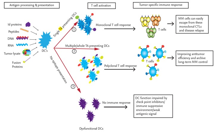
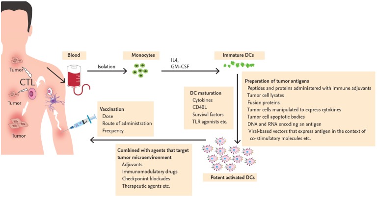
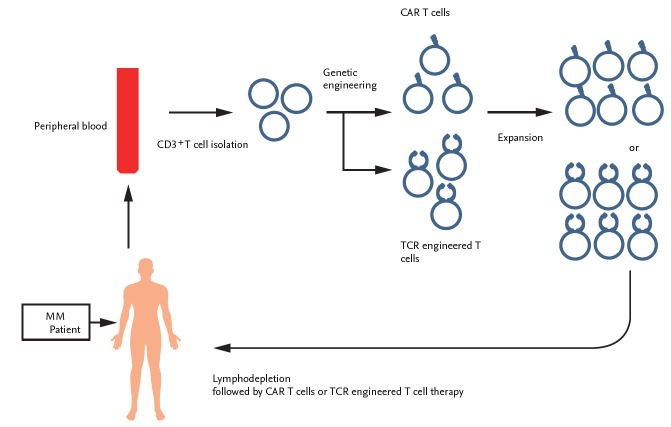
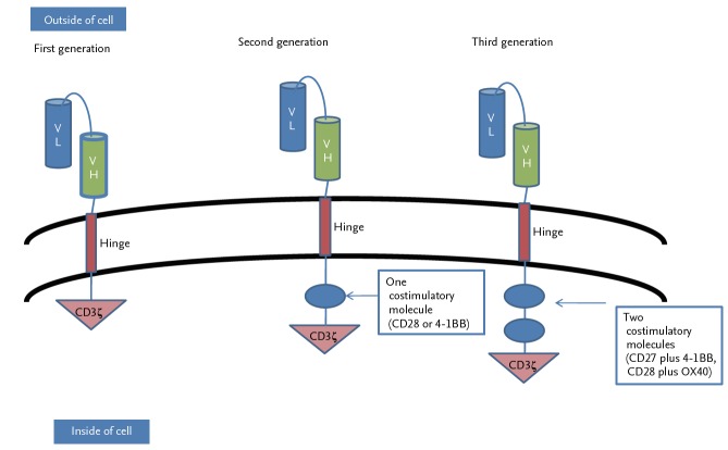
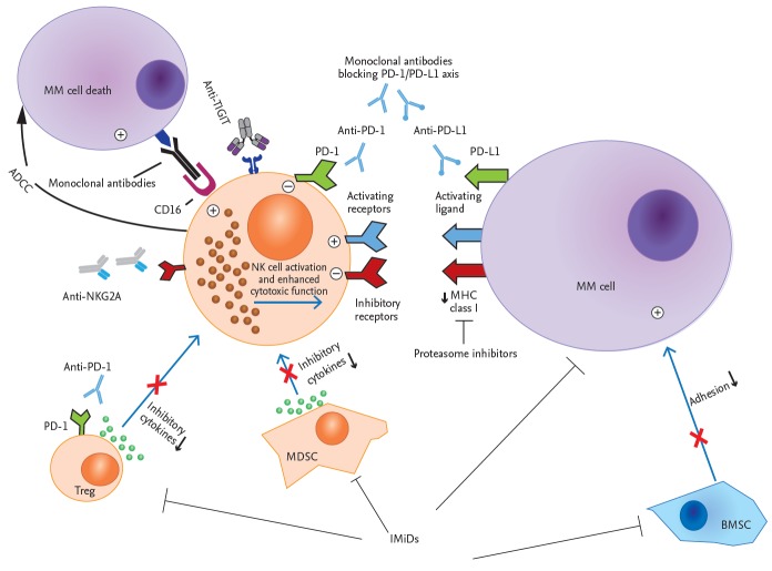
In multiple myeloma (MM), the impaired function of several types of immune cells favors the tumor's escape from immune surveillance and, therefore, its growth and survival. Tremendous improvements have been made in the treatment of MM over the past decade but cellular immunotherapy using dendritic cells, natural killer cells, and genetically engineered T-cells represent a new therapeutic era. The application of these treatments is growing rapidly, based on their capacity to eradicate MM. In this review, we summarize recent progress in cellular immunotherapy for MM and its future prospects.

Keywords: Cellular immunotherapy; Dendritic cells; Engineered effector T cell; Immunomodulatory drug; Killer cells, natural; Multiple myeloma.


Conflict of interest statement

No potential conflict of interest relevant to this article was reported.

Figures

Figure 1.
Figure 1.
Immune response induced by different modes of tumor antigen-pulsed dendritic cells (DCs). The source of the antigen significantly influences DC function, as evidenced by the activation of tumor-specific T-cells. When DCs are pulsed with only a single tumor antigen, in the form of a single peptide, a DNA or RNA fragment encoding a single antigen, or a single idiotype (Id) protein, they induce the expansion of active monoclonal cytotoxic T lymphocytes (CTLs) that may exert cytotoxic effects on multiple myeloma (MM) cells. However, MM cells can elicit immunosuppressive and inhibitory signals or express different tumor antigens, which lead to the escape of the tumor from these monoclonal CTLs. To overcome these limitations, DCs pulsed with whole tumor lysates, multi-peptides, a cocktail of tumor-associated antigens (TAAs), fusion proteins, or whole tumor-derived DNA or RNA are used to induce the activation of multiple tumor-antigen-specific polyclonal CTLs covering almost all tumor-specific antigen targets. The goal is improved antitumor efficiency and long-term MM control. In some cases, DCs are not fully mature or are not fully activated by tumor antigens and are thus unable to induce an immune response. TA, tumor antigen.
Figure 2.
Figure 2.
Process of clinical dendritic cell (DC) vaccination in patients with multiple myeloma. The DCs of patients with multiple myeloma (MM) are functionally impaired because of the tumor microenvironment. DCs cultured ex vivo and used to vaccinate MM patients can overcome the immune dysregulation. Monocytes obtained from patients with MM are differentiated into immature DCs during their in vitro culture with interleukin 4 (IL-4) and granulocyte-macrophage colony-stimulating factor (GM-CSF). Immature DCs are then maturated with various stimuli (cytokines, cluster of differentiation 40 ligand [CD40L], survival factors or toll-like receptor [TLR] agonist) and loaded with various tumor-associated antigens using techniques such as the administration of peptides and proteins with immune adjuvants, tumor cell lysates, fusion protein, tumor cells manipulated to express cytokines, tumor cell apoptotic bodies, DNA and RNA encoding an antigen, or viral-based vectors to express antigen in the context of co-stimulatory molecules. Multiple modalities with adjuvants, immunomodulatory drugs, checkpoint blockades, and other therapeutic agents are necessary to enhance the efficacy of DC vaccination and, thus, suppress the tumor microenvironment. Numerous variables, such as dose, frequency, and route of DC vaccination also need to be optimized to induce an MM specific immune response effectively in both primary and secondary lymphoid organs. CTL, cytotoxic T lymphocyte.
Figure 3.
Figure 3.
Scheme of genetically engineered T-cell therapy in patients with multiple myeloma (MM). T-cells were isolated from the peripheral blood of patients with MM via apheresis and then transfected with the genes containing chimeric antigen receptor (CAR)-based tumor antigen by lentiviral, gammaretroviral or transposon/transposase approaches. Adoptive transfer of in vitro generated autologous CAR T-cells was conducted in patients with or without prior lymphodepletion. TCR, T-cell receptor.
Figure 4.
Figure 4.
The generations of chimeric antigen receptor T-cells. Chimeric antigen receptors (CARs) target tumor antigen independently of major histocompatibility complex I (MHC-I). They consist of an ectodomain, a hinge domain, a transmembrane domain, and an endodomain. First-generation CARs consisted of single chain variable fragment (scFv) (light chain variable region [VL] and heavy chain variable region [VH]) and cluster of differentiation 3ζ (CD3ζ) alone. Second-generation CARs were generated to mediate T-cell activation by the immunoreceptor tyrosine-based activation motif (ITAM) of the CD3ζ chain with a single costimulatory molecule, either CD28 or 4-1BB. Improved third-generation CARs were generated by combining the ITAM of CD3ζ chain with two costimulatory molecules, such as CD27 plus 4-1BB or CD28 plus OX40.
Figure 5.
Figure 5.
Recent therapeutic approaches to enhance natural killer (NK) cytotoxicity against multiple myeloma (MM). This schematic shows how various therapeutic agents modulate NK cell-mediated cytotoxicity to target myeloma cells. Suppressive immune cells as well as bone marrow stromal cells (BMSCs) inside the tumor microenvironment are negative regulators of NK cell activation. The MM cells themselves develop several strategies to evade NK-cell-mediated killing. New immune modulators, including immunomodulatory drugs (iMiDs), immune checkpoint inhibitors, such as anti-programmed cell death 1 (PD-1)/PD-L1, anti-NKG2A, anti-TIGIT blocking antibodies, and proteasome inhibitors, target and suppress immunosuppressive factors in the MM microenvironment and enhance the cytotoxic effect of NK cells to kill MM cells. In addition, the antibodies used to treat MM, such as elotuzumab and daratumumab, induce NK cell-mediated antibody-dependent cellular cytotoxicity (ADCC). TIGIT, T cell immunoreceptor with Ig and ITIM domains; CD16, cluster of differentiation 16; NKG2A, natural killer G2A; MDSC, myeloid-derived suppressor cell; MHC, major histocompatibility complex.

Similar articles

Cited by

References

    1. Sirohi B, Powles R. Multiple myeloma. Lancet. 2004;363:875–887. - PubMed
    1. Kyle RA, Rajkumar SV. Multiple myeloma. N Engl J Med. 2004;351:1860–1873. - PubMed
    1. Hong J, Lee JH. Recent advances in multiple myeloma: a Korean perspective. Korean J Intern Med. 2016;31:820–834. - PMC - PubMed
    1. Lonial S, Cavenagh J. Emerging combination treatment strategies containing novel agents in newly diagnosed multiple myeloma. Br J Haematol. 2009;145:681–708. - PubMed
    1. Attal M, Harousseau JL. The role of high-dose therapy with autologous stem cell support in the era of novel agents. Semin Hematol. 2009;46:127–132. - PubMed

MeSH terms

Substances