. 2019 Apr 17;10(4):2070-2079.
doi: 10.1021/acschemneuro.9b00033. Epub 2019 Feb 20.

Direct, Real-Time Detection of Adenosine Triphosphate Release from Astrocytes in Three-Dimensional Culture Using an Integrated Electrochemical Aptamer-Based Sensor

Affiliations

Direct, Real-Time Detection of Adenosine Triphosphate Release from Astrocytes in Three-Dimensional Culture Using an Integrated Electrochemical Aptamer-Based Sensor

Mirelis Santos-Cancel et al. ACS Chem Neurosci. .

Abstract

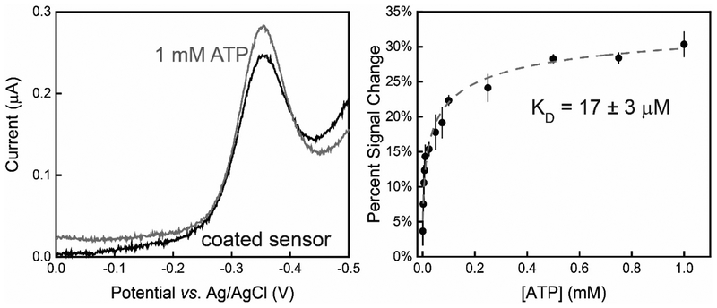
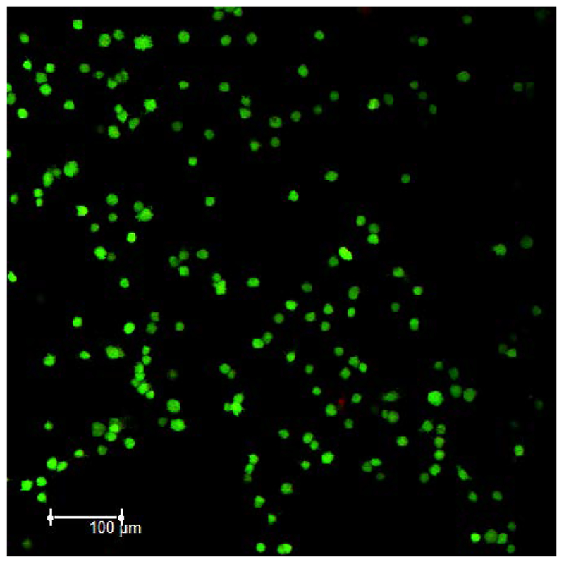
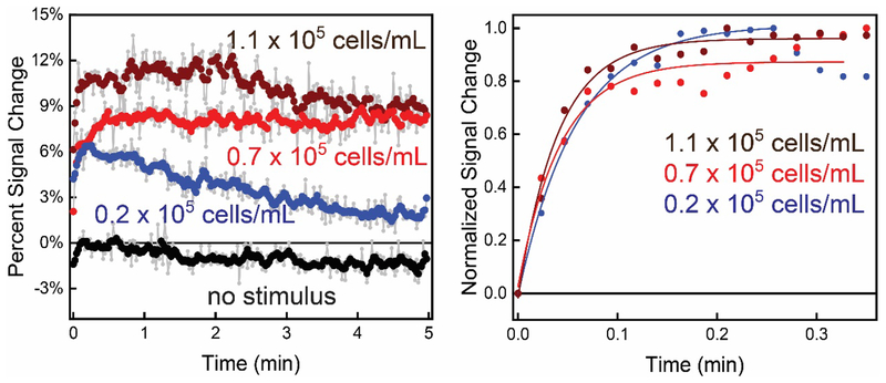
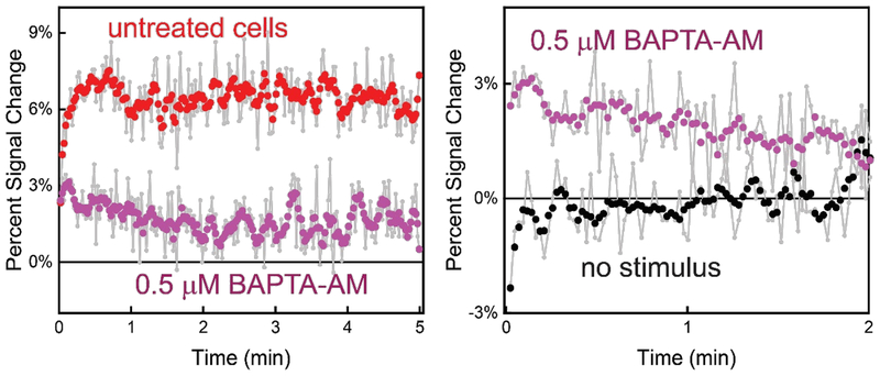
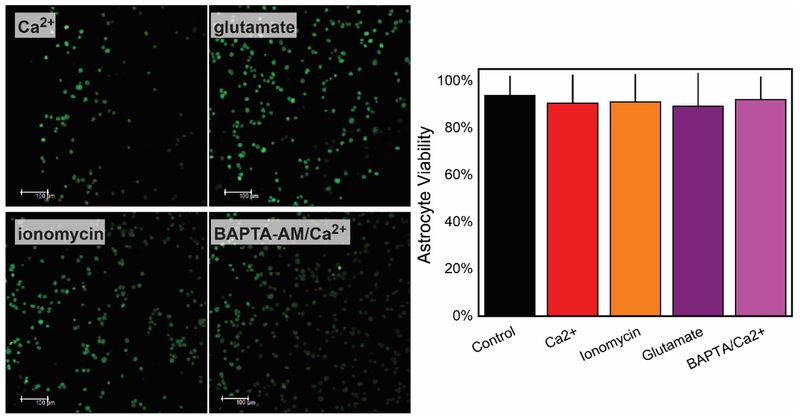
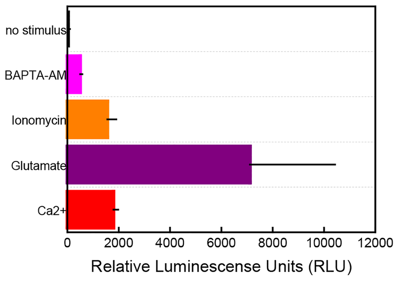
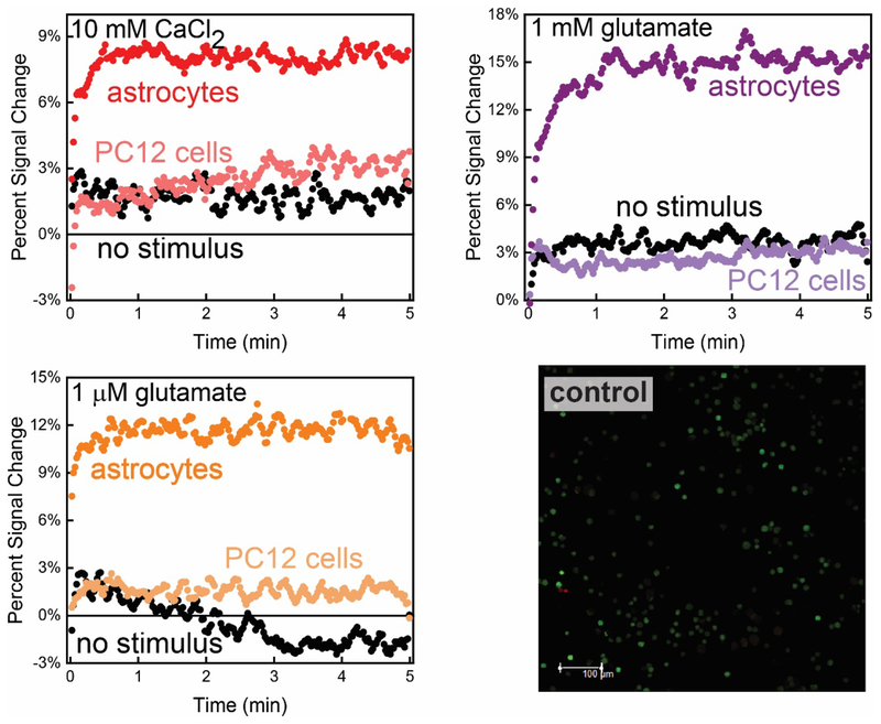
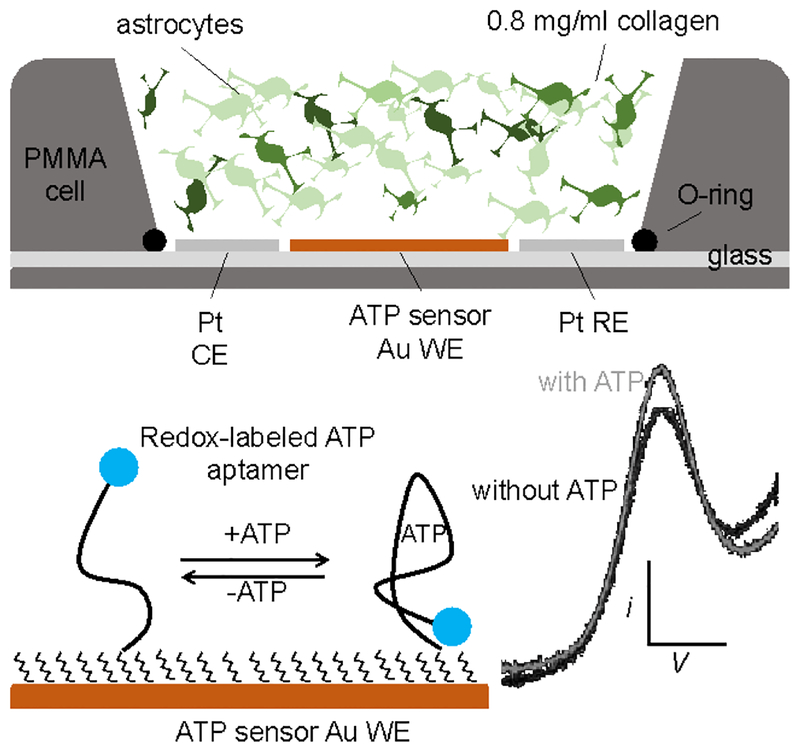
In this manuscript, we describe the development and application of electrochemical aptamer-based (E-AB) sensors directly interfaced with astrocytes in three-dimensional (3D) cell culture to monitor stimulated release of adenosine triphosphate (ATP). The aptamer-based sensor couples specific detection of ATP, selective performance directly in cell culture media, and seconds time resolution using squarewave voltammetry for quantitative ATP-release measurements. More specifically, we demonstrate the ability to quantitatively monitor ATP release into the extracellular environment after stimulation by the addition of calcium (Ca2+), ionomycin, and glutamate. The sensor response is confirmed to be specific to ATP and requires the presence of astrocytes in culture. For example, PC12 cells do not elicit a sensor response after stimulation with the same stimulants. In addition, we confirmed cell viability in the collagen matrix for all conditions tested. Our hydrogel-sensor interface offers the potential to study the release of small molecule messengers in 3D environments. Given the generality of electrochemical aptamer-based sensors and the demonstrated successful interfacing of sensors with tissue scaffold material, in the long term, we anticipate our sensors will be able to translate from in vitro to in vivo small molecule recordings.

Keywords: 3D cell culture; ATP; aptamer-based sensor; astrocytes; electrochemistry; gliotransmission.


Figures

Figure 1.
Figure 1.
Collagen-coated, structure-switching E-AB sensors specific to ATP exhibited a quantitative increase in faradaic current in the presence of ATP directly in cell culture media (84% DMEM-high glucose, 15% FBS, and 1% 10,000 U/mL Pen/Strep). (Left) Squarewave voltammograms exhibit an increase in peak current of ~30% after the addition of 1 mM ATP. (Right) The collagen-coated sensor exhibited an observed binding affinity (KD) = 17 ± 3 μM to ATP when the calibration curve was fit to a Langmuir-like isotherm. Data showed and error bars represent the mean and standard deviation of three independently fabricated E-AB sensors using the same experimental conditions.
Figure 2.
Figure 2.
Fluorescence images demonstrates 94% astrocyte viability (green fluorescent calcein AM dye) of an 8 × 10⁵ astrocyte cell/mL cell density encapsulated in a 0.8 mg/mL collagen I hydrogel when polymerized at RT for 30 mins and equilibrated in warm culture media for 1 hr.
Figure 3.
Figure 3.
The interface between E-AB sensors and 3D cell culture allows rapid (s) quantification of purinergic activity from astrocyte cells. (Left) More specifically, plotting the percent signal change in voltammetric peak current, we observe an increase in sensor signal upon Ca2+-evoked ATP release (at t=0) that is quantitatively related to the astrocyte population in the collagen hydrogel (1.1, 0.7, and 0.2 × 10⁵ cells/mL (wine, red, and blue, respectively)). The increase in signal is contrasted against a control signal when no Ca2+ is added. All raw data (light grey) was weight averaged to n=5 for ease of viewing. (Right) Normalized signal change of the E-AB sensor to ATP shows an exponential and rapid increase after the addition of stimulus.
Figure 4.
Figure 4.
Treatment with the ([Ca2+]i) chelator BAPTA-AM significantly reduces extracellular ATP after stimulation with Ca2+ (at t=0) as indicated by a lower sensor signal change. (Left) For example, an untreated cell population density of 0.7 × 10⁵ cells/mL exhibits a signal increase of ~ 7% with the addition of Ca2+ (untreated). Conversely, cells treated with BAPTA-AM only exhibited a ~3% signal increase indicating a blocking of ATP release from astrocytes (treated). (Right) Treated cells, however, still released some ATP after evocation with Ca2+. Sensor signal increased ~3% (Ca2+-triggered) compared signal without the addition of Ca2+.
Figure 5.
Figure 5.
Glutamate and ionomycin also stimulate ATP release from astrocytes. For example, the signal increase observed for glutamate, and ionomycin stimulation (t=0) are ~14% and ~12%, respectively. This change in signal is contrasted to the lack of signal change observed without the addition of stimulus (as also shown in Figure 3). (Right) The incorporation of ionomycin to cell culture media results in a faster rise time in extracellular ATP compared to glutamate and Ca2+. All raw data (light grey) were adjacently averaged to n=5. Individual experiments were carried out stimulating 0.7 × 10⁵ astrocyte cells/mL on top of individual E-AB sensors.
Figure 6.
Figure 6.
Viability for an 8 × 10⁵ cell/mL cell density encapsulated in a 0.8 mg/mL collagen hydrogel remains minimally disturbed after chemical studies performed after the stimulation with 5 mM CaCl2, 1 mM glutamate, 1 μM ionomycin. Incorporation of BAPTA-AM did not cause detrimental effects, and cell remained viable after the incorporation of Ca2+ (Left). All images and viability test were acquired after the incubation (> 1 min) with all triggers. Green and red fluorescent (viable and dead, respectively) images when acquired with a p-value of 0.945 indicate there is no significant difference between the cell viabilities with stimulus and the control.
Figure 7.
Figure 7.
Luciferin/luciferase bioluminescence assay confirms the presence of ATP in a 0.8 mg/mL collagen hydrogel after stimuli addition. Similar to the electrochemical recordings, the incorporation of glutamate induces the most substantial ATP response when compared with other stimuli (purple bar). The cells respond similarly when Ca2+ and ionomycin are added (red and orange bars, respectively). Treatment with the intracellular chelator, BAPTA-AM significantly reduces ATP signal when incubated for 1.5 hr to the collagen-containing astrocytes (pink bar) and stimulated with Ca2+. The negative demonstrates that no ATP is released when no stimulus is added to a 0.7 X10⁵ cell/mL density (black bar). All luminescence results were acquired and averaged n=3.
Figure 8.
Figure 8.
The ATP E-AB sensor exhibited no appreciable response to catecholamine release from PC12 cells cultured in the sensor 3D cell culture array. (Top left) Stimulation with Ca2+ shows baseline sensor response (light red), comparable to electrochemical recordings when no stimulus is added to the cell culture media (black line trace). (Top right and bottom leftSimilarly, the sensor did not respond to catecholamine release upon the addition of glutamate and ionomycin (traces). comparison of the signal change generated by astrocytic Ca2+, glutamate and ionomycin-triggered ATP release (dark red, purple and orange time traces, respectively) confirms the specificity of the E-AB sensor to extracellular ATP presence after same stimuli were exerted in astrocytes cultured in same conditions. These figures encompass adjacent averaged (n=5) raw data for the E-AB sensor response. (Bottom right) Control viability studies helped to visualize the stability of PC12 cells under current experimental conditions. Fluorescent images were acquired with a p-value of 0.183 indicating there is no significant difference between the cell viabilities with stimulus and the control.
Scheme 1.
Scheme 1.
Crossectional representation of the E-AB sensor, 3D astrocyte cell culture hybrid interface. The three-electrode setup is placed inside a commercial holder (Micrux Technologies, Oviedo, Spain) with a PMMA holder on top where the 3D cell culture was polymerized, equilibrated, stimulated, and tested. Structure-switching E-AB sensors fabricated on the 1 mm working electrode allowed continuous quantification of ATP release from astrocytes.

Similar articles

Cited by

References

    1. Conditions P; Fiacco TA; Mccarthy KD; Savtchouk XI; Volterra XA Gliotransmission: Beyond Black-and-White. J. Neurosci 2018, 38, 14–25. - PMC - PubMed
    1. Fiacco XTA; Mccarthy KD Multiple Lines of Evidence Indicate That Gliotransmission Does Not Occur under Physiological Conditions. J. Neurosci 2018, 38 (1), 3–13. - PMC - PubMed
    1. Verderio C; Matteoli M ATP in Neuron-Glia Bidirectional Signalling. Brain Res. Rev 2011, 66, 106–114. - PubMed
    1. Franke H; Verkhratsky A; Burnstock G; Illes P Pathophysiology of Astroglial Purinergic Signalling. Purinergic Signal 2012, 8, 629–657. - PMC - PubMed
    1. Bodin P; Burnstock G Purinergic Signalling: ATP Release. Neurochem. Res 2001, 26, 959–969. - PubMed

Publication types

Substances