

Enterovirus Persistence in Cardiac Cells of Patients With Idiopathic Dilated Cardiomyopathy Is Linked to 5' Terminal Genomic RNA-Deleted Viral Populations With Viral-Encoded Proteinase Activities
- PMID: 30755025
- PMCID: PMC6517084
- DOI: 10.1161/CIRCULATIONAHA.118.035966
Enterovirus Persistence in Cardiac Cells of Patients With Idiopathic Dilated Cardiomyopathy Is Linked to 5' Terminal Genomic RNA-Deleted Viral Populations With Viral-Encoded Proteinase Activities
Abstract
Background: Group B enteroviruses are common causes of acute myocarditis, which can be a precursor of chronic myocarditis and dilated cardiomyopathy, leading causes of heart transplantation. To date, the specific viral functions involved in the development of dilated cardiomyopathy remain unclear.
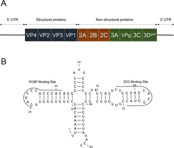
Methods: Total RNA from cardiac tissue of patients with dilated cardiomyopathy was extracted, and sequences corresponding to the 5' termini of enterovirus RNAs were identified. After next-generation RNA sequencing, viral cDNA clones mimicking the enterovirus RNA sequences found in patient tissues were generated in vitro, and their replication and impact on host cell functions were assessed on primary human cardiac cells in culture.
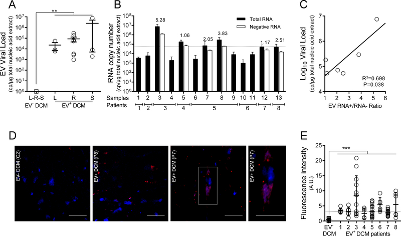
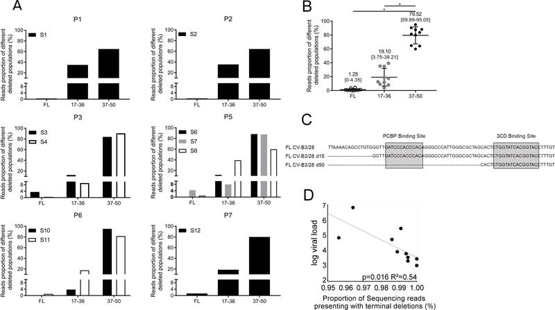
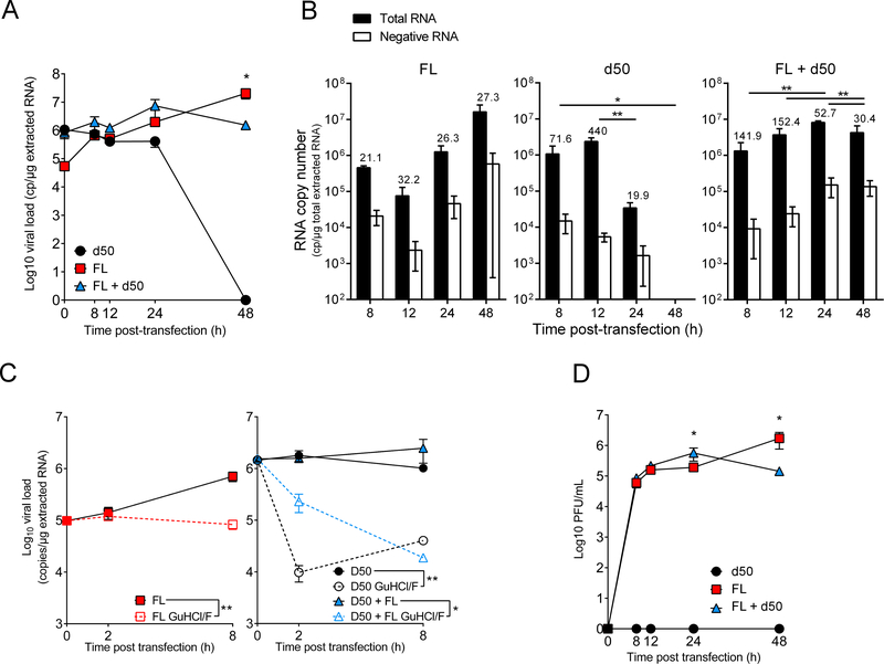
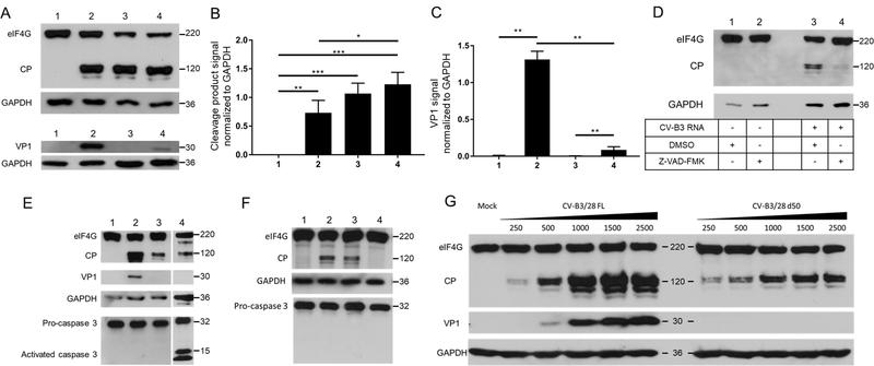
Results: Major enterovirus B populations characterized by 5' terminal genomic RNA deletions ranging from 17 to 50 nucleotides were identified either alone or associated with low proportions of intact 5' genomic termini. In situ hybridization and immunohistological assays detected these persistent genomes in clusters of cardiomyocytes. Transfection of viral RNA into primary human cardiomyocytes demonstrated that deleted forms of genomic RNAs displayed early replication activities in the absence of detectable viral plaque formation, whereas mixed deleted and complete forms generated particles capable of inducing cytopathic effects at levels distinct from those observed with full-length forms alone. Moreover, deleted or full-length and mixed forms of viral RNA were capable of directing translation and production of proteolytically active viral proteinase 2A in human cardiomyocytes.
Conclusions: We demonstrate that persistent viral forms are composed of B-type enteroviruses harboring a 5' terminal deletion in their genomic RNAs and that these viruses alone or associated with full-length populations of helper RNAs could impair cardiomyocyte functions by the proteolytic activity of viral proteinase 2A in cases of unexplained dilated cardiomyopathy. These results provide a better understanding of the molecular mechanisms that underlie the persistence of EV forms in human cardiac tissues and should stimulate the development of new therapeutic strategies based on specific inhibitors of the coxsackievirus B proteinase 2A activity for acute and chronic cardiac infections.
Keywords: dilated cardiomyopathy; enterovirus; genomic RNA deletion; genomics; infection; persistent infection; viral 2A protease; viruses.
Conflict of interest statement
Disclosure
None of the authors of the present manuscript have a commercial or other association that might pose a conflict of interest (
Figures
Comment in
-
Dilated Cardiomyopathy.Circulation. 2019 May 14;139(20):2339-2341. doi: 10.1161/CIRCULATIONAHA.119.040037. Circulation. 2019. PMID: 31082299 No abstract available.
Similar articles
-
Enterovirus-Cardiomyocyte Interactions: Impact of Terminally Deleted Genomic RNAs on Viral and Host Functions.J Virol. 2023 Jan 31;97(1):e0142622. doi: 10.1128/jvi.01426-22. Epub 2022 Dec 8. J Virol. 2023. PMID: 36475766 Free PMC article.
-
Functional Consequences of RNA 5'-Terminal Deletions on Coxsackievirus B3 RNA Replication and Ribonucleoprotein Complex Formation.J Virol. 2017 Jul 27;91(16):e00423-17. doi: 10.1128/JVI.00423-17. Print 2017 Aug 15. J Virol. 2017. PMID: 28539455 Free PMC article.
-
Major 5'terminally deleted enterovirus populations modulate type I IFN response in acute myocarditis patients and in human cultured cardiomyocytes.Sci Rep. 2020 Jul 20;10(1):11947. doi: 10.1038/s41598-020-67648-5. Sci Rep. 2020. PMID: 32686697 Free PMC article.
-
Persistent coxsackievirus infection: enterovirus persistence in chronic myocarditis and dilated cardiomyopathy.Curr Top Microbiol Immunol. 2008;323:275-92. doi: 10.1007/978-3-540-75546-3_13. Curr Top Microbiol Immunol. 2008. PMID: 18357775 Review.
-
CVB infection and mechanisms of viral cardiomyopathy.Curr Top Microbiol Immunol. 2008;323:315-35. doi: 10.1007/978-3-540-75546-3_15. Curr Top Microbiol Immunol. 2008. PMID: 18357777 Review.
Cited by
-
Viral proteases activate the CARD8 inflammasome in the human cardiovascular system.J Exp Med. 2022 Oct 3;219(10):e20212117. doi: 10.1084/jem.20212117. Epub 2022 Sep 21. J Exp Med. 2022. PMID: 36129453 Free PMC article.
-
Persistent Enterovirus Infection: Little Deletions, Long Infections.Vaccines (Basel). 2022 May 12;10(5):770. doi: 10.3390/vaccines10050770. Vaccines (Basel). 2022. PMID: 35632526 Free PMC article. Review.
-
Early Emergence of 5' Terminally Deleted Coxsackievirus-B3 RNA Forms Is Associated with Acute and Persistent Infections in Mouse Target Tissues.Vaccines (Basel). 2022 Jul 28;10(8):1203. doi: 10.3390/vaccines10081203. Vaccines (Basel). 2022. PMID: 36016091 Free PMC article.
-
Severe Myocardial Involvement and Persistent Supraventricular Arrhythmia in a Premature Infant Due to Enterovirus Infection: Case Report and Literature Review.J Cardiovasc Dev Dis. 2025 Jun 14;12(6):228. doi: 10.3390/jcdd12060228. J Cardiovasc Dev Dis. 2025. PMID: 40558663 Free PMC article.
-
Enterovirus-Cardiomyocyte Interactions: Impact of Terminally Deleted Genomic RNAs on Viral and Host Functions.J Virol. 2023 Jan 31;97(1):e0142622. doi: 10.1128/jvi.01426-22. Epub 2022 Dec 8. J Virol. 2023. PMID: 36475766 Free PMC article.
References
-
- Racaniello VR. Picornaviridae : The viruses and Their Replication In: Fields Virology. New York: Knipe & Howley; 2007. p. 795–838.
-
- Li Y, Bourlet T, Andreoletti L, Mosnier JF, Peng T, Yang Y, Archard LC, Pozzetto B, Zhang H. Enteroviral capsid protein VP1 is present in myocardial tissues from some patients with myocarditis or dilated cardiomyopathy. Circulation. 2000;101:231–234. - PubMed
-
- Nguyen Y, Renois F, Leveque N, Giusti D, Picard-Maureau M, Bruneval P, Fornes P, Andreoletti L. Virus detection and semiquantitation in explanted heart tissues of idiopathic dilated cardiomyopathy adult patients by use of PCR coupled with mass spectrometry analysis. J Clin Microbiol. 2013;51:2288–2294. - PMC - PubMed
