. 2019 Feb 19;8(4):e011704.
doi: 10.1161/JAHA.118.011704.

Inositol 1,4,5-Trisphosphate Receptors in Endothelial Cells Play an Essential Role in Vasodilation and Blood Pressure Regulation

Affiliations

Inositol 1,4,5-Trisphosphate Receptors in Endothelial Cells Play an Essential Role in Vasodilation and Blood Pressure Regulation

Qingsong Lin et al. J Am Heart Assoc. .

Abstract

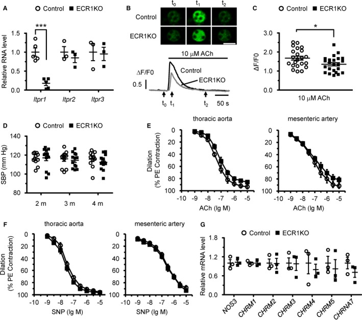
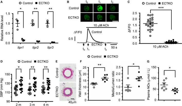
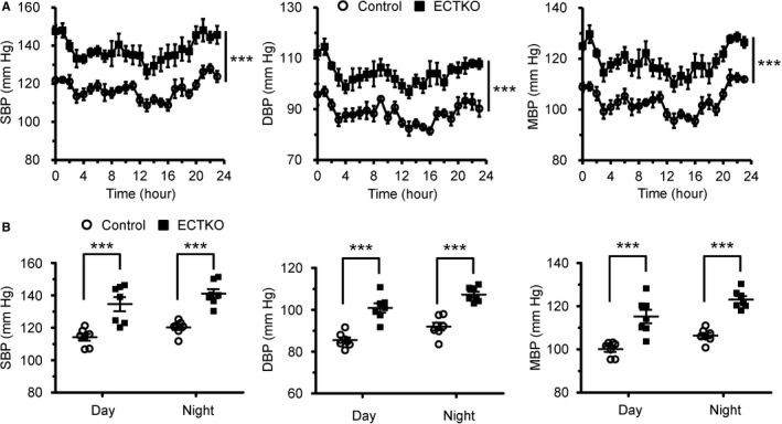
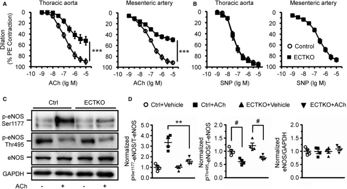
Background Endothelial NO synthase plays a central role in regulating vasodilation and blood pressure. Intracellular Ca2+ mobilization is a critical modulator of endothelial NO synthase function, and increased cytosolic Ca2+ concentration in endothelial cells is able to induce endothelial NO synthase phosphorylation. Ca2+ release mediated by 3 subtypes of inositol 1,4,5-trisphosphate receptors ( IP 3Rs) from the endoplasmic reticulum and subsequent Ca2+ entry after endoplasmic reticulum Ca2+ store depletion has been proposed to be the major pathway to mobilize Ca2+ in endothelial cells. However, the physiological role of IP 3Rs in regulating blood pressure remains largely unclear. Methods and Results To investigate the role of endothelial IP 3Rs in blood pressure regulation, we first generated an inducible endothelial cell-specific IP 3R1 knockout mouse model and found that deletion of IP 3R1 in adult endothelial cells did not affect vasodilation and blood pressure. Considering all 3 subtypes of IP 3Rs are expressed in mouse endothelial cells, we further generated inducible endothelial cell-specific IP 3R triple knockout mice and found that deletion of all 3 IP 3R subtypes decreased plasma NO concentration and increased basal blood pressure. Furthermore, IP 3R deficiency reduced acetylcholine-induced vasodilation and endothelial NO synthase phosphorylation at Ser1177. Conclusions Our results reveal that IP 3R-mediated Ca2+ release in vascular endothelial cells plays an important role in regulating vasodilation and physiological blood pressure.

Keywords: blood pressure; calcium; calcium signaling; endothelial cell; hypertension.


Figures

Figure 1
Figure 1
Endothelial cell–specific deletion of inositol 1,4,5‐trisphosphate receptor 1 (IP 3R1) in adult mice had no major effects on blood pressure and vasodilation. A, mRNA levels of IP 3Rs in isolated endothelial cells from control and endothelial cell–specific IP 3R1 knockout (ECR1KO) mice. n=3 to 5 (with endothelial cells from 3 mice pooled as 1 sample) per group. B, Confocal Ca2+ imaging of isolated endothelial cells using Fluo‐4‐AM. Intracellular Ca2+ mobilization was elicited by 10 μmol/L acetylcholine. Top, Sequential confocal images of endothelial cells at the time points of time 0, time 1, and time 2, as indicated by the arrows at the bottom. Bar=20 μm. Bottom, Representative traces of Ca2+ signals in control (black) and ECR1KO (gray) endothelial cells. F0, the Fluo‐4 fluorescence at rest. F/F0, normalized fluorescence. C, The amplitude of Ca2+ signals induced by 10 μmol/L acetylcholine. n=20 to 30 cells from 3 independent experiments per group. D, Systolic blood pressure (SBP) measured in control and ECR1KO mice at 2, 3, and 4 months after tamoxifen injection using the tail‐cuff system. n=11 to 12 mice per group. E, Vascular reactivity in response to endothelium‐dependent agonist acetylcholine in ECR1 aortas and mesenteric arteries showed a slight trend toward reduced vasodilation that was not statistically significant. F, Vascular reactivity in response to endothelium‐independent agonist sodium nitroprusside (SNP) in aortas and mesenteric arteries. The vessels were preconstricted by 10 μmol/L phenylephrine, and the vasorelaxing effects of acetylcholine and sodium nitroprusside (SNP) were presented as a percentage of phenylephrine‐induced contraction. n=6 mice per group. G, Quantitative real‐time polymerase chain reaction analysis of the expression of NOS3 and major acetylcholine receptors, including CHRM1,CHRM2,CHRM3,CHRM4,CHRM5, and CHRNA7, in control and ECR1KO endothelial cells. n=3 (with endothelial cells from 3 mice pooled as 1 sample) per group. Significance was determined using a 2‐tailed, unpaired, Student t test or 2‐way ANOVA analysis with Bonferroni post hoc test. Error bars represent mean±SEM. *P<0.05, ***P<0.001 vs control.
Figure 2
Figure 2
Endothelial cell–specific deletion of all 3 subtypes of inositol 1,4,5‐trisphosphate receptors (IP 3Rs) in adult mice increased systolic blood pressure (SBP). A, Quantitative real‐time polymerase chain reaction analysis of the 3 IP 3R subtypes in isolated endothelial cells from control and endothelial cell–specific IP 3R triple knockout (ECTKO) mice. n=3 (with endothelial cells from 3 mice pooled as 1 sample) per group. B, Confocal Ca2+ imaging of endothelial cells isolated from control and ECTKO mice. Top, Sequential confocal images of endothelial cells at the time point of time 0, time 1, and time 2, as indicated by the arrows at the bottom. Bar=20 μm. Bottom, Representative traces of Ca2+ signals in control (black) and ECTKO (gray) endothelial cells. F0, the Fluo‐4 fluorescence at rest. F/F0, normalized fluorescence. C, The amplitude of Ca2+ signals induced by 10 μmol/L acetylcholine in control and ECTKO endothelial cells. n=20 to 30 cells from 3 independent experiments per group. D, SBP measured in control and ECTKO mice at 2, 3, and 4 months after tamoxifen administration using the tail‐cuff system. n=13 to 17 mice per group. E, Representative hematoxylin and eosin–stained sections of the second‐order mesenteric arteries isolated from control and ECTKO mice 4 months after tamoxifen administration. Bar=40 μm. F, The wall thickness and the ratio of medial/luminal area were calculated in control and ECTKO mice. n=5 to 6 mice per group. G, The concentration of nitrite and nitrate (NOx) in the serum measured in control and ECTKO mice 4 months after tamoxifen administration. n=6 mice per group. Significance was determined using a 2‐tailed, unpaired, Student t test. Error bars represent mean±SEM. *P<0.05, **P<0.01, ***P<0.001 vs control.
Figure 3
Figure 3
Basal blood pressure was significantly elevated in endothelial cell–specific inositol 1,4,5‐trisphosphate receptor triple knockout (ECTKO) mice. Blood pressures were measured in unstrained mice using an implantable telemetry system. A, Systolic blood pressure (SBP), diastolic blood pressure (DBP), and mean blood pressure (MBP) in control and ECTKO mice. The data were presented at times indicated on a 24‐hour scale, and data shown at each point represent 1‐hour rolling averages of data sampled each minute in 3 consecutive days. n=7 mice per group. B, Mean values for SBP, DBP, and MBP were calculated during the day (8 am–8 pm) and night (8 pm–8 am). n=7 mice per group. Significance was determined using a 2‐tailed, unpaired, Student t test or 2‐way ANOVA analysis with Bonferroni post hoc test. Error bars represent mean±SEM. ***P<0.001 vs control.
Figure 4
Figure 4
Deficiency of inositol 1,4,5‐trisphosphate receptors (IP 3Rs) in endothelial cells affected acetylcholine‐induced vasodilation and endothelial NO synthase (eNOS) phosphorylation. A and B, Vascular reactivity in response to endothelium‐dependent agonist acetylcholine (A) and endothelium‐independent agonist sodium nitroprusside (SNP) (B) in aortas and mesenteric arteries. The vessels were preconstricted by 10 μmol/L phenylephrine, and the vasorelaxing effects of acetylcholine and SNP were presented as a percentage of phenylephrine‐induced contraction. n=6 to 12 mice per group. C, Expression and phosphorylation of eNOS measured by Western blot. Thoracic aortas isolated from control and endothelial cell–specific IP 3R triple knockout (ECTKO) mice were treated with or without 10 μmol/L acetylcholine for 10 minutes. D, The levels of phosphorylated eNOS at Ser1177 and Thr495 were normalized to total eNOS. The levels of total eNOS were normalized to GAPDH. n=4 mice per group. Significance was determined using a 2‐tailed, unpaired, Student t test or 2‐way ANOVA analysis with Bonferroni post hoc test. Error bars represent mean±SEM. **P<0.01, ***P<0.001 vs control; # P<0.05 vs vehicle.

References

    1. Blacher J, Levy BI, Mourad JJ, Safar ME, Bakris G. From epidemiological transition to modern cardiovascular epidemiology: hypertension in the 21st century. Lancet. 2016;388:530–532. - PubMed
    1. Kokubo Y, Iwashima Y. Higher blood pressure as a risk factor for diseases other than stroke and ischemic heart disease. Hypertension. 2015;66:254–259. - PubMed
    1. Feletou M, Kohler R, Vanhoutte PM. Endothelium‐derived vasoactive factors and hypertension: possible roles in pathogenesis and as treatment targets. Curr Hypertens Rep. 2010;12:267–275. - PMC - PubMed
    1. Balligand JL, Feron O, Dessy C. eNOS activation by physical forces: from short‐term regulation of contraction to chronic remodeling of cardiovascular tissues. Physiol Rev. 2009;89:481–534. - PubMed
    1. Palmer RM, Ashton DS, Moncada S. Vascular endothelial cells synthesize nitric oxide from L‐arginine. Nature. 1988;333:664–666. - PubMed

Publication types

MeSH terms