. 2019 Feb 12;14(1):47.
doi: 10.1186/s13018-019-1086-0.

L-shaped corticotomy with bone flap sliding in the management of chronic tibial osteomyelitis: surgical technique and clinical results

Affiliations

L-shaped corticotomy with bone flap sliding in the management of chronic tibial osteomyelitis: surgical technique and clinical results

Teng-Fei Lou et al. J Orthop Surg Res. .

Abstract

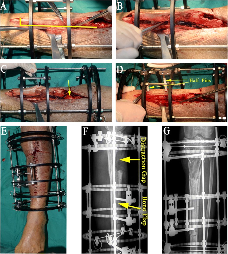
Background: We described the use of the technique of L-shaped corticotomy with bone flap sliding to treat chronic osteomyelitis of the tibia in eight patients and presented the preliminary results.

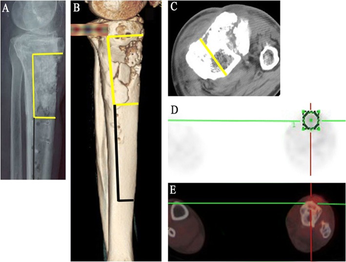
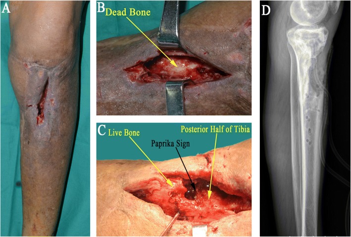
Methods: L-shaped corticotomy with bone flap sliding was performed in eight patients between 2007 and 2014. All patients had chronic tibial osteomyelitis involving the anterior tibial cortex with intact and healthy posterior cortex. The size of bone defects following sequestrectomy and radical debridement was 8.1 cm on average. One patient required a latissimus dorsi flap. The mean follow-up period was 34.1 months. The functional and bone results were evaluated at the time of the latest follow-up.

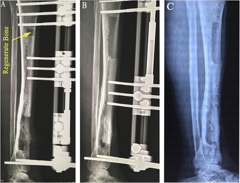
Results: Complete eradication of infection and union of docking sites were achieved in all patients. Functional results were judged excellent in five patients and good in the rest three patients. Bone results were graded as excellent in all cases. The mean external fixation time was 169.9 days and external fixation index was 21.2 days/cm. Pain was the most common complaint that we faced during lengthening. Pin tract infections were observed in four patients, and mild transient stiffness of ankle joint was observed in three patients.

Conclusions: We have found this technique to be safe and effective, significantly diminishing the external fixation index. The earlier removal of the external fixator may result in increased patient comfort, a reduced complication rate, and a rapid and convenient rehabilitation.

Keywords: Bone flap; Bone transport; L-shaped corticotomy; Tibial osteomyelitis.


Conflict of interest statement

Ethics approval and consent to participate

This study are approved by the Ethics Committee of Shanghai Jiao Tong University Affiliated Sixth People’s Hospital. Informed consent for participation was obtained from all participants in this study.

Consent for publication

All consents to publish from the patients who took part in this study were obtained.

Competing interests

The authors declare that they have no competing interests.

Publisher’s Note

Springer Nature remains neutral with regard to jurisdictional claims in published maps and institutional affiliations.

Figures

Fig. 1
Fig. 1
ag A 60-year-old man with chronic osteomyelitis of the left tibia. Imaging examinations are taken to assist in determining the extension of infection and resection. a A preoperative lateral radiograph shows the extension of the infection that compromises the proximal diaphysis and margins of resection (yellow line) and corticotomy (black line) is determined. b, c The CT scans show the extension of bone destruction and margins of resection (yellow line) and corticotomy (black line) is determined on lateral (3D) and coronal images. d, e The PET-CT scan shows increased uptake in the area of infection
Fig. 2
Fig. 2
Debridement technique. a Clinical photograph of the affected leg. b, c During debridement, the infected or necrotic bone is removed radically until the “paprika sign” (cortical bone bleeding area) appears. It is of great importance to make sure that the posterior half of tibia retained following the radical debridement is uninfected and intact. d Lateral radiograph shows bone defect following resection of dead bone
Fig. 3
Fig. 3
Application of external fixator and L-shaped osteotomy. a The holes drilled by Kirschner wires form an L-shaped configuration (yellow line), and osteotome is used to separate the bone flap. b The bone flap is separated from the posterior half of tibia. c The bone flap is not separated from the soft tissue it attaches to, thus being provided with good blood supply from the anterior and lateral soft tissue (black arrow), and remaining posterior cortex after debridement and sequestrectomy keeps the periosteal attachment intact and also ensures a good blood supply (yellow arrow). d Two 5.0-mm half pins are used for unilateral cortical fixation of the bone flap. e The incision is closed with drainage tube. f, g Immediate postoperative latera and anteroposterior radiographs
Fig. 4
Fig. 4
A 33-year-old man with chronic osteomyelitis of the left tibia. a Radiograph shows bone defect following resection of dead bone. b Soft tissue defect following debridement. c Planning on the harvest of free latissimus dorsi flap. d The latissimus dorsi flap is harvested. e The donor site is sutured directly drainage tubes. f The bone flap is separated from the posterior half of tibia, and one tip of the flap is sewed up with the wound edge. g The latissimus dorsi flap completely covers the soft tissue defect, the external fixator is assembled, and two half pins are used to fix the bone flap. h Immediate postoperative lateral radiograph shows distraction gap and bone flap
Fig. 5
Fig. 5
Postoperative lateral radiograph of the patient treated with monolateral external fixator
Fig. 6
Fig. 6
a Twenty-five days after operation with partial bone flap sliding. b Ninety days after operation, bone ends had contacted with each other and the regenerate bone had commenced to be mineralized. c One hundred eighty-five days after operation, consolidation of the newly formed bone and union of the docking site were presented, and the fixator was removed
Fig. 7
Fig. 7
The possible advantages of L-shaped corticotomy with bone flap sliding in the management of chronic tibial osteomyelitis

References

    1. Lin CC, Chen CM, Chiu FY, Su YP, Liu CL, Chen TH. Staged protocol for the treatment of chronic tibial shaft osteomyelitis with Ilizarov’s technique followed by the application of intramedullary locked nail. Orthopedics. 2012;35(12):e1769. doi: 10.3928/01477447-20121120-23. - DOI - PubMed
    1. Simpson AH, Deakin M, Latham JM. Chronic osteomyelitis. The effect of the extent of surgical resection on infection-free survival. J Bone Joint Surg Br. 2001;83(3):403–407. doi: 10.1302/0301-620X.83B3.10727. - DOI - PubMed
    1. Cierny G, 3rd, Mader JT, Penninck JJ. A clinical staging system for adult osteomyelitis. Clin Orthop Relat Res. 2003;414(414):7–24. doi: 10.1097/01.blo.0000088564.81746.62. - DOI - PubMed
    1. Selhi HS, Mahindra P, Yamin M, Jain D, De Long WG, Jr, Singh J. Outcome in patients with an infected nonunion of the long bones treated with a reinforced antibiotic bone cement rod. J Orthop Trauma. 2012;26(3):184–188. doi: 10.1097/BOT.0b013e318225f77c. - DOI - PubMed
    1. Sun Y, Zhang C, Jin D, Sheng J, Cheng X, Liu X. Free vascularised fibular grafting in the treatment of large skeletal defects due to osteomyelitis. Int Orthop. 2010;34(3):425–430. doi: 10.1007/s00264-009-0761-x. - DOI - PMC - PubMed

LinkOut - more resources