. 2019 Feb 12;16(1):34.
doi: 10.1186/s12974-019-1411-x.

AMPK activation attenuates inflammatory pain through inhibiting NF-κB activation and IL-1β expression

Affiliations

AMPK activation attenuates inflammatory pain through inhibiting NF-κB activation and IL-1β expression

Hong-Chun Xiang et al. J Neuroinflammation. .

Abstract

Background: Chronic pain is a major clinical problem with limited treatment options. Previous studies have demonstrated that activation of adenosine monophosphate-activated protein kinase (AMPK) can attenuate neuropathic pain. Inflammation/immune response at the site of complete Freund's adjuvant (CFA) injection is known to be a critical trigger of the pathological changes that produce inflammatory pain. However, whether activation of AMPK produces an analgesic effect through inhibiting the proinflammatory cytokines, including interleukin-1β (IL-1β), in inflammatory pain remains unknown.

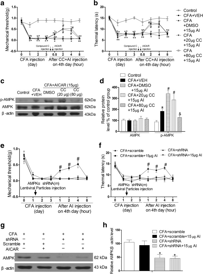
Methods: Inflammatory pain was induced in mice injected with CFA. The effects of AICAR (5-aminoimidazole-4-carboxyamide ribonucleoside, an AMPK activator), Compound C (an AMPK inhibitor), and IL-1ra (an IL-1 receptor antagonist) were tested at day 4 after CFA injection. Inflammatory pain was assessed with von Frey filaments and hot plate. Immunoblotting, hematoxylin and eosin (H&E) staining, and immunofluorescence were used to assess inflammation-induced biochemical changes.

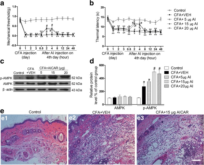
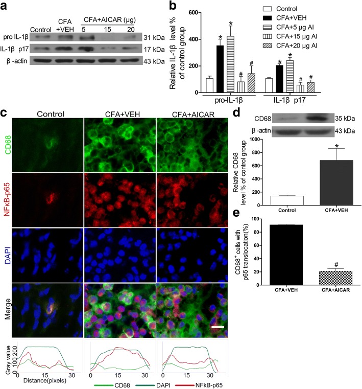
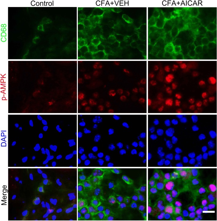
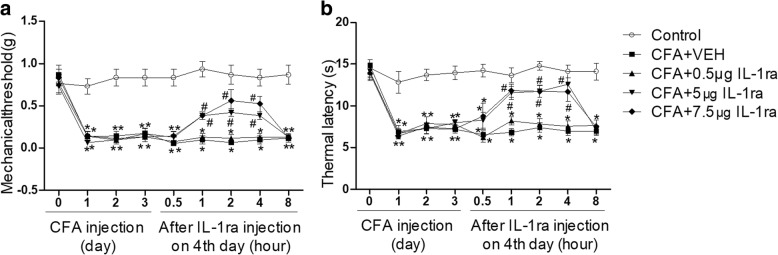
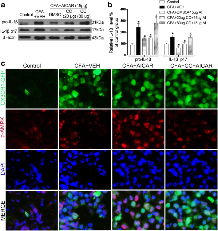
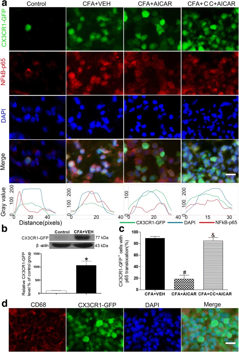
Results: The AMPK activator AICAR produced an analgesic effect and inhibited the level of proinflammatory cytokine IL-1β in the inflamed skin in mice. Moreover, activation of AMPK suppressed CFA-induced NF-κB p65 translocation from the cytosol to the nucleus in activated macrophages (CD68+ and CX3CR1+) of inflamed skin tissues. Subcutaneous injection of IL-1ra attenuated CFA-induced inflammatory pain. The AMPK inhibitor Compound C and AMPKα shRNA reversed the analgesic effect of AICAR and the effects of AICAR on IL-1β and NF-κB activation in inflamed skin tissues.

Conclusions: Our study provides new information that AMPK activation produces the analgesic effect by inhibiting NF-κB activation and reducing the expression of IL-1β in inflammatory pain.

Keywords: AMPK; IL-1β; Inflammatory pain; NF-κB.


Conflict of interest statement

Ethics approval and consent to participate

All animal experiments were approved by the Animal Care Committee at Huazhong University of Science and Technology and were designed to minimize suffering and the number of animals used. All procedures were strictly carried out in accordance with the ethical guidelines of the International Association for the Study of Pain.

Consent for publication

Not applicable.

Competing interests

The authors declare that they have no competing interests.

Publisher’s Note

Springer Nature remains neutral with regard to jurisdictional claims in published maps and institutional affiliations.

Figures

Fig. 1
Fig. 1
AICAR attenuates CFA-induced pain behavior and local inflammation and promotes phosphorylation of AMPK in mice. Effects of a single hypodermic injection of AICAR (5, 15, or 20 μg/20 μl) on mechanical allodynia (a) and thermal hyperalgesia (b) in CFA-injected mice. The panels show the hindpaw withdrawal threshold in response to mechanical stimulation as assessed with von Frey hairs in a or thermal stimulation as assessed with hot plate in b (data are expressed as mean ± SEM, n = 8 mice/group). Two-way ANOVA revealed a significant difference at *P < 0.05 vs. Control group and #P < 0.05 vs. CFA+VEH group. c, d Subcutaneous administration of AICAR (5, 15, or 20 μg /20 μl) activated AMPK in the inflamed skin tissues in a dose-dependent manner. One-way ANOVA revealed a significant difference at *P < 0.05 vs. Control group and #P < 0.05 vs. CFA+VEH group. e H&E staining. (e1) Control mice with no inflammation in the skin, (e2) inflammation with abundant lymphocytes and sparse neutrophilic granulocytes in an CFA mouse with hypodermic injection of vehicle (20 μl sterilized PBS, the vehicle of AICAR), and (e3) low-grade inflammation in an CFA mouse with hypodermic injection of AICAR (15 μg/20 μl). Scale bar represents 100 μm. Tissues were collected at 2 h after AICAR treatment for Western blotting and HE. The abbreviations used here are VEH (vehicle of AICAR, sterilized double steamed H2O) and AI (AICAR)
Fig. 2
Fig. 2
AICAR downregulates expression of IL-1β and suppresses CFA-induced NF-κB activation in macrophages of the inflamed skin. AICAR (5, 15, 20 μg/20 μl) was administrated at CFA injection day 4. Skin tissues were collected at 2 h after AICAR treatment for Western blotting and immunofluorescence. Representative gels and quantification for pro-IL-1β and IL-1β p17 bands (a and b) are shown. One-way ANOVA revealed a significant difference at *P < 0.05 vs. Control group and #P < 0.01 vs. CFA+VEH group. c AICAR inhibited the CFA-induced NF-κB translocation from the cytosol to the nucleus, as demonstrated by the immunofluorescence of p65 in CD68 marked macrophage cells. Scale bar represents 50 μm. The bottom line of the panels shows the merged profiles of the fluorescent intensity of CD68, NF-κB p65 and DAPI signals along the lines drawn through the axis of immunostaining macrophage cells. d Quantification for CD68 protein level of skin tissues. e Quantification for cells with p65 translocation of c. Non-paired Student’s t test revealed a significant difference at *P < 0.05 vs. Control group and #P < 0.01 vs. CFA + VEH group (n = 4 mice/group). The abbreviations used here are VEH (vehicle of AICAR) and AI (AICAR)
Fig. 3
Fig. 3
Co-localization of CD68+ cells and p-AMPK at day 4 after CFA injection in mice. Immunostaining of skin sections showing the co-localization of CD68+ cells and p-AMPK in inflamed skins at day 4 after AICAR administration. Scale bar represents 50 μm. Skin tissues were collected at 2 h after AICAR treatment for immunofluorescence
Fig. 4
Fig. 4
Subcutaneous injection of IL-1ra attenuates CFA-induced inflammatory pain of mice. a Effects of a single injection of IL-1ra (0.5, 5, or 7.5 μg/20 μl) on mechanical allodynia induced by CFA in mice. b Effects of a single injection of IL-1ra (0.5, 5, or 7.5 μg/20 μl) on thermal hyperalgesia induced by CFA in mice. Data are expressed as mean ± SEM, n = 8 mice/group. Two-way ANOVA revealed a significant difference at *P < 0.05 vs. Control group and #P < 0.05 vs. CFA + VEH (vehicle of IL-1ra, sterilized PBS) group. AI, AICAR
Fig. 5
Fig. 5
AMPK activation is required for AICAR to attenuate CFA-induced inflammatory pain in mice. Compound C (20 μg, 80 μg/20 μl) was administrated at post-injection of CFA day 4 before AICAR (15 μg/20 μl) treatment for 30 min. ad AMPK inhibitor Compound C (80 μg/20 μl) reversed the effects of AICAR (15 μg/20 μl). Effects of a single injection of Compound C (20 μg or 80 μg/20 μl) on mechanical allodynia (a) and thermal hyperalgesia (b) in CFA-injected mice with 15 μg/20 μl AICAR administration. Two-way ANOVA revealed a significant difference at *P < 0.05 vs. Control group and #P < 0.05 vs. CFA+VEH group (n = 8 each group). Skin tissues were collected at 2 h after AICAR treatment for Western blotting. Representative gels and quantification for p-AMPK and AMPK bands are shown (c, d). One-way ANOVA revealed a significant difference at *P < 0.05 vs. Control group and #P < 0.05 vs. CFA+VEH group. eh Effects of AMPKα shRNA on CFA-induced mechanical allodynia, thermal hyperalgesia, and AICAR (15 μg/20 μl) treatment in mice (n = 5 mice/group). After the local administration of AMPKα shRNA, AICAR could not attenuate mechanical allodynia (e) and thermal hyperalgesia (f) and downregulated the level of AMPKα in inflamed tissues (g, h). Two-way ANOVA revealed a significant difference at #P < 0.05 vs. CFA + shRNA + 15μg AI group (e and f). One-way ANOVA revealed a significant difference at *P < 0.05 vs. CFA +scramble group (h). The abbreviations used here are VEH (vehicle of AICAR) and CC (Compound C, dissolve in DMSO (dimethyl sulphoxide))
Fig. 6
Fig. 6
AMPK inhibitor Compound C reverses the effects of AICAR. Compound C (20 μg, 80 μg/20 μl) was administrated at post-injection of CFA day 4 before AICAR (15 μg/20 μl) treatment 30 min. Skin tissues were collected at 2 h after AICAR treatment. Representative gels and quantification for pro-IL-1β and IL-1β p17 bands are shown (a, b). Data are expressed as mean ± SEM. One-way ANOVA revealed a significant difference at *P < 0.05 vs. Control group and #P < 0.05 vs. CFA+VEH group (n = 4 mice/group). The abbreviations used here are VEH (vehicle of AICAR), AI (AICAR) and CC (Compound C). c Immunostaining of skin sections showing the co-localization of CX3CR1-GFP + cells and p-AMPK in inflamed skins on the fourth day (c). Scale bar represents 50 μm
Fig. 7
Fig. 7
AMPK activation is required for inhibiting NF-κB p65 translocation. Compound C reverses the effects of AICAR to suppress CFA-induced NF-κB p65 translocation in CX3CR1-GFP mice. After a pre-treatment of Compound C (80 μg/20 μl), mice were treated with AICAR (15 μg/20 μl). Skin tissues from CX3CR1-GFP mice were collected at 2 h after AICAR treatment for immunofluorescence. a AICAR inhibited the CFA-induced NF-κB translocation from the cytosol to the nucleus, as demonstrated by the immunofluorescence of NF-κB p65 in CX3CR1-GFP-marked macrophage cells. Scale bars represent 50 μm. The same experimental results were repeated in four mice of each group. The bottom line of the panels shows the merged profiles of the fluorescent intensity of CX3CR1-GFP, NF-κB p65, and DAPI signals along the lines drawn through the axis of CX3CR1-GFP-positive macrophage cells. b Quantification for CX3CR1-GFP protein level of skin tissues. c Quantification for CX3CR1-GFP macrophages with p65 translocation of a. Non-paired Student’s t test revealed a significant difference at *P < 0.05 vs. Control group, #P < 0.01 vs. CFA + VEH group, and &P < 0.05 vs. CFA + AICAR group, (n = 4 mice/group). d The panels are images showing double-labeled cells in CX3CR1-GFP mice (after CFA injection at day 4) with CD68 and DAPI. The abbreviations used here are VEH (vehicle of AICAR) and CC (Compound C)

References

    1. Basbaum AI, Bautista DM, Scherrer G, Julius D. Cellular and molecular mechanisms of pain. Cell. 2009;139:267–284. doi: 10.1016/j.cell.2009.09.028. - DOI - PMC - PubMed
    1. Hucho T, Levine JD. Signaling pathways in sensitization: toward a nociceptor cell biology. Neuron. 2007;55:365–376. doi: 10.1016/j.neuron.2007.07.008. - DOI - PubMed
    1. Geboes L, De Klerck B, Van Balen M, Kelchtermans H, Mitera T, Boon L, De Wolf-Peeters C, Matthys P. Freund’s complete adjuvant induces arthritis in mice lacking a functional interferon-gamma receptor by triggering tumor necrosis factor alpha-driven osteoclastogenesis. Arthritis Rheum. 2007;56:2595–2607. doi: 10.1002/art.22791. - DOI - PubMed
    1. Ahmed O, Fahim H, Mahmoud A, Ahmed E. Bee venom and hesperidin effectively mitigate complete Freund’s adjuvant-induced arthritis via immunomodulation and enhancement of antioxidant defense system. Arch Rheumatol. 2018;33:198–212. doi: 10.5606/ArchRheumatol.2018.6519. - DOI - PMC - PubMed
    1. Hardie DG. AMP-activated protein kinase: an energy sensor that regulates all aspects of cell function. Genes Dev. 2011;25:1895–1908. doi: 10.1101/gad.17420111. - DOI - PMC - PubMed

MeSH terms

LinkOut - more resources