. 2019 Mar 5;116(5):860-873.
doi: 10.1016/j.bpj.2019.01.016. Epub 2019 Jan 25.

Gramicidin Increases Lipid Flip-Flop in Symmetric and Asymmetric Lipid Vesicles

Affiliations

Gramicidin Increases Lipid Flip-Flop in Symmetric and Asymmetric Lipid Vesicles

Milka Doktorova et al. Biophys J. .

Abstract

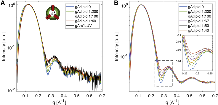
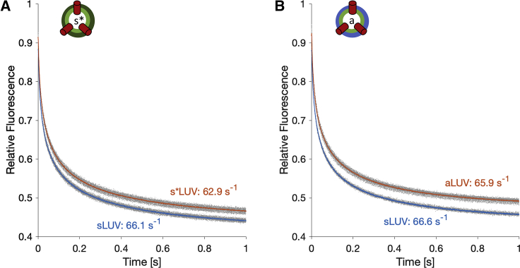
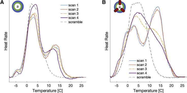
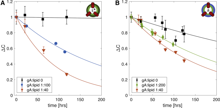
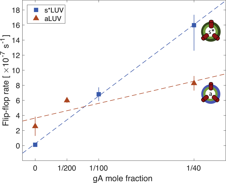
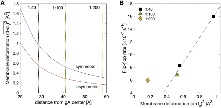
Unlike most transmembrane proteins, phospholipids can migrate from one leaflet of the membrane to the other. Because this spontaneous lipid translocation (flip-flop) tends to be very slow, cells facilitate the process with enzymes that catalyze the transmembrane movement and thereby regulate the transbilayer lipid distribution. Nonenzymatic membrane-spanning proteins with unrelated primary functions have also been found to accelerate lipid flip-flop in a nonspecific manner and by various hypothesized mechanisms. Using deuterated phospholipids, we examined the acceleration of flip-flop by gramicidin channels, which have well-defined structures and known functions, features that make them ideal candidates for probing the protein-membrane interactions underlying lipid flip-flop. To study compositionally and isotopically asymmetric proteoliposomes containing gramicidin, we expanded a recently developed protocol for the preparation and characterization of lipid-only asymmetric vesicles. Channel incorporation, conformation, and function were examined with small angle x-ray scattering, circular dichroism, and a stopped-flow spectrofluorometric assay, respectively. As a measure of lipid scrambling, we used differential scanning calorimetry to monitor the effect of gramicidin on the melting transition temperatures of the two bilayer leaflets. The two calorimetric peaks of the individual leaflets merged into a single peak over time, suggestive of scrambling, and the effect of the channel on the transbilayer lipid distribution in both symmetric 1-palmitoyl-2-oleoyl-sn-glycero-3-phosphocholine and asymmetric 1-palmitoyl-2-oleoyl-sn-glycero-3-phosphocholine/1,2-dimyristoyl-sn-glycero-3-phosphocholine vesicles was quantified from proton NMR measurements. Our results show that gramicidin increases lipid flip-flop in a complex, concentration-dependent manner. To determine the molecular mechanism of the process, we used molecular dynamics simulations and further computational analysis of the trajectories to estimate the extent of membrane deformation. Together, the experimental and computational approaches were found to constitute an effective means for studying the effects of transmembrane proteins on lipid distribution in both symmetric and asymmetric model membranes.


Figures

Figure 1
Figure 1
Gramicidin incorporation is similar in symmetric and asymmetric liposomes. (A) Experimental SAXS form factors of a series of POPC gA-sLUVs with an increasing concentration of gA and isotopically asymmetric LUVs, gA-sLUV, composed of deuterated variants of POPC and prepared with a nominal gA/lipid ratio of 1:40. The cartoon schematic next to the figure legend represents an isotopically asymmetric gA-sLUV. All measurements were performed at 25°C. (B) SAXS form factors calculated from Monte Carlo simulations of a POPC vesicle with different concentrations of gA (see Supporting Materials and Methods). The increase in the intensity at the minimum between the first and second scattering lobes with increasing gA concentration (shown in an expanded view in the inset) is caused by the electron density contrast between the lipid bilayer and the membrane-spanning gA dimers. To see this figure in color, go online.
Figure 2
Figure 2
Gramicidin channel function remains intact in asymmetric liposomes. Changes in ANTS fluorescence over time for the isotopically (A) and compositionally (B) asymmetric samples (in red) and their corresponding symmetric acceptor vesicles (in blue). Replicate traces are shown in gray; their averages are shown in color. The fluorescence signal was normalized to the fluorescence measured before the addition of TI+. The average rates calculated from the traces are indicated next to each trace. The cartoon schematic in (B) represents a compositionally asymmetric gA-aLUV. All measurements were performed at an ambient temperature of ∼22°C. To see this figure in color, go online.
Figure 3
Figure 3
Gramicidin scrambles lipids in compositionally asymmetric vesicles. DSC data of compositionally asymmetric LUVs with DMPC-d54 and POPC and without (A) or with (B) gramicidin at gA/lipid ratio of 1:40 are shown. Four consecutive cooling scans were performed as follows: after the asymmetric vesicle preparation (scan 1, thin blue line), after scan 1 (scan 2, dotted red line), after subsequent incubation at 20°C for 12 h followed by incubation at 45°C for 5 h (scan 3, dash-dotted yellow line), and after another set of incubations at 20°C for 12 h and 45°C for 5 h (scan 4, thick purple line). Also shown for comparison with gray dashed lines are data for the symmetric samples (scramble) with the same overall composition as the asymmetric vesicles (Table S1). To see this figure in color, go online.
Figure 4
Figure 4
Gramicidin increases the rate of lipid flip-flop in isotopically and compositionally asymmetric vesicles. The time evolution of the interleaflet distribution of POPC-d31 in sLUVs (A) and DMPC-d54 in aLUVs (B) either without gA (black squares) or with gA at a gA/lipid ratio of 1:40 (red triangles), 1:100 (blue circles), and 1:200 (green diamonds). Both plots show the time-dependent changes in the distribution of POPC-31 between the outer and inner leaflets, relative to the first time point measured after vesicle preparation (ΔC). See text for details. Error bars represent SDs of at least three consecutive Pr3+ additions. Each time trace in (A) is from a single sample. The time traces of the compositionally asymmetric vesicles in (B) are from one (gA/lipid 1:40), two (no gA), and three (gA/lipid 1:200) separately prepared samples, respectively. The kinetics reported here corresponds to sample behavior at an ambient temperature of ∼22°C. To see this figure in color, go online.
Figure 5
Figure 5
The rate of lipid flip-flop shows a complex relationship with gA mole fraction. Flip-flop rates were calculated from the data in Fig. 4 for the compositionally symmetric (blue squares) and asymmetric (red triangles) LUVs (see Table 2) as a function of the nominal gA mole fraction in the samples. Error bars represent 95% confidence intervals. To see this figure in color, go online.
Figure 6
Figure 6
The gA-mediated lipid flip-flop rate at high gA concentrations correlates with membrane deformation. (A) Average squared deviations in bilayer thickness as a function of distance from gA center for a symmetric POPC bilayer and an asymmetric bilayer as in the NMR experiments in Fig. 4. The thickness deviations were calculated from the membrane deformation profiles around a single gA channel obtained by a CTMD-guided free-energy minimization utilizing information from MD simulations (see Materials and Methods). Dotted lines indicate the approximated half distances between gA channels on the surface of the LUVs at three different gA/lipid ratios: 1:40 (black), 1:100 (green), and 1:200 (yellow). (B) gA-mediated lipid flip-flop rate from Fig. 5 as a function of the corresponding membrane deformation from (A). The data points denote different gA/lipid mole ratios: 1:40 (black square), 1:100 (green triangle), and 1:200 (yellow diamond). To see this figure in color, go online.

References

    1. Collins M.D., Keller S.L. Tuning lipid mixtures to induce or suppress domain formation across leaflets of unsupported asymmetric bilayers. Proc. Natl. Acad. Sci. USA. 2008;105:124–128. - PMC - PubMed
    1. Kiessling V., Wan C., Tamm L.K. Domain coupling in asymmetric lipid bilayers. Biochim. Biophys. Acta. 2009;1788:64–71. - PMC - PubMed
    1. Perillo V.L., Peñalva D.A., Antollini S.S. Transbilayer asymmetry and sphingomyelin composition modulate the preferential membrane partitioning of the nicotinic acetylcholine receptor in Lo domains. Arch. Biochem. Biophys. 2016;591:76–86. - PubMed
    1. Vitrac H., MacLean D.M., Dowhan W. Dynamic membrane protein topological switching upon changes in phospholipid environment. Proc. Natl. Acad. Sci. USA. 2015;112:13874–13879. - PMC - PubMed
    1. Perlmutter J.D., Sachs J.N. Interleaflet interaction and asymmetry in phase separated lipid bilayers: molecular dynamics simulations. J. Am. Chem. Soc. 2011;133:6563–6577. - PubMed

Publication types