. 2019 Feb 12;10(1):e02273-18.
doi: 10.1128/mBio.02273-18.

A Genome-Wide Analysis of Adhesion in Caulobacter crescentus Identifies New Regulatory and Biosynthetic Components for Holdfast Assembly

Affiliations

A Genome-Wide Analysis of Adhesion in Caulobacter crescentus Identifies New Regulatory and Biosynthetic Components for Holdfast Assembly

David M Hershey et al. mBio. .

Abstract

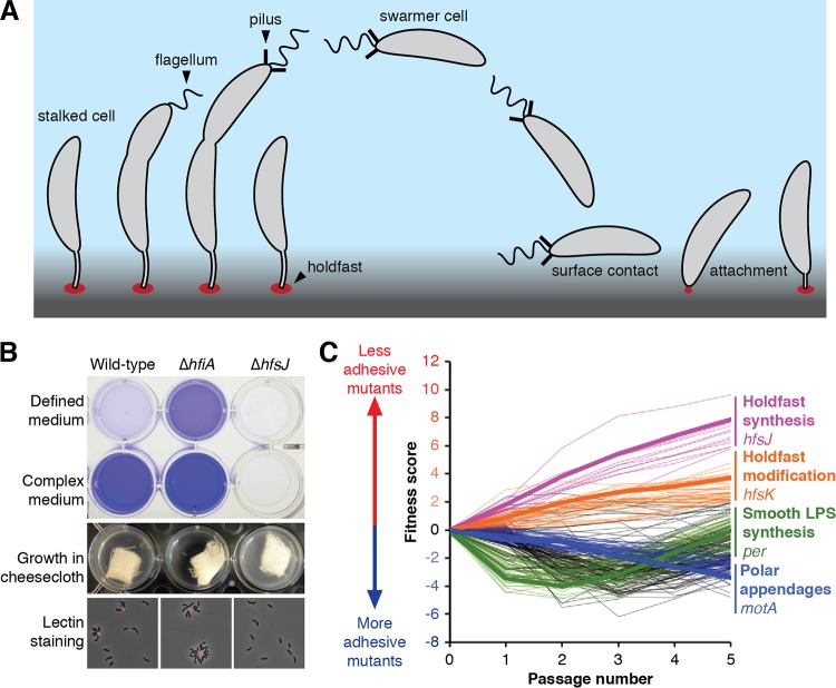
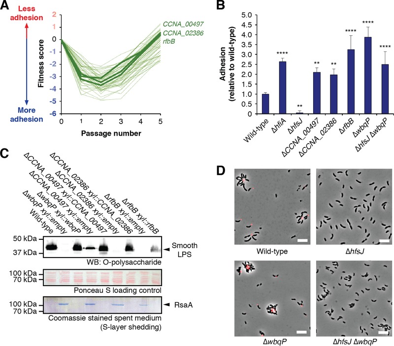
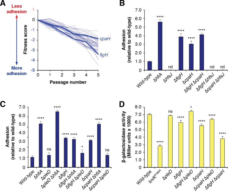
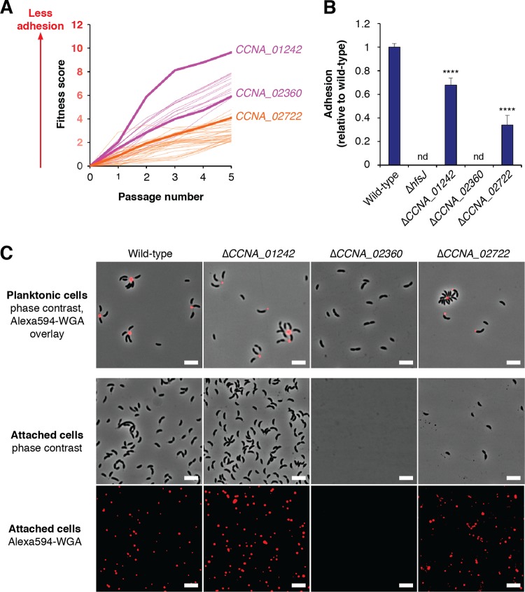
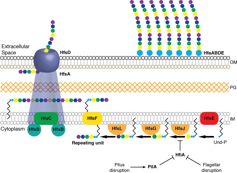
Due to their intimate physical interactions with the environment, surface polysaccharides are critical determinants of fitness for bacteria. Caulobacter crescentus produces a specialized structure at one of its cell poles called the holdfast that enables attachment to surfaces. Previous studies have shown that the holdfast is composed of carbohydrate-based material and identified a number of genes required for holdfast development. However, incomplete information about its chemical structure, biosynthetic genes, and regulatory principles has limited progress in understanding the mechanism of holdfast synthesis. We leveraged the adhesive properties of the holdfast to perform a saturating screen for genes affecting attachment to cheesecloth over a multiday time course. Using similarities in the temporal profiles of mutants in a transposon library, we defined discrete clusters of genes with related effects on cheesecloth colonization. Holdfast synthesis, flagellar motility, type IV pilus assembly, and smooth lipopolysaccharide (SLPS) production represented key classes of adhesion determinants. Examining these clusters in detail allowed us to predict and experimentally define the functions of multiple uncharacterized genes in both the holdfast and SLPS pathways. In addition, we showed that the pilus and the flagellum control holdfast synthesis separately by modulating the holdfast inhibitor hfiA. This report defines a set of genes contributing to adhesion that includes newly discovered genes required for holdfast biosynthesis and attachment. Our data provide evidence that the holdfast contains a complex polysaccharide with at least four monosaccharides in the repeating unit and underscore the central role of cell polarity in mediating attachment of C. crescentus to surfaces.IMPORTANCE Bacteria routinely encounter biotic and abiotic materials in their surrounding environments, and they often enlist specific behavioral programs to colonize these materials. Adhesion is an early step in colonizing a surface. Caulobacter crescentus produces a structure called the holdfast which allows this organism to attach to and colonize surfaces. To understand how the holdfast is produced, we performed a genome-wide search for genes that contribute to adhesion by selecting for mutants that could not attach to cheesecloth. We discovered complex interactions between genes that mediate surface contact and genes that contribute to holdfast development. Our genetic selection identified what likely represents a comprehensive set of genes required to generate a holdfast, laying the groundwork for a detailed characterization of the enzymes that build this specialized adhesin.

Keywords: BarSeq; Caulobacter; adhesion; holdfast; pilus; polysaccharide.


Figures

FIG 1
FIG 1
A genome-wide screen for holdfast biosynthesis genes identified multiple classes of mutants affecting adhesion. (A) During the dimorphic C. crescentus life cycle, each cell division produces a motile swarmer cell and a sessile stalked cell. Swarmer cells stop swimming, shed their flagellum and pili, and develop into stalked cells before dividing. Stalked cells can adhere strongly to exogenous surfaces using a specialized material called the holdfast. (B) The holdfast can be visualized by staining with fluorescently labeled wheat germ agglutinin (fWGA), and biofilm formation can be quantified by crystal violet staining of attached cells. The ΔhfiA cells overproduced holdfast and were hyperadhesive. The ΔhfsJ cells did not produce holdfasts and were nonadhesive. Attachment was reduced in defined medium due to high levels of hfiA expression. (C) Fitness profiles for the 250 genes with the strongest adhesion phenotypes in the cheesecloth passaging experiment. Lines are drawn to connect mean fitness scores at days 0 through 5 for each of the 250 genes. Genes in the four major fitness clusters are colored as indicated in the legend, with a specific example listed. Genes shown in black do not fit into any of the four clusters.
FIG 2
FIG 2
Disrupting SLPS production leads to ectopic adhesion. (A) Fitness profiles for genes in the SLPS cluster. A full list of these genes and their annotations is provided in Table S5. wbqP was not characterized in our library due to low insertion density in this region. (B) Surface attachment of SLPS mutants measured by CV staining. Cultures were grown for 24 h in M2X medium before staining of surface-attached cells. Disruption of SLPS led to increased adhesion in a holdfast-independent fashion. The graph shows averages ± standard deviations of results from five biological replicates. Statistical significance was assessed by analysis of variance (ANOVA) with a pairwise Dunnett’s posttest to determine which samples differed from the wild type. **, P < 0.01; ****, P < 0.0001. (C) Smooth LPS production was disrupted in mutants ΔCCNA_00497, ΔCCNA_02386, and ΔrfbB. (Top) Western blot (WB) to detect SLPS. (Middle) Total protein stained with Ponceau S as a loading control for SLPS blotting. (Bottom) Coomassie staining of spent medium from the cultures. Each mutant showed a loss of or a decrease in SLPS production by Western blotting and released the S-layer protein RsaA into the spent medium. The cell-surface defects can be complemented by ectopic expression of the appropriate gene. “Empty” refers to plasmid control strains.
FIG 3
FIG 3
Disrupting polar appendages stimulates holdfast production. (A) Fitness profiles for genes in the polar appendage cluster. A full list of these genes and their annotations is provided in Table S5. (B) Surface attachment of motility mutants measured by CV staining. Cultures were grown for 17 h in M2X medium before staining of surface-attached cells. Deletion of the genes for either the outer membrane flagellar base protein FlgH or the inner membrane type IV pilus component CpaH caused increased adhesion. In both mutant backgrounds, hfsJ is required for attachment. The graph shows averages ± standard deviations of results from five biological replicates. (C) Effect of hfiA and pleD deletions on surface attachment of cpaH and flgH mutants. The ΔhfiA mutation did not affect adhesion in the ΔflgH background and increased adhesion in the ΔcpaH background. The increased adhesion in both the ΔflgH and ΔcpaH mutants can be eliminated by deletion of pleD. The graph shows averages ± standard deviations of results from four biological replicates. (D) PhfiA-lacZ reporter activity in polar appendage mutants. The chart shows averages ± standard deviations of results from four biological replicates. Statistical significance was assessed by ANOVA with a pairwise Dunnett’s posttest to determine which samples differed from the wild type. nd, not detected; ns, not significant; *, P < 0.05; ****, P < 0.0001. Where appropriate, P values for additional pairwise comparisons pertinent to interpretation are indicated in the text.
FIG 4
FIG 4
Opposing effects of pilus mutants on adhesion. (A) Fitness profiles for genes at the pilus assembly locus. A full list of these genes and their annotations is provided in Table S5. (B) Surface attachment of pilus mutants measured by CV staining. Cultures were grown for 17 h in M2X medium before staining. Deletion of the gene for the main pilin subunit (PilA) reduced adhesion. The ΔpilA mutant was epistatic with respect to the ΔcpaH mutant but not to the ΔflgH mutant. The graph shows averages ± standard deviations of results from seven biological replicates. (C) Effect of hfiA and pleD deletions on surface attachment in the ΔpilA background. Staining is slightly lower for the ΔhfiA ΔpilA mutant than for the ΔhfiA mutant, reflecting the holdfast-independent defect in surface attachment that occurred when the pilus was disrupted. pleD has no effect on adhesion in the ΔpilA mutant. The graph shows averages ± standard deviations of results from six biological replicates. (D) PhfiA-lacZ reporter activity in various pilA mutants. The chart shows averages ± standard deviations of results from four biological replicates. Statistical significance was assessed by ANOVA with a pairwise Dunnett’s posttest to determine which samples differed from the wild type. ns, not significant; **, P < 0.01; ****, P < 0.0001. Where appropriate, P values for additional pairwise comparisons pertinent to interpretation are indicated in the text.
FIG 5
FIG 5
New holdfast biosynthesis factors. (A) Fitness profiles for genes in the hfs (magenta) and holdfast modification (orange) clusters. Full lists of these genes and their annotations are provided in Table S5. (B) Surface attachment of putative holdfast mutants measured by CV staining. Cultures were grown for 24 h in PYE medium before being stained. The ΔCCNA_01242 and ΔCCNA_02722 mutants displayed reduced staining, and the ΔCCNA_02360 mutant was nonadhesive. The graph shows averages ± standard deviations of results from five biological replicates. Statistical significance was assessed by ANOVA with a pairwise Dunnett’s posttest to determine which samples differed from the wild type. nd, not detected; ns, not significant; ****, P < 0.0001. (C) Analysis of holdfast phenotypes by fWGA staining. The top panels show overlays of phase-contrast and fluorescence images after staining of planktonic cells was performed as described in Materials and Methods. Adherent cells from the slide attachment assay are shown as phase-contrast images in the middle set of panels, and the fluorescence channel showing attached holdfast material from the same slides is represented in the bottom panels. The ΔCCNA_01242 mutant did not have an apparent holdfast defect, the ΔCCNA_02722 mutant had a holdfast attachment defect, and the ΔCCNA_02360 mutant did not produce holdfasts. The scale bars represent 5 μm.
FIG 6
FIG 6
Updated model for holdfast biosynthesis. The model shows a wzy-type polysaccharide biosynthesis pathway. Four glycosyltransferases, HfsE, HfsJ, HfsG, and newly described HfsL, add monsaccharides sequentially onto the UPP carrier to produce a glycolipid repeating unit. This intermediate is flipped across the membrane by HfsF, polymerized by HfsC, and exported by a putative HfsABD transevelope complex. Attachment of the holdfast matrix is mediated by the Hfa proteins, including newly identified HfaE, reported here. Disruptions to the flagellum or the pilus activate holdfast production by relieving the inhibition of HfsJ by HfiA. OM, outer membrane; PG, peptidoglycan; IM, inner membrane.

Similar articles

Cited by

References

    1. Silhavy TJ, Kahne D, Walker S. 2010. The bacterial cell envelope. Cold Spring Harb Perspect Biol 2:a000414. doi:10.1101/cshperspect.a000414. - DOI - PMC - PubMed
    1. Whitfield C. 2006. Biosynthesis and assembly of capsular polysaccharides in Escherichia coli. Annu Rev Biochem 75:39–68. doi:10.1146/annurev.biochem.75.103004.142545. - DOI - PubMed
    1. Joiner KA. 1988. Complement evasion by bacteria and parasites. Annu Rev Microbiol 42:201–230. doi:10.1146/annurev.mi.42.100188.001221. - DOI - PubMed
    1. Roberson EB, Firestone MK. 1992. Relationship between desiccation and exopolysaccharide production in a soil Pseudomonas sp. Appl Environ Microbiol 58:1284–1291. - PMC - PubMed
    1. Geno KA, Gilbert GL, Song JY, Skovsted IC, Klugman KP, Jones C, Konradsen HB, Nahm MH. 2015. Pneumococcal capsules and their types: past, present, and future. Clin Microbiol Rev 28:871–899. doi:10.1128/CMR.00024-15. - DOI - PMC - PubMed

Publication types

MeSH terms

Substances