. 2019 Feb 12;10(1):e02510-18.
doi: 10.1128/mBio.02510-18.

Human Cytomegalovirus Immediate Early 86-kDa Protein Blocks Transcription and Induces Degradation of the Immature Interleukin-1β Protein during Virion-Mediated Activation of the AIM2 Inflammasome

Affiliations

Human Cytomegalovirus Immediate Early 86-kDa Protein Blocks Transcription and Induces Degradation of the Immature Interleukin-1β Protein during Virion-Mediated Activation of the AIM2 Inflammasome

Sara Botto et al. mBio. .

Abstract

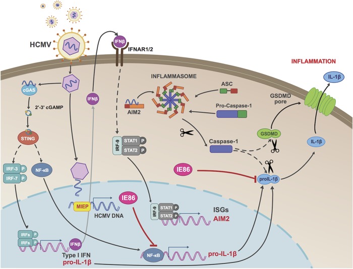
Secretion of interleukin-1β (IL-1β) represents a fundamental innate immune response to microbial infection that, at the molecular level, occurs following activation of proteolytic caspases that cleave the immature protein into a secretable form. Human cytomegalovirus (HCMV) is the archetypal betaherpesvirus that is invariably capable of lifelong infection through the activity of numerous virally encoded immune evasion phenotypes. Innate immune pathways responsive to cytoplasmic double-stranded DNA (dsDNA) are known to be activated in response to contact between HCMV and host cells. Here, we used clustered regularly interspaced short palindromic repeat (CRISPR)-CRISPR-associated protein 9 (Cas9) genome editing to demonstrate that the dsDNA receptor absent in melanoma 2 (AIM2) is required for secretion of IL-1β following HCMV infection. Furthermore, dsDNA-responsive innate signaling induced by HCMV infection that leads to activation of the type I interferon response is also shown, unexpectedly, to play a contributory role in IL-1β secretion. Importantly, we also show that rendering virus particles inactive by UV exposure leads to substantially increased IL-1β processing and secretion and that live HCMV can inhibit this, suggesting the virus encodes factors that confer an inhibitory effect on this response. Further examination revealed that ectopic expression of the immediate early (IE) 86-kDa protein (IE86) is actually associated with a block in transcription of the pro-IL-1β gene and, independently, diminishment of the immature protein. Overall, these results reveal two new and distinct phenotypes conferred by the HCMV IE86 protein, as well as an unusual circumstance in which a single herpesviral protein exhibits inhibitory effects on multiple molecular processes within the same innate immune response.IMPORTANCE Persistent infection with HCMV is associated with the operation of diverse evasion phenotypes directed at antiviral immunity. Obstruction of intrinsic and innate immune responses is typically conferred by viral proteins either associated with the viral particle or expressed immediately after entry. In line with this, numerous phenotypes are attributed to the HCMV IE86 protein that involve interference with innate immune processes via transcriptional and protein-directed mechanisms. We describe novel IE86-mediated phenotypes aimed at virus-induced secretion of IL-1β. Intriguingly, while many viruses target the function of the molecular scaffold required for IL-1β maturation to prevent this response, we find that HCMV and IE86 target the IL-1β protein specifically. Moreover, we show that IE86 impairs both the synthesis of the IL-1β transcript and the stability of the immature protein. This indicates an unusual phenomenon in which a single viral protein exhibits two molecularly separate evasion phenotypes directed at a single innate cytokine.

Keywords: AIM2; DNA sensor; HCMV; IE2; IE86; STING; cGAS; cytomegalovirus; inflammasome; innate immunity; interferons.


Figures

FIG 1
FIG 1
Exposure of differentiated THP-1 cells to live and UV-inactivated HCMV induces secretion of IL-1β. THP-1 cells were differentiated using PMA for 24 h and either left untreated (mock) or exposed to live or UV-inactivated HCMV-TB40/E (MOI of 3) for 18 h. (A) IL-1β secretion in culture medium was measured by ELISA. Values are averages ± standard errors of the means (SEM) from three biological replicates. **, P < 0.01; ***, P < 0.001. (B) Immunoblot (IB) assay showing results for pro-IL-1β, mature processed IL-1β (supernatant), HCMV IE72/IE86, pp65, and GAPDH. (C) Primary monocytes were differentiated using 2.5 mg/ml M-CSF for 6 days and then stimulated with IFN-γ and LPS for 24 h and either left untreated (mock) or exposed to live or UV-inactivated HCMV-TB40/E (MOI of 10) for 18 h. IL-1β secretion in culture medium was measured by ELISA. Values are averages ± SEM from three biological replicates. One-way analysis of variance (ANOVA) with Bonferroni correction was performed. *, P < 0.05; ***, P < 0.001.
FIG 2
FIG 2
Secretion of IL-1β in response to live and UV-inactivated HCMV requires the AIM2 inflammasome. (A) Immunoblot assay for caspase-1, ASC, NLRP3, IFI16, AIM2, and GAPDH in THP-1 cells transduced with Cas9 and guide RNA (gRNA) targeting HUMCYC pseudogene or in indicated CRISPR-generated knockout cells. (B) IL-1β secretion in THP-1 cells with the HUMCYC pseudogene or gene encoding an indicated protein deleted was measured following mock treatment (white) or treatment with live (gray) or UV-inactivated (black) HCMV (HCMV-TB40/E; MOI of 3) for 18 h. Values are averages ± SEM from three biological replicates. (C) IL-1β secretion in differentiated THP-1 cells following mock treatment or treatment with UV-HCMV (UV) alone or in combination with caspase-1 inhibitor (YVAD), caspase-4 inhibitor (LEVD), caspase-5 inhibitor (WEHD), or caspase-8 inhibitor (IETD) for 18 h. Values are averages ± SEM from three replicates. One-way ANOVA with Bonferroni correction was performed. **, P < 0.01.
FIG 3
FIG 3
The cGAS-STING-IFN pathway contributes to IL-1β secretion in response to UV-inactivated HCMV. (A) Immunoblot assay for cGAS, STING, IFNAR1, IRF3/IRF7, and GAPDH in THP-1 cells transduced with Cas9 and gRNA targeting HUMCYC pseudogene or in indicated CRISPR-generated knockout cells. WT, wild type. (B, C) Pro-IL-1β (B) and AIM2 (C) mRNA levels in THP-1 cells transduced with Cas9 and gRNA targeting the HUMCYCPS3 (ΔHUMCYC) pseudogene or indicated cellular gene(s) following treatment with UV-inactivated HCMV-TB40/E (MOI of 3) for 18 h. Transcript fold changes are expressed as relative to the levels in untreated cells (PMA−). (D) IL-1β secretion in THP-1 cells lacking indicated protein(s) following exposure to UV-inactivated HCMV-TB40/E (MOI of 3) for 18 h. Values are averages ± SEM from three biological replicates. One-way ANOVA with Bonferroni correction was performed. *, P < 0.05; **, P < 0.01; ***, P < 0.001.
FIG 4
FIG 4
Infection of THP-1 cells with live HCMV inhibits IL-1β secretion in response to UV-HCMV. (A) Schematic depicting five experimental treatments of differentiated THP-1 cells, including mock treatment (condition M), UV-inactivated HCMV for 24 h (condition 1), live HCMV for 24 h (condition 2), UV-inactivated HCMV for 12 h and then retreatment with UV-inactivated virus for 12 h (condition 3), or live HCMV for 12 h and then UV-HCMV virus for 12 h (condition 4). (B) IL-1β secretion measured by ELISA following indicated treatments. (C, D) mRNA levels for IE86 (C) and pro-IL-1β (D) measured by qPCR following indicated treatments. Transcript fold changes are expressed as relative to levels in untreated cells (mock). Values are averages ± SEM from three biological replicates. One-way ANOVA with Bonferroni correction was performed. *, P < 0.05; ***, P < 0.001. (E) Differentiated wild-type (WT) or ΔCASP-1 THP-1 cells were infected with HCMV-TB40/E (MOI of 3) for 6 days, and titers in supernatants from these cells were determined on human fibroblasts. Values are averages ± SEM from three biological replicates. ffu, focus-forming units. (F) Differentiated ΔHUMCYC or ΔIL-1β THP-1 cells were infected with HCMV-TB40/E (MOI of 3) for 6 days, and titers in supernatants from these cells were determined on human fibroblasts. Values are averages ± SEM from three biological replicates. Immunoblot assay shows successful knockout of IL-1β in ΔIL-1β cells. (G) WT or ΔCASP-1 THP-1 cells were infected with HCMV-TB40/E (MOI of 10) for 5 days and then treated with PMA to induce HCMV reactivation. Supernatants were collected 7 days post-PMA treatment, and titers determined on human fibroblasts. Values are averages ± SEM from three biological replicates. (H) Telomerized human fibroblasts (THF) were treated with 2 ng/ml of recombinant IL-1β or with 1,000 U of IFN-β overnight and then infected with HCMV-TB40/E (MOI of 3) for 5 days. Titers in supernatants from THF were next determined on human fibroblasts. Values are averages ± SEM from three biological replicates. One-way ANOVA with Bonferroni correction was performed. *, P < 0.05.
FIG 5
FIG 5
UV-HCMV-induced IL-1β secretion is blocked in the presence of HCMV IE86 but not IE72 protein. (A) Immunoblot assay showing results for IE72, IE86, and GAPDH in THP-1 cells ectopically expressing IE72 (THP-1-IE72) or IE86 (THP-1-IE86) under the control of a doxycycline (DOX)-inducible promoter in the presence but not absence of DOX. (B) IL-1β secretion from THP-1-IE72 and THP-1-IE86 cells left untreated (mock) or following exposure to UV-inactivated HCMV-TB40/E (MOI of 3) for 18 h in the presence (gray) or absence (black) of DOX. Values are averages ± SEM from three biological replicates. One-way ANOVA with Bonferroni correction was performed. n.s., not significant; **, P < 0.01.
FIG 6
FIG 6
HCMV IE72 and IE86 do not inhibit UV-HCMV-induced caspase-1 cleavage, IL-18 secretion, or GSDMD cleavage. (A) Immunoblot assay for pro-IL-1β, pro-caspase-1, active caspase-1, IE72/IE86, and GAPDH in THP-1-IE72 and THP-1-IE86 cells either left untreated (mock) or treated with UV-inactivated HCMV-TB40/E (MOI of 3) for 18 h in the presence or absence of doxycycline. (B) IL-18 secretion in THP-1-IE86 cells left untreated (mock) or infected with UV-inactivated HCMV-TB40/E (MOI of 3) for 18 h in the presence (gray) or absence (black) of doxycycline. Values are averages ± SEM from three biological replicates. One-way ANOVA with Bonferroni correction was performed. n.s., not significant. (C) Immunoblot assay for total and cleaved (*) GSDMD and GAPDH in THP-1-IE86 cells either untreated (M) or infected with UV-inactivated (UV) HCMV-TB40/E (MOI of 3) for 18 h in the absence (−) or presence (+) of doxycycline.
FIG 7
FIG 7
HCMV IE86 impairs transcription of endogenous but not constitutively expressed ectopic pro-IL-1β. (A) Pro-IL-1β mRNA levels in THP-1-IE72 or THP-1-IE86 cells left untreated (mock) or infected with UV-inactivated HCMV-TB40/E (MOI of 3) for 18 h in the presence (gray) or absence (black) of doxycycline. (B) Immunoblot assay for NF-κB subunits P65 and P50, as well as GAPDH, from parental THP-1 cells or those from which the indicated proteins were deleted using CRISPR-Cas9. (C) Pro-IL-1β mRNA levels in control (ΔHUMCYC) THP-1 cells or those from which P65/P50 have been deleted, as indicated, following treatment with PMA or UV-HCMV (MOI of 3). Values presented are mean mRNA fold changes ± SEM calculated relative to levels in control cells not treated with PMA (PMA−). (D) THP-1 NF-κB Quanti-Blue reporter cells were transduced with recombinant adenovirus expressing IE72 or IE86 (AdIE72 and AdIE86) overnight and then mock treated or treated with UV-HCMV (MOI of 3) for an additional 18 h. Values are averages ± SEM from three replicates. One-way ANOVA with Bonferroni correction was performed. n.s., not significant; **, P < 0.01. (E) Levels of Myc-specific mRNA in THP-1-IE86 cells ectopically expressing Myc-tagged pro-IL-1β that were either left untreated (mock) or infected with UV-inactivated HCMV-TB40/E (MOI of 3) for 18 h in the presence (gray) or absence (black) of doxycycline. Data are expressed as average levels of transcript ± SEM relative to levels in untreated cells (mock) from three biological replicates. (F) Constitutive ectopic pro-IL-1β mRNA degradation was examined by quantifying transcript levels at indicated times after the addition of actinomycin D using qPCR in the presence (gray) or absence (black) of IE86. Transcript levels are expressed relative to levels in DMSO-treated cells. (G) IL-1β secretion in THP-1-IE86 cells ectopically expressing Myc-tagged pro-IL-1β that were either left untreated (mock) or infected with UV-inactivated HCMV (MOI of 3) for 18 h in the presence (gray) or absence (black) of doxycycline. (H) Immunoblot assay for pro-IL-1β (ectopic and endogenous, as indicated), Myc (ectopic pro-IL-1β), IE86, and GAPDH in THP-1-IE86 cells ectopically expressing Myc-tagged pro-IL-1β under a constitutive promoter. (I) Immunoblot assay for pro-IL-18, pro-IL-1β, IE86, and GAPDH in THP-1-IE86 cells ectopically expressing pro-IL-18 under a constitutive promoter.
FIG 8
FIG 8
Levels of pro-IL-1β are reduced in HCMV-positive cells. (A) Immunofluorescence microscopy showing HCMV IE expression in THP-1 cells infected at the indicated MOI overnight. Yellow markers indicate DAPI-stained cells that lack detectable nuclear IE protein. (B) Immunoblot assay for GSDMD in ΔHUMCYC and ΔGSDMD THP-1 cells. (C, D) Intracellular staining for IL-1β in ΔGSDMD EF1α-pro-IL-1β THP-1 cells either mock infected or infected with HCMV-TB40/E (MOI of 3) for 18 h (C) and gated for virally encoded GFP (D). GFP-positive (GFP+) population depicts the number of HCMV-infected cells in the sample. The bar graphs show the mean fluorescence intensities (MFI) for IL-1β staining. The insets show IL-1β staining in the indicated populations. Values are averages ± SEM from two biological replicates. Student’s t test was performed. *, P < 0.05; **, P < 0.01.
FIG 9
FIG 9
Proposed model of IFN and inflammasome activation by HCMV and IL-1β inhibition by IE86. Cellular entry of HCMV particles into THP-1 cells is detected by the dsDNA-reactive PRR cGAS, which synthesizes 2′,3′-cGAMP and triggers STING-mediated activation of IRFs and NF-κB. These then induce transcription of type I IFN and pro-IL-1β, and the IFN-dependent pathway drives AIM2 expression, enabling virus-mediated activation of the inflammasome, formation of GSDMD pores, and IL-1β secretion. Expression of the HCMV IE86 protein counteracts this response both by inhibiting pro-IL-1β transcription and by leading to degradation of the pro-IL-1β protein. MIEP, major immediate-early promoter; ISGs, interferon-stimulated genes.

Similar articles

Cited by

References

    1. Brubaker SW, Bonham KS, Zanoni I, Kagan JC. 2015. Innate immune pattern recognition: a cell biological perspective. Annu Rev Immunol 33:257–290. doi:10.1146/annurev-immunol-032414-112240. - DOI - PMC - PubMed
    1. Thompson MR, Kaminski JJ, Kurt-Jones EA, Fitzgerald KA. 2011. Pattern recognition receptors and the innate immune response to viral infection. Viruses 3:920–940. doi:10.3390/v3060920. - DOI - PMC - PubMed
    1. Mathur A, Hayward JA, Man SM. 2018. Molecular mechanisms of inflammasome signaling. J Leukoc Biol 103:233–257. doi:10.1189/jlb.3MR0617-250R. - DOI - PubMed
    1. Broz P, Dixit VM. 2016. Inflammasomes: mechanism of assembly, regulation and signalling. Nat Rev Immunol 16:407–420. doi:10.1038/nri.2016.58. - DOI - PubMed
    1. Wen H, Miao EA, Ting JPY. 2013. Mechanisms of NOD-like receptor-associated inflammasome activation. Immunity 39:432–441. doi:10.1016/j.immuni.2013.08.037. - DOI - PMC - PubMed