

Structural, functional, and behavioral insights of dopamine dysfunction revealed by a deletion in SLC6A3
- PMID: 30755521
- PMCID: PMC6397532
- DOI: 10.1073/pnas.1816247116
Structural, functional, and behavioral insights of dopamine dysfunction revealed by a deletion in SLC6A3
Abstract
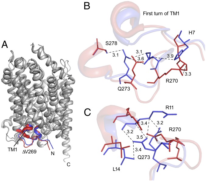
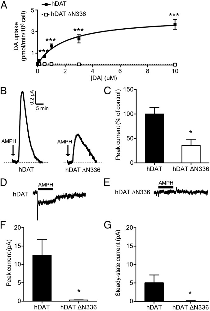
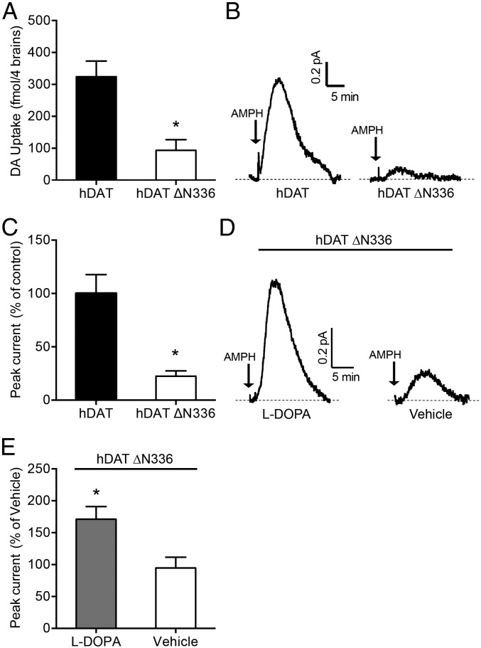
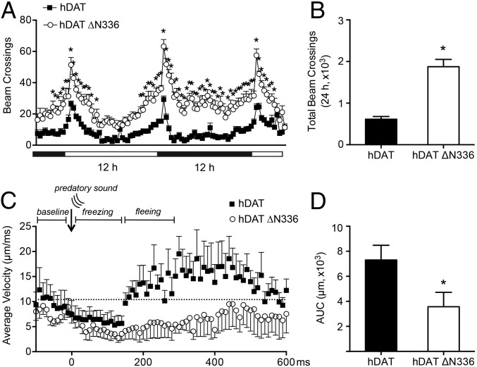
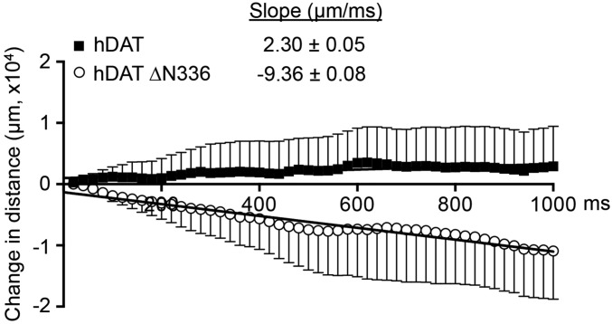
The human dopamine (DA) transporter (hDAT) mediates clearance of DA. Genetic variants in hDAT have been associated with DA dysfunction, a complication associated with several brain disorders, including autism spectrum disorder (ASD). Here, we investigated the structural and behavioral bases of an ASD-associated in-frame deletion in hDAT at N336 (∆N336). We uncovered that the deletion promoted a previously unobserved conformation of the intracellular gate of the transporter, likely representing the rate-limiting step of the transport process. It is defined by a "half-open and inward-facing" state (HOIF) of the intracellular gate that is stabilized by a network of interactions conserved phylogenetically, as we demonstrated in hDAT by Rosetta molecular modeling and fine-grained simulations, as well as in its bacterial homolog leucine transporter by electron paramagnetic resonance analysis and X-ray crystallography. The stabilization of the HOIF state is associated both with DA dysfunctions demonstrated in isolated brains of Drosophila melanogaster expressing hDAT ∆N336 and with abnormal behaviors observed at high-time resolution. These flies display increased fear, impaired social interactions, and locomotion traits we associate with DA dysfunction and the HOIF state. Together, our results describe how a genetic variation causes DA dysfunction and abnormal behaviors by stabilizing a HOIF state of the transporter.
Keywords: amphetamine; autism; dopamine transporter; efflux; leucine transporter.
Conflict of interest statement
The authors declare no conflict of interest.
Figures


Similar articles
-
Psychomotor impairments and therapeutic implications revealed by a mutation associated with infantile Parkinsonism-Dystonia.Elife. 2021 May 18;10:e68039. doi: 10.7554/eLife.68039. Elife. 2021. PMID: 34002696 Free PMC article.
-
De novo mutation in the dopamine transporter gene associates dopamine dysfunction with autism spectrum disorder.Mol Psychiatry. 2013 Dec;18(12):1315-23. doi: 10.1038/mp.2013.102. Epub 2013 Aug 27. Mol Psychiatry. 2013. PMID: 23979605 Free PMC article.
-
A juxtamembrane mutation in the N terminus of the dopamine transporter induces preference for an inward-facing conformation.Mol Pharmacol. 2009 Mar;75(3):514-24. doi: 10.1124/mol.108.048744. Epub 2008 Dec 19. Mol Pharmacol. 2009. PMID: 19098122 Free PMC article.
-
ADHD and the dopamine transporter: are there reasons to pay attention?Handb Exp Pharmacol. 2006;(175):373-415. doi: 10.1007/3-540-29784-7_17. Handb Exp Pharmacol. 2006. PMID: 16722244 Review.
-
The dopamine transporter role in psychiatric phenotypes.Am J Med Genet B Neuropsychiatr Genet. 2018 Mar;177(2):211-231. doi: 10.1002/ajmg.b.32578. Epub 2017 Aug 2. Am J Med Genet B Neuropsychiatr Genet. 2018. PMID: 28766921 Review.
Cited by
-
Dopamine Transporter Deficient Rodents: Perspectives and Limitations for Neuroscience.Biomolecules. 2023 May 9;13(5):806. doi: 10.3390/biom13050806. Biomolecules. 2023. PMID: 37238676 Free PMC article. Review.
-
The State of the Dopaminergic and Glutamatergic Systems in the Valproic Acid Mouse Model of Autism Spectrum Disorder.Biomolecules. 2022 Nov 15;12(11):1691. doi: 10.3390/biom12111691. Biomolecules. 2022. PMID: 36421705 Free PMC article.
-
Identifying dominant-negative actions of a dopamine transporter variant in patients with parkinsonism and neuropsychiatric disease.JCI Insight. 2021 Sep 22;6(18):e151496. doi: 10.1172/jci.insight.151496. JCI Insight. 2021. PMID: 34375312 Free PMC article.
-
Gingerol inhibits cisplatin-induced acute and delayed emesis in rats and minks by regulating the central and peripheral 5-HT, SP, and DA systems.J Nat Med. 2020 Mar;74(2):353-370. doi: 10.1007/s11418-019-01372-x. Epub 2019 Nov 25. J Nat Med. 2020. PMID: 31768887 Free PMC article.
-
Heterogeneity of dopamine release sites in health and degeneration.Neurobiol Dis. 2020 Feb;134:104633. doi: 10.1016/j.nbd.2019.104633. Epub 2019 Nov 5. Neurobiol Dis. 2020. PMID: 31698055 Free PMC article. Review.
References
-
- Koob GF, Bloom FE. Cellular and molecular mechanisms of drug dependence. Science. 1988;242:715–723. - PubMed
Publication types
MeSH terms
Substances
Associated data
- Actions
Grants and funding
- R01 DA035263/DA/NIDA NIH HHS/United States
- R01 MH070039/MH/NIMH NIH HHS/United States
- R01 GM080403/GM/NIGMS NIH HHS/United States
- T32 NS007491/NS/NINDS NIH HHS/United States
- R01 DA038058/DA/NIDA NIH HHS/United States
- S10 RR027091/RR/NCRR NIH HHS/United States
- U54 HD083211/HD/NICHD NIH HHS/United States
- R25 GM062459/GM/NIGMS NIH HHS/United States
- U54 GM087519/GM/NIGMS NIH HHS/United States
- R01 HL122010/HL/NHLBI NIH HHS/United States
- R37 MH070039/MH/NIMH NIH HHS/United States
- F31 MH114316/MH/NIMH NIH HHS/United States
LinkOut - more resources
Full Text Sources
Medical
Molecular Biology Databases
