. 2019 Feb 26;116(9):3893-3898.
doi: 10.1073/pnas.1814015116. Epub 2019 Feb 12.

PILS6 is a temperature-sensitive regulator of nuclear auxin input and organ growth in Arabidopsis thaliana

Affiliations

PILS6 is a temperature-sensitive regulator of nuclear auxin input and organ growth in Arabidopsis thaliana

Elena Feraru et al. Proc Natl Acad Sci U S A. .

Abstract

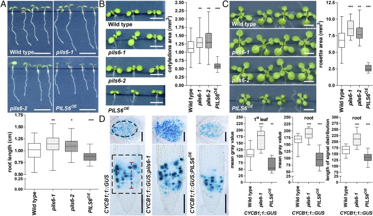
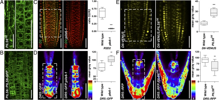
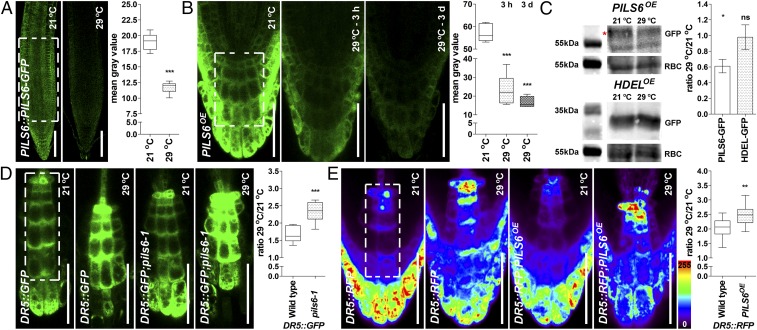
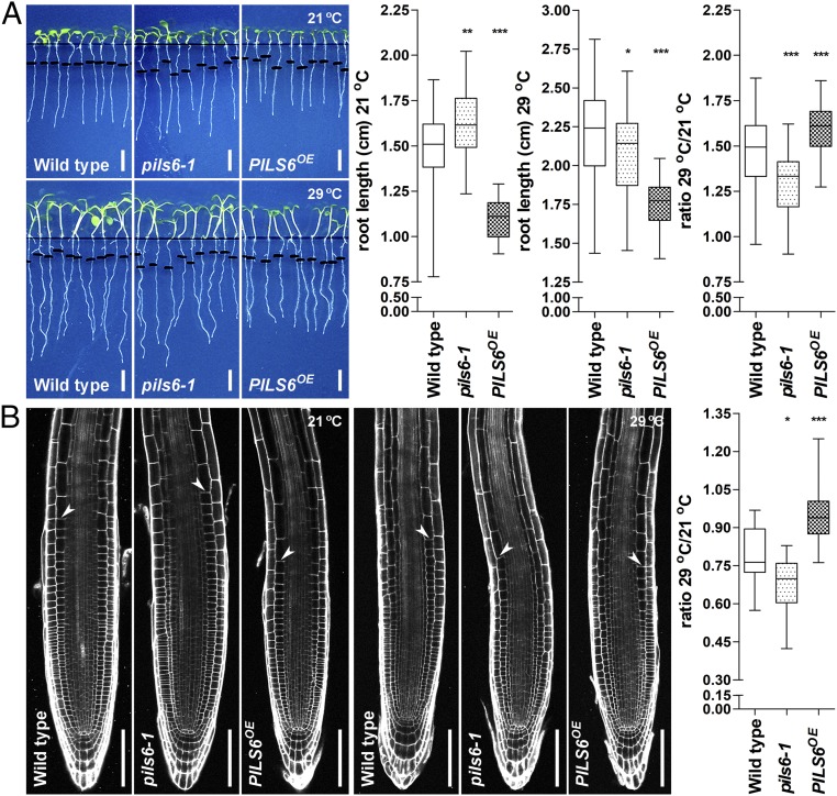
Temperature modulates growth and development throughout the entire lifecycle of a plant. High temperature (HT) triggers the auxin biosynthesis-dependent growth in aerial tissues. On the other hand, the contribution of auxin to HT-induced root growth is currently under debate. Here we show that the putative intracellular auxin carrier PIN-LIKES 6 (PILS6) is a negative regulator of organ growth and that its abundance is highly sensitive to HT. PILS6 localizes to the endoplasmic reticulum and limits the nuclear availability of auxin, consequently reducing the auxin signaling output. HT represses the PILS6 protein abundance, which impacts on PILS6-dependent auxin signaling in roots and root expansion. Accordingly, we hypothesize that PILS6 is part of an alternative mechanism linking HT to auxin responses in roots.

Keywords: PILS proteins; auxin signaling; endoplasmic reticulum; high temperature; root growth.


Conflict of interest statement

The authors declare no conflict of interest.

Figures

Fig. 1.
Fig. 1.
PILS6 is a negative regulator of organ growth. (AC) PILS6 affects organ growth. Scanned images and measurements show that total root length (A), cotyledon area (B), and rosette area (C) are affected in the pils6 mutants and PILS6OE compared with the wild-type control. (D) PILS6 affects cell division. GUS images and measurements of CYCB1;1::GUS expression (first leaf in Upper images, root in Lower images) and root meristem distribution show that cell division is affected in the pils6 mutant and PILS6OE compared with the wild-type control. The black dashed boxes represent the ROIs used to quantify CYCB1;1::GUS signal intensity. The red dashed line shows how the length of CYCB1;1::GUS distribution in the meristem was quantified. [Scale bars, 500 μm (AC) and 100 μm (D).] n = 49–55 roots (A), 50–56 cotyledons (B), 28 rosettes (C), 10–12 first leaves (D), and 28 roots (D). *P = 0.01–0.05, **P = 0.001–0.01, ***P < 0.001, one-way ANOVA.
Fig. 2.
Fig. 2.
ER-localized PILS6 affects the nuclear availability of auxin. (A and B) PILS6 localizes to the ER. Confocal images of PILS6-GFP in the elongated (Left image) and meristematic (Right image) cells of PILS6OE (A) and meristematic cells of PILS6::PILS6-GFP (B) show ER-like distribution in roots. (CF) PILS6 affects auxin signaling. Merged confocal images of R2 (red) and D2 (green) expressing R2D2 marker and quantification of D2/R2 ratio (C) show enhanced degradation of D2 signal in the pils6-1 mutant. Pseudocolored confocal images and fluorescence intensity quantification of DR5rev::GFP (D) or DR5rev::mRFP1er (F) in the root tip show stronger signal in the pils6-1 (D) and weaker signal in the PILS6OE (F) compared with the wild type. Note that in the pseudocolored visualization, red indicates high and blue indicates low intensity (see the color code bar). Confocal images and quantification of DII-VENUS (E) show enhanced fluorescence in the PILS6OE compared with the wild type. The white dashed boxes represent defined ROIs used to quantify the respective signal intensities. [Scale bars, 50 μm (AF).] n = 9 (C), 6 (D), 7 (E), and 6 (F) roots. **P = 0.001–0.01, ***P < 0.001, Student’s t test.
Fig. 3.
Fig. 3.
HT affects PILS6 protein level and PILS6-dependent auxin signaling. (AC) HT down-regulates PILS6 protein level. Confocal images and quantification of PILS6-GFP fluorescence show that HT reduces the PILS6 protein levels in roots of PILS6::PILS6-GFP (A) and PILS6OE (B). Note that the response of PILS6 protein to HT is fast, showing dramatic reduction (about 60%) already after 3 h. Immunoblot with anti-GFP antibody and quantification of signal intensity (C) showing that HT down-regulates protein levels in PILS6OE seedlings, but not in 35S::HDEL-GFP (HDELOE) control line. RuBisCo (RBC) antibody was used for normalization. The red asterisk marks the PILS6-GFP, which runs above a nonspecific band. Please note that the 55kDa molecular weight marker for RBC is duplicated because the loading controls originated from the same protein immunoblot. The statistical evaluation shows the differences between the respective 21 °C and 29 °C values. (D and E) HT affects PILS6-dependent auxin signaling. Confocal images and quantification of DR5::GFP (D) or DR5::RFP (E), showing a relative increase in auxin response in roots of pils6-1 (D) and PILS6OE (E) when exposed to HT. The white dashed boxes represent defined ROIs in the most representative regions of the root, used to quantify the respective signal intensities. [Scale bars, 100 μm (A) and 50 μm (B, D, and E).] n = 7 (A), 6 (B), 9 (D), and 12 (E) roots. ns, not significant; *P = 0.01–0.05, **P = 0.001–0.01, ***P < 0.001, Student’s t test (A and C–E) or one-way ANOVA (B).
Fig. 4.
Fig. 4.
PILS6 regulates root growth under HT. (A and B) PILS6 affects root growth under HT. Scanned images, quantification of the root segment grown after 3-d exposure to 21 °C (control) or 29 °C (HT), and calculated ratio showing that pils6-1 and PILS6OE affect root growth under HT compared with the wild type (A). Confocal images and (relative) quantification of wild type, pils6-1, and PILS6OE root meristems exposed for 3 d to 21 °C or 29 °C (B). Root meristem is depicted as the distance between the quiescent center and the uppermost first cortical cell that is twice as long as it is wide. [Scale bars, 500 μm (A) and 100 μm (B).] n = 32–34 roots (A) and 20 root meristems (B). *P = 0.01–0.05, **P = 0.001–0.01, ***P < 0.001, one-way ANOVA.

References

    1. Rivero L, et al. Handling Arabidopsis plants: Growth, preservation of seeds, transformation, and genetic crosses. Methods Mol Biol. 2014;1062:3–25. - PubMed
    1. Liu J, Feng L, Li J, He Z. Genetic and epigenetic control of plant heat responses. Front Plant Sci. 2015;6:267. - PMC - PubMed
    1. Davies PJ. The Plant Hormones: Their Nature, Occurrence, and Functions. Plant Hormones. Springer; Dordrecht: 2010. pp. 1–15.
    1. Shibasaki K, Rahman A. Auxin and temperature stress: Molecular and cellular perspectives. In: Chen R, Baluška F, editors. Polar Auxin Transport. Springer; Berlin: 2013. pp. 295–310.
    1. Ahammed GJ, Li X, Zhou J, Zhou Y-H, Yu J-Q. Role of hormones in plant adaptation to heat stress. In: Ahammed GJ, Yu J-Q, editors. Plant Hormones Under Challenging Environmental Factors. Springer Netherlands; Dordrecht: 2016. pp. 1–21.

Publication types

MeSH terms