. 2019 Feb 26;116(9):3614-3623.
doi: 10.1073/pnas.1803794116. Epub 2019 Feb 12.

Stage-dependent cardiac regeneration in Xenopus is regulated by thyroid hormone availability

Affiliations

Stage-dependent cardiac regeneration in Xenopus is regulated by thyroid hormone availability

Lindsey N Marshall et al. Proc Natl Acad Sci U S A. .

Abstract

Despite therapeutic advances, heart failure is the major cause of morbidity and mortality worldwide, but why cardiac regenerative capacity is lost in adult humans remains an enigma. Cardiac regenerative capacity widely varies across vertebrates. Zebrafish and newt hearts regenerate throughout life. In mice, this ability is lost in the first postnatal week, a period physiologically similar to thyroid hormone (TH)-regulated metamorphosis in anuran amphibians. We thus assessed heart regeneration in Xenopus laevis before, during, and after TH-dependent metamorphosis. We found that tadpoles display efficient cardiac regeneration, but this capacity is abrogated during the metamorphic larval-to-adult switch. Therefore, we examined the consequence of TH excess and deprivation on the efficiently regenerating tadpole heart. We found that either acute TH treatment or blocking TH production before resection significantly but differentially altered gene expression and kinetics of extracellular matrix components deposition, and negatively impacted myocardial wall closure, both resulting in an impeded regenerative process. However, neither treatment significantly influenced DNA synthesis or mitosis in cardiac tissue after amputation. Overall, our data highlight an unexplored role of TH availability in modulating the cardiac regenerative outcome, and present X. laevis as an alternative model to decipher the developmental switches underlying stage-dependent constraint on cardiac regeneration.

Keywords: Xenopus; cardiac regeneration; extracellular matrix; metamorphosis; thyroid hormone.


Conflict of interest statement

The authors declare no conflict of interest.

Figures
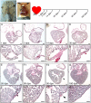
Fig. 1.
Fig. 1.
Transient fibrotic response followed by complete ventricle rebuilding after cardiac resection in tadpole heart. (Top) Cardiac resection was performed on prometamorphic NF57 X. laevis tadpoles by dissecting the abdominal skin and opening the pericardium, then removing ∼10–15% of the ventricle toward the apex. Hearts were collected at 1, 3, 14, 30, 50, 90, and 180 dpa. (AG) PSR histological labeling reveals an intensification of fibrotic connective tissue up to 14 dpa at the site of amputation (labeled in dark red). From 30 dpa up to 90 dpa, the fibrotic zone regressed and progressively disappeared as the ventricle apex regained a normal morphology, and was strongly reduced or undetectable after 90 (F2 and F1, respectively) or 180 dpa (G). (A′G′) Higher magnifications, with dotted lines indicating amputated area and arrows indicating the amputation site. (Scale bars, 100 μm.)
Fig. 2.
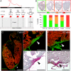
Fig. 2.
Cardiac regenerative capacity is reduced then permanently lost after metamorphosis. (A) Cardiac resection was performed at different developmental stages before, during, and after TH-dependent metamorphosis: prometamorphic stages (NF57–58), climax (NF61–62), end (NF65–66), and just after metamorphosis (froglet), and in 6-mo-old juveniles. Hearts were collected 6 mpa for metamorphic stages (NF57 to froglet), and ≥1 ypa for juveniles. (B) Using PSR histological staining, hearts were categorized and quantified according to different heart phenotypes observed: fully regenerated (green: no fibrotic tissue present at the amputated site), partially regenerated (orange: fibrotic tissue remaining present at the amputated site), and those displaying an absence of regeneration (red: strong appearance of a fibrotic scar at the amputated site); arrowheads indicate the amputation site. All samples were obtained from more than four independent experiments: NF57–58, n = 4; NF61–62, n = 15; NF65–66, n = 8; froglet, n = 6, juvenile, n = 7. Immunolabeling (C and D) fibronectin (green), tropomyosin (red), and PSR staining (E) of a juvenile heart at 1 ypa (n = 5) displaying a clear presence of invasive fibrotic tissue at the amputation site (arrowhead). Immunolabeling (F) and PSR staining (G) of a juvenile heart at 3.3 ypa (n = 2) still showing a strong presence of fibrotic tissue and, hence, a persistent scar at the amputation site (arrowhead). (Scale bars, 1 mm inC and 0.5 mm in DG.)
Fig. 3.
Fig. 3.
Increased DNA synthesis is observed in tadpole and juvenile cardiomyocyte nuclei after heart amputation. (A) DNA synthesis in cardiomyocytes was assessed in amputated tadpole and juvenile hearts using EdU treatment for 1 wk (pulse), followed by a 1-wk chase period. (B and C) Immunolabeling with Mef2 antibody (green) and EdU detection (red) at 14 dpa. DNA synthesis in cardiomyocytes, characterized by colabeled Mef2+/EdU+ (yellow) nuclei, are observed in both tadpole (B, and magnification B′) and juvenile hearts (C, and magnification C′); arrowheads indicate the areas magnified in B′ and C′. (DJ) Mef2+ (converted to purple dots) and Mef2+/EdU+ (converted to yellow dots) nuclei were quantified in one-third of the ventricle at the apex for tadpoles (DF) and juveniles (GI): white lines define the corresponding one-third area analyzed for the tadpole (B) and the juvenile (JUV) (C) (see SI Appendix, Fig. S5 for remaining two-thirds quantification). Analyses were performed for nonoperated animals (CTRL, D and G), animals operated without (SHAM, E and H) and with ventricular resection (AMP, F and I). The ratio of colabeled nuclei (Mef2+EdU+) over total number of cardiomyocyte nuclei (Mef2+) was calculated (J). All samples were obtained from two independent experiments. n ≥ 3 for each group and ≥3 sections were quantified for each sample, and graphed, representing mean with SEM. NF57 values were normally distributed, therefore a one-way ANOVA followed by Sidak’s multiple comparisons test was performed. A normal distribution was not observed for JUV; therefore a nonparametric Kruskal–Wallis test followed by a Dunn’s multiple comparisons test was performed (*P < 0.05; **P < 0.01). (Scale bars, 100 μm.)
Fig. 4.
Fig. 4.
Short TH exposure or deprivation before tadpole heart amputation hinders the regenerative capacity. (A) A ventricular resection was performed on tadpoles following a 3-d TH-pretreatment (T3, 10−10 M) or treated with the goitrogen perchlorate for more than 2 mo. Hearts were analyzed at 30, 90, and 150 dpa and compared with nontreated amputated tadpoles as a control (CTRL). (B) Survival curves for all groups. Significantly increased mortality was observed for T3-treated tadpoles compared with CTRL following amputation, whereas no significant difference in survival up to 150 dpa was seen with perchlorate-treated tadpoles. The time period corresponding to the metamorphosis of the experimented tadpoles are indicated. (C) Fibrosis extent drastically diminished between 30 and 90 dpa in CTRL, but remained significantly present for both T3-treated and perchlorate-treated tadpoles at 90 dpa. (D) Representative immunolabelings for cardiomyocytes (tropomyosin, red), fibrous tissue (fibronectin, green), and nuclei (DAPI, blue) focusing on the cardiac amputation site for untreated CTRL, T3-treated, and perchlorate-treated tadpoles at 30 dpa. Black and white pictures showing only the tropomyosin channel are shown for each heart, facilitating myocardial wall continuity assessment. No gaps in the myocardial wall were observed for CTRL ventricles at 30 dpa, whereas the ventricles of T3-treated and perchlorate-treated tadpoles presented an evident absence of myocardium closure, with numerous gaps observed in the myocardial wall at the amputated site for the T3-treated heart (white arrowheads). (E) Immunolabeling for tropomyosin (red), fibronectin (green), and DAPI-labeled nuclei (blue) showed that compared with untreated CTRL, an invasive fibrotic zone remained detectable at the amputation site at 90 dpa in T3-treated tadpoles and perchlorate-treated tadpoles (arrowheads indicate the amputation site). All data were obtained from more than three independent experiments. n = 74 (CTRL), n = 58 (T3), and n = 47 (Perchlo) and a log-rank (Mantel–Cox) test was used to compare survival curves (ns, nonsignificant, P > 0.05; ****P < 0.0001). For fibrosis extent and fibronectin immunolabeling: 30 dpa, n = 16 (CTRL), n = 12 (T3), and n = 16 (Perchlo); 90 dpa, n = 11 (CTRL), n = 8 (T3), and n = 11 (Perchlo), and graph represented as box plot showing median with interquartile range. A nonparametric Mann–Whitney test was performed for fibrosis extent comparison (ns, nonsignificant, P > 0.05; *P < 0.05; **P < 0.01; ****P < 0.0001). (Scale bars, 100 μm in D and 50 μm in E.)
Fig. 5.
Fig. 5.
Short exposure or TH deprivation prior tadpole heart amputation alters extracellular matrix components and not DNA synthesis following injury. (A) Control nontreated and tadpoles either T3-treated or perchlorate-treated were amputated at day 0 and followed the same EdU protocol, as in Fig. 3. Ventricles were analyzed at 14 dpa. (BE) Mef2+ and Mef2+/EdU+ nuclei quantification was performed for nontreated (B, CTRL), T3-treated (C, T3), and perchlorate-treated (D, Perchlo), for nonamputated (NON AMP) and amputated (AMP) hearts, and the ratio (%) Mef2+EdU+/Mef2+ given for each condition (E). Samples were obtained from three independent experiments. n ≥ 3 per group and >3 sections were quantified per sample, and graph represented as mean with SEM. One-way ANOVA followed by Sidak’s multicomparisons test was performed (*P < 0.05; ***P < 0.001). (F) Control untreated, T3-treated, and perchlorate-treated tadpoles were collected at day 0 without amputation and ECM gene expression was quantified. (G) The TH-responsive gene thibz was a positive control, showing a strong induction of its expression in heart treated with T3. (H) The expression of cardiac ECM genes col1a1, fapα, mmp2, mmp9, mmp11, mmp13, and mmp14 showed a differential regulation in T3-treated and perchlorate-treated hearts, excepted tnc and fn1, compared with control hearts. Samples were obtained from >3 independent experiments (CTRL, n = 8; T3, n = 8; Perchlo, n = 6). Fold-change (FC) is shown in log2 scale, respective to CTRL. Data are represented as an aligned dot plot showing median with interquartile range. For normally distributed values (thibz, fn1, col1a1, tnc, mmp1, mmp2, mmp11), a one-way ANOVA followed by Sidak’s multiple comparisons test was performed. For not normal distributions (fapα, mmp9, mmp13, mmp14) a nonparametric Kruskal–Wallis test followed by a Dunn’s multiple comparisons test was performed. (ns, nonsignificant, P > 0.05; *P < 0.05; **P < 0.01; ***P < 0.001; ****P < 0.0001). (IP) Impact of pharmacological treatments on TnC presence at the site of amputation at 30 dpa (IL) and 90 dpa (MP). Representative images are shown for CTRL untreated, T3-treated, and perchlorate-treated tadpoles as well as a quantification of the staining intensity for TnC with the following color scheme: Absence (−, dark blue), slight staining at the ventricle surface (−/+, blue), weak (+, light orange), average (++, medium orange), and strong presence (+++, dark orange). Arrowheads (IO) indicate the site of amputation, magnified in (I′–O′). Data were obtained from more than three independent experiments: 30 dpa, n = 16 (CTRL), n = 12 (T3), and n = 16 (Perchlo); 90 dpa, n = 11 (CTRL), n = 8 (T3), and n = 11 (Perchlo). [Scale bars: 100 μm (BD) 200 μm (I, J, K, M, N, and O) and 50 μm (I′, J′, K′, M′, N′, and O′).]

Similar articles

Cited by

References

    1. World Health Organisation 2018 The top 10 causes of death: Fact sheet. Available at https://www.who.int/en/news-room/fact-sheets/detail/the-top-10-causes-of.... Accessed February 1, 2019.
    1. Tzahor E, Poss KD. Cardiac regeneration strategies: Staying young at heart. Science. 2017;356:1035–1039. - PMC - PubMed
    1. Laflamme MA, Murry CE. Heart regeneration. Nature. 2011;473:326–335. - PMC - PubMed
    1. Gamba L, Harrison M, Lien CL. Cardiac regeneration in model organisms. Curr Treat Options Cardiovasc Med. 2014;16:288. - PMC - PubMed
    1. Vivien CJ, Hudson JE, Porrello ER. Evolution, comparative biology and ontogeny of vertebrate heart regeneration. NPJ Regen Med. 2016;1:16012. - PMC - PubMed

Publication types

MeSH terms

Substances