. 2019 Feb 12;10(2):137.
doi: 10.1038/s41419-019-1352-4.

Long noncoding RNA LINC01234 promotes serine hydroxymethyltransferase 2 expression and proliferation by competitively binding miR-642a-5p in colon cancer

Affiliations

Long noncoding RNA LINC01234 promotes serine hydroxymethyltransferase 2 expression and proliferation by competitively binding miR-642a-5p in colon cancer

Changwei Lin et al. Cell Death Dis. .

Retraction in

Abstract

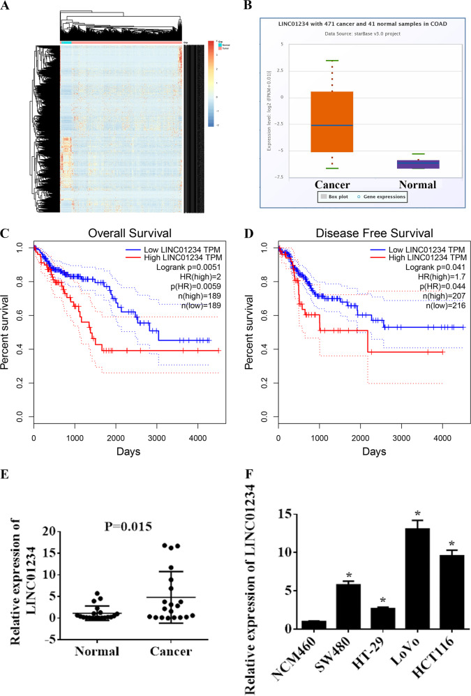
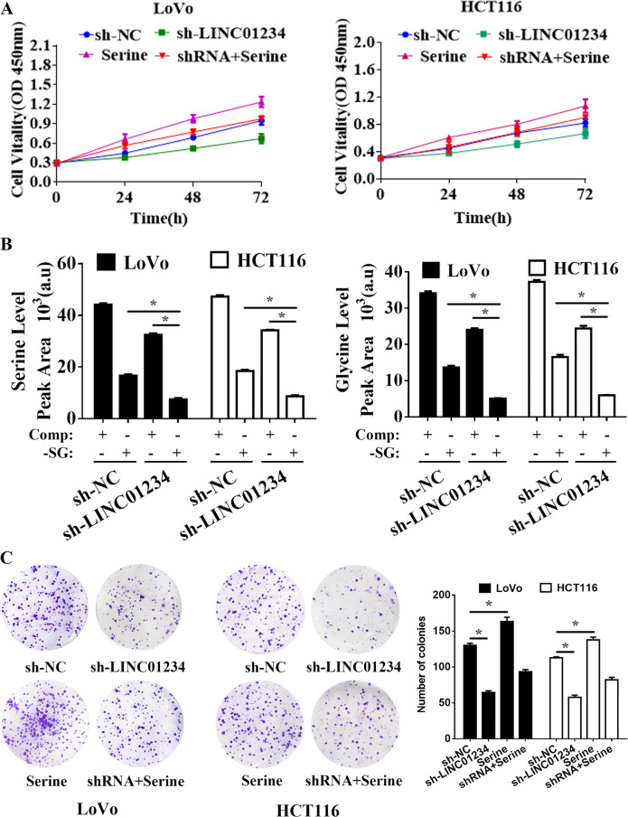
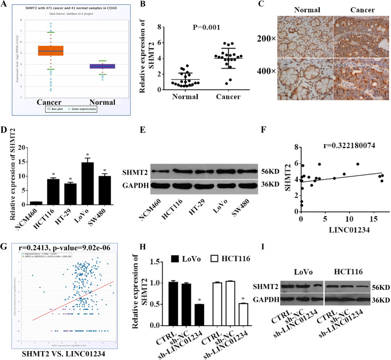
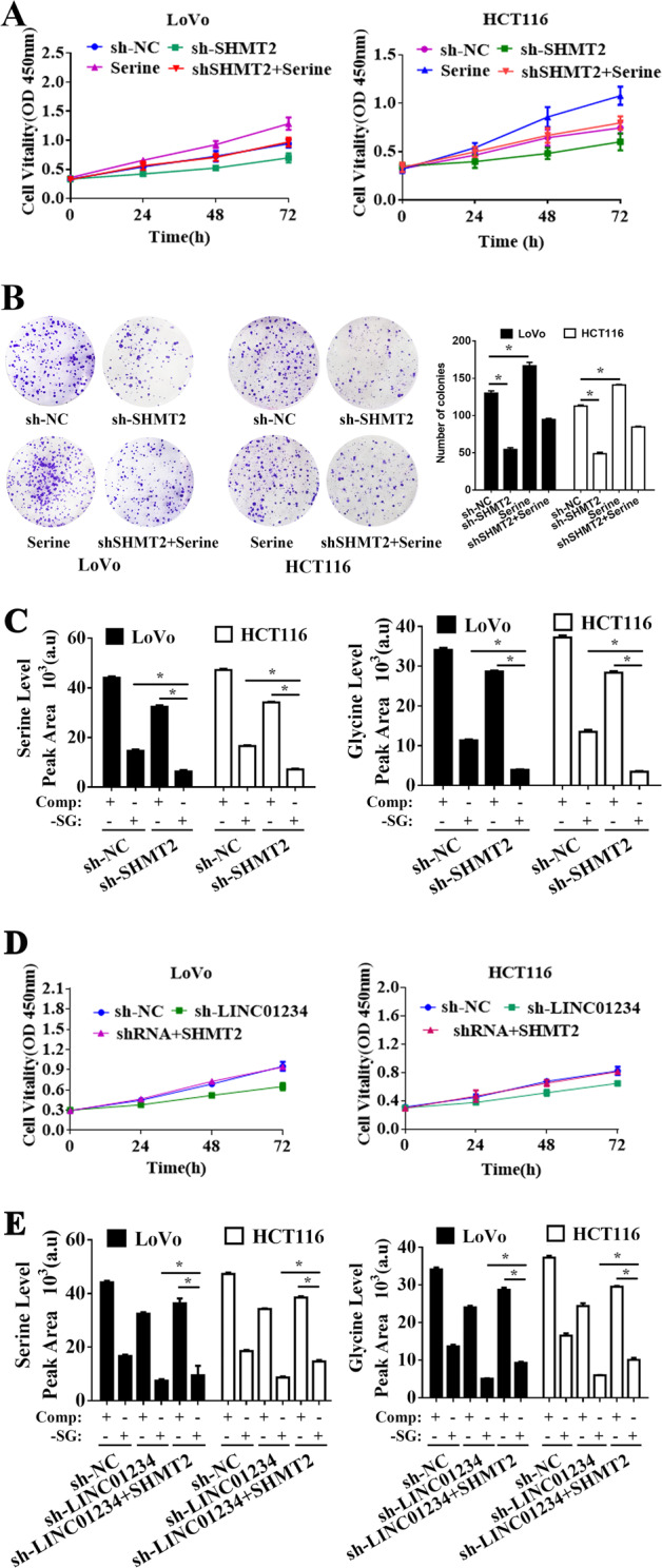
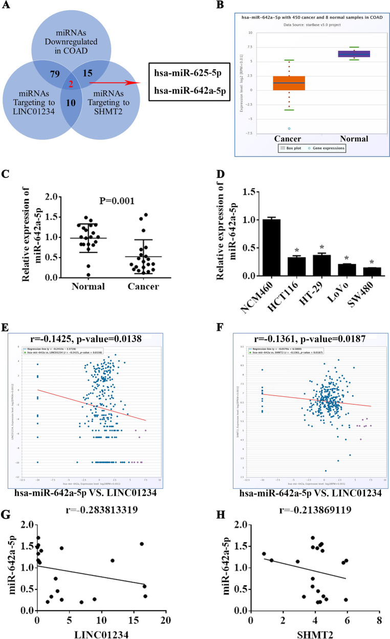
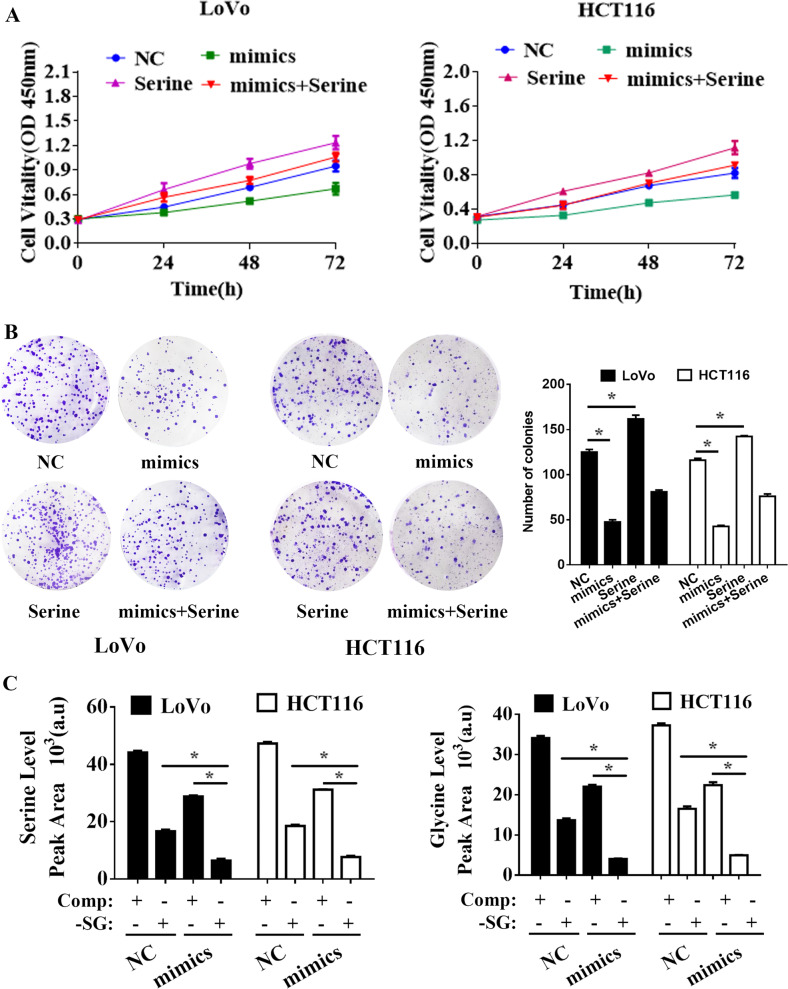
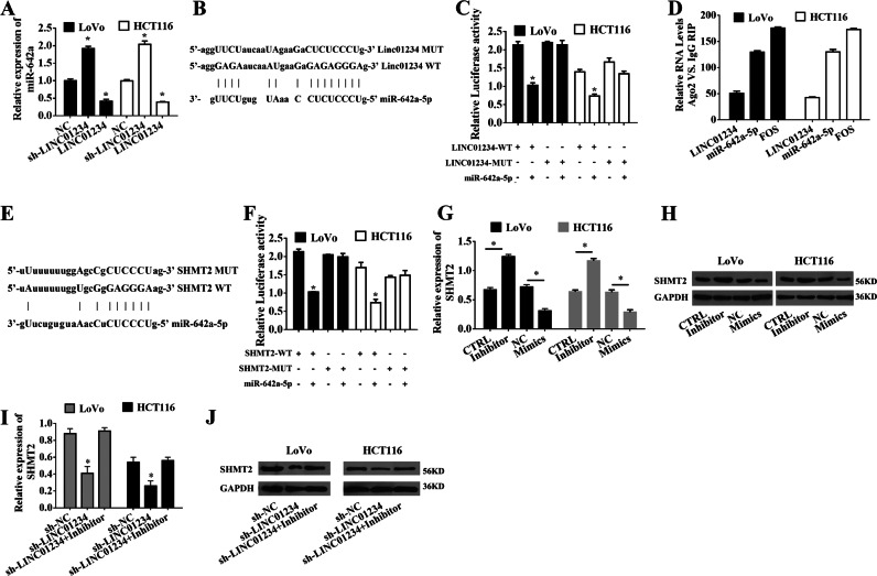
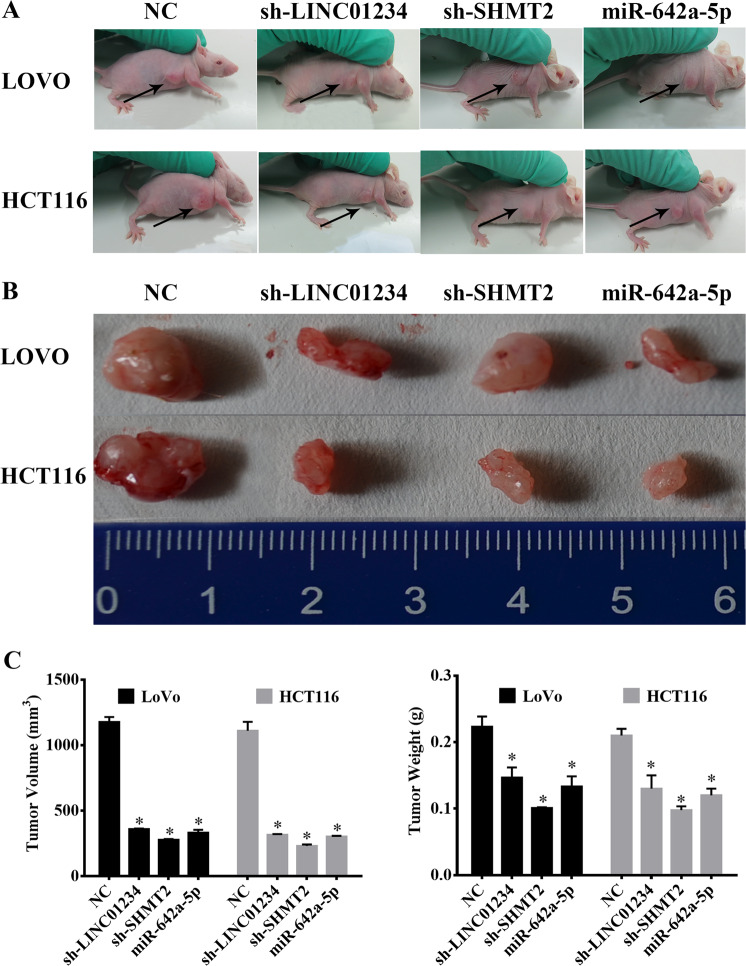
Long noncoding RNAs (lncRNAs) have been indicated as important regulators in various human cancers. However, the overall biological roles and clinical significance of most lncRNAs in colon carcinogenesis are not fully understood. Hence, we investigated the clinical significance, biological function and mechanism of LINC01234 in colon cancer. First, we analyzed LINC01234 alterations in colon cancer tissues and corresponding paracancerous tissues through the analysis of sequencing data obtained from The Cancer Genome Atlas and colon cancer patients. Next, we evaluated the effect of LINC01234 on colon cancer cell proliferation and its regulatory mechanism of serine hydroxymethyltransferase 2 (SHMT2) by acting as a competing endogenous RNA (ceRNA). We found that LINC01234 expression was significantly upregulated in colon cancer tissues and was associated with a shorter survival time. Furthermore, the knockdown of LINC01234 induced proliferation arrest via suppressing serine/glycine metabolism. Mechanistic investigations have indicated that LINC01234 functions as a ceRNA for miR-642a-5p, thereby leading to the derepression of its endogenous target serine hydroxymethyltransferase 2 (SHMT2). LINC01234 is significantly overexpressed in colon cancer, and the LINC01234-miR642a-5p-SHMT2 axis plays a critical role in colon cancer proliferation. Our findings may provide a potential new target for colon cancer diagnosis and therapy.


Conflict of interest statement

The authors declare that they have no conflict of interest.

Figures

Fig. 1
Fig. 1
LINC01234 expression is upregulated in colon cancer and is associated with poor prognosis. a Hierarchically clustered heatmap of upregulated and downregulated lncRNAs in TCGA COAD. b The relative expression of LINC01234 in colon cancer tissues compared with normal tissue was analyzed using TCGA COAD. c, d Kaplan–Meier overall survival and disease-free survival curves according to LINC01234 expression levels. e LINC01234 expression was analyzed by qRT-PCR in colon cancer tissues and corresponding adjacent nontumor tissues, and the data were presented as 2-△CT values (n = 20). f LINC01234 expression was analyzed by qRT-PCR in 4 human colon cancer cell lines (HCT116, HT-29, LoVo, and SW480) and normal human colonic epithelial NCM460 cells. The experiments were performed in triplicate. The data are represented as means ± SD from three independent experiments. *P < 0.05
Fig. 2
Fig. 2
Knockdown of LINC01234 inhibits colon cancer cell proliferation via suppressing serine/glycine metabolism. a MTT assays were used to determine the viability of negative control (NC), sh-LINC01234, serine treatment or serine treatment sh-LINC01234-transfected LoVo cells and HCT116 cells. b Stable isotope tracing experiments. LoVo cells and HCT116 cells expressing sh-NC or sh-LINC01234 were cultured in complete medium (Comp) or serine/glycine-deprived medium (-SG) for 24 h. GC-MS was used to detect the relative intracellular levels of serine (left) or glycine (right). Histobars represent the mean value of the peak area ± SD (arbitrary unit) corresponding to serine and glycine peaks on the MS chromatogram. c Colony formation assays were performed to determine the proliferation of negative control (NC), sh-LINC01234, serine treatment or serine treatment sh-LINC01234-transfected LoVo cells and HCT116 cells. The experiments were performed in triplicate. The data are represented as means ± SD from three independent experiments. *P < 0.05
Fig. 3
Fig. 3
LINC01234 promotes SHMT2 expression in colon cancer. a Relative expression of SHMT2 in colon cancer tissues compared with that of normal tissue was analyzed using TCGA COAD. b SHMT2 expression was analyzed by qRT-PCR in colon cancer tissues and corresponding adjacent nontumor tissues, and the data were presented as 2−△CT values (n = 20). c SHMT2 expression was analyzed by IHC in colon cancer tissues and corresponding adjacent nontumor tissues. d, e. SHMT2 expression was analyzed by qRT-PCR and Western blotting in 4 human colon cancer cell lines (HCT116, HT-29, LoVo, and SW480) and normal human colonic epithelial NCM460 cells. f Association analysis of the relationship between LINC01234 and SHMT2 expression levels in 20 paired colon cancer tissues. g Association analysis of the relationship between LINC01234 and SHMT2 expression levels in TCGA COAD. h, i Relative mRNA and protein levels of SHMT2 in LoVo cells and HCT116 cells transfected with control, sh-NC or sh-LINC01234. The experiments were performed in triplicate. The data are represented as means ± SD from three independent experiments. *P < 0.05
Fig. 4
Fig. 4
LINC01234 activity is partially mediated by the positive regulation of SHMT2. a MTT assays were used to determine the viability of sh-SHMT2-transfected LoVo cells and HCT116 cells. b Colony formation assays were performed to determine the proliferation of sh-NC, sh-SHMT2, serine treatment or serine treatment sh-SHMT2-transfected LoVo cells and HCT116 cells. c Stable isotope tracing experiments. LoVo cells and HCT116 cells expressing sh-NC or sh-SHMT2 were cultured in complete medium (Comp) or serine/glycine-deprived medium (-SG) for 24 h. GC-MS was used to detect the relative intracellular levels of serine (left) or glycine (right). Histobars represent the mean value of the peak area ± SD (arbitrary unit) corresponding to the serine and glycine peaks on the MS chromatogram. d MTT assays were used to determine the viability of LoVo cells and HCT116 cells transfected with NC, sh-SHMT2 or cotransfected sh-LINC01234 and SHMT2-expressing vector. e Stable isotope-tracing experiments. LoVo cells and HCT116 cells transfected with NC, sh-SHMT2 or cotransfected sh-LINC01234 and SHMT2-expressing vector were cultured in complete medium (Comp) or serine/glycine-deprived medium (-SG) for 24 h. GC-MS was used to detect the relative intracellular levels of serine (left) or glycine (right). Histobars represent the mean value of the peak area ± SD (arbitrary unit) corresponding to the serine and glycine peaks on the MS chromatogram. The experiments were performed in triplicate. The data are represented as means ± SD from three independent experiments. *P < 0.05
Fig. 5
Fig. 5
miR-642a-5p may mediate a ceRNA network with LINC01234 and SHMT2. a The number of significantly downregulated miRNAs in TCGA COAD is 290. The numbers of predicted miRNAs of LINC01234 is 332 according to LncBase Predicted v.2. The number of experimentally validated miRNAs of SHMT2 is 120 according to TarBase v7.0. The number of overlapped miRNAs in the above three intersections is 2, indicating that LINC01234 and SHMT2 may share 2 common microRNAs with experimental evidence. b The relative expression of miR-642a-5p in colon cancer tissues compared with normal tissue was analyzed using TCGA COAD. c miR-642a-5p expression was analyzed by qRT-PCR in colon cancer tissues and corresponding adjacent nontumor tissues, and the data are presented as 2-△CT values (n = 20). d LINC01234 expression was analyzed by qRT-PCR in 4 human colon cancer cell lines (HCT116, HT-29, LoVo, and SW480) and normal human colonic epithelial NCM460 cells. e Association analysis of the relationship between miR-642a-5p and LINC01234 expression levels in TCGA COAD. f Association analysis of the relationship between miR-642a-5p and SHMT2 expression levels in TCGA COAD. g Association analysis of the relationship between miR-642a-5p and LINC01234 expression levels in 20 paired colon cancer tissues. h Association analysis of the relationship between miR-642a-5p and SHMT2 expression levels in 20 paired colon cancer tissues. The experiments were performed in triplicate. The data are represented as means ± SD from three independent experiments. *P < 0.05
Fig. 6
Fig. 6
Effects of miR-642a-5p on colon cancer cell proliferation in vitro. a MTT assays were used to determine the viability of negative control (NC), miR-642a-5p, serine treatment or serine treatment miR-642a-5p-transfected LoVo cells and HCT116 cells. b Colony formation assays were performed to determine the proliferation of negative control (NC), miR-642a-5p, serine treatment or serine treatment miR-642a-5p-transfected LoVo cells and HCT116 cells. c Stable isotope tracing experiments. LoVo cells and HCT116 cells transfected with NC or miR-642a-5p were cultured in complete medium (Comp) or serine/glycine-deprived medium (-SG) for 24 h. GC-MS was used to detect the relative intracellular levels of serine (left) or glycine (right). Histobars represent the mean value of the peak area ± SD (arbitrary unit) corresponding to the serine and glycine peaks on the MS chromatogram. The experiments were performed in triplicate. The data are represented as means ± SD from three independent experiments. *P < 0.05
Fig. 7
Fig. 7
LINC01234 upregulates SHMT2 expression by competitively binding to miR-642a-5p. a qRT-PCR analysis of miR-642a-5p expression in LoVo cells and HCT116 cells transfected with negative control (NC), sh-LINC01234 or LINC01234-expressing vectors. b Schematic view of the miR-642a-5p putative targeting site in the WT and MUT of LINC01234. c The luciferase reporter plasmid containing wild-type (WT) or mutant (MUT) LINC01234 was cotransfected into LoVo cells and HCT116 cells with miR-642a-5p. d The RNA levels in RNA immunoprecipitates are presented as the fold enrichment in Ago2 relative to IgG immunoprecipitates. e Schematic view of the miR-642a-5p putative targeting site in the WT and MUT 3’-UTR of SHMT2. f The luciferase reporter plasmid containing wild-type (WT) or mutant (MUT) 3’-UTR of SHMT2 was cotransfected into LoVo cells and HCT116 cells with miR-642a-5p. g qRT-PCR analysis of SHMT2 expression in LoVo cells and HCT116 cells transfected with control, miR-642a-5p inhibitor, negative control (NC) or miR-642a-5p mimic. h Western blot analysis of SHMT2 expression in LoVo cells and HCT116 cells transfected with control, miR-642a-5p inhibitor, negative control (NC) or miR-642a-5p mimic. i qRT-PCR analysis of SHMT2 expression in LoVo cells and HCT116 cells transfected with sh-NC, sh-LINC01234, or cotransfected sh-LINC01234 and miR-642a-5p inhibitor. j Western blot analysis of SHMT2 expression in LoVo cells and HCT116 cells transfected with sh-NC, sh-LINC01234, or cotransfected sh-LINC01234 and miR-642a-5p inhibitor. The experiments were performed in triplicate. The data are represented as means ± SD from three independent experiments. *P < 0.05
Fig. 8
Fig. 8
Effect of LINC01234, SHMT2 and miR-642a-5p on tumor growth in vivo. a Tumors formed in nude mice. LoVo cells and HCT116 cells stably transfected with negative control (NC), sh-LINC01234, sh-SHMT2 or miR-642a-5p-expressing vectors were injected into the flanks of nude mice (n = 5), and the mice were sacrificed after 4 weeks. b Images of whole tumors from the nude mice injected with LoVo cells and HCT116 cells stably transfected with negative controls (NC), sh-LINC01234, sh-SHMT2 or miR-642a-5p-expressing vector. c The average volume and weight of the tumors from the mice injected with LoVo cells and HCT116 cells stably transfected with sh-LINC01234, sh-SHMT2 or miR-642a-5p-expressing vector were significantly lower than those injected with negative control cells. The data are expressed as the mean ± sd. *P < 0.05

References

    1. Siegel RL, et al. Colorectal cancer statistics, 2017. CA Cancer J. Clin. 2017;67:177–193. doi: 10.3322/caac.21395. - DOI - PubMed
    1. Arnold, M. etal. Global patterns and trends in colorectal cancer incidence and mortality. Gut66, 683–391 (2016). - PubMed
    1. Chen HY, et al. miR-103/107 promote metastasis of colorectal cancer by targeting the metastasis suppressors DAPK and KLF4. Cancer Res. 2012;72:3631. doi: 10.1158/0008-5472.CAN-12-0667. - DOI - PubMed
    1. Kato T, et al. Therapeutic results for hepatic metastasis of colorectal cancer with special reference to effectiveness of hepatectomy - Analysis of prognostic factors for 763 cases recorded at 18 institutions. Dis. Colon & Rectum. 2003;46:22–31. - PubMed
    1. Lin J, et al. Long Noncoding RNA BC032913 as a novel therapeutic target for colorectal cancer that suppresses metastasis by upregulating TIMP3. Mol. Ther. Nucleic Acids. 2017;8:469–481. doi: 10.1016/j.omtn.2017.07.009. - DOI - PMC - PubMed

Publication types

MeSH terms

LinkOut - more resources