. 2019 Feb 12;9(1):87.
doi: 10.1038/s41398-019-0422-9.

Immunoglobulin G modulation of the melanocortin 4 receptor signaling in obesity and eating disorders

Affiliations

Immunoglobulin G modulation of the melanocortin 4 receptor signaling in obesity and eating disorders

Nicolas Lucas et al. Transl Psychiatry. .

Abstract

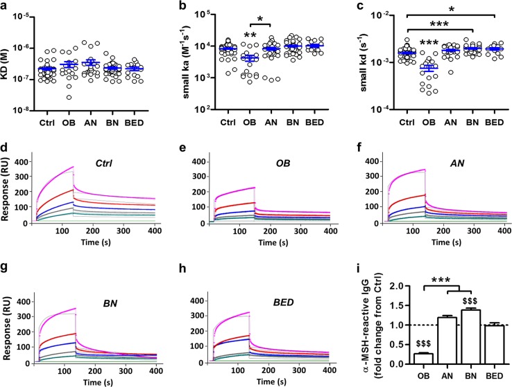
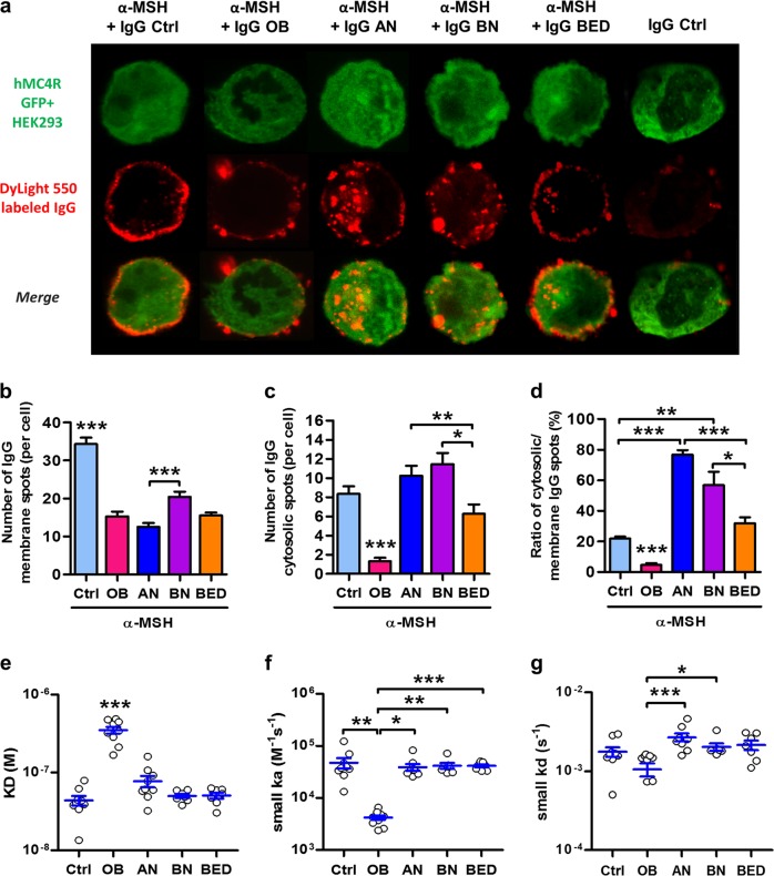
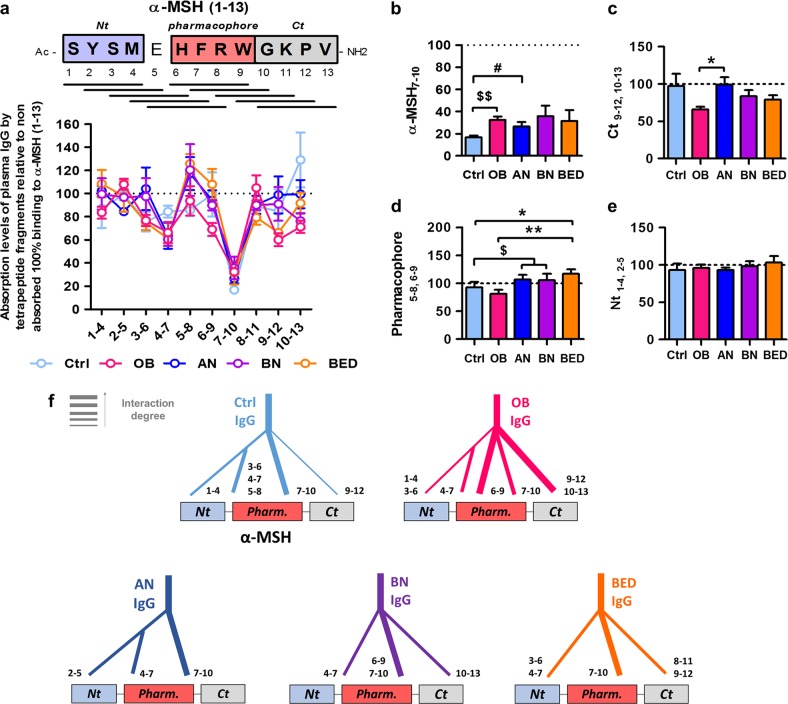
Melanocortin 4 receptor (MC4R) plays a key role in regulation of appetite activated by its main ligand α-melanocyte-stimulating hormone (α-MSH) in both central and peripheral targets. α-MSH also binds to circulating immunoglobulins (Igs) but the functional significance of such immune complexes (ICs) in MC4R signaling in normal and pathological conditions of altered appetite has remained unknown. To address this question, we analyzed plasma levels, affinity kinetics, and binding epitopes of α-MSH-reactive IgG extracted from plasma samples of female patients with hyperphagic obesity, anorexia nervosa, bulimia nervosa, binge-eating disorder, and healthy controls. Ability of α-MSH/IgG IC to bind and activate human MC4R were studied in vitro and to influence feeding behavior in vivo in rodents. We found that α-MSH-reactive IgG were low in obese but increased in anorectic and bulimic patients and displayed different epitope and kinetics of IC formation. Importantly, while α-MSH/IgG IC from all subjects were binding and activating MC4R, the receptor binding affinity was decreased in obesity. Additionally, α-MSH/IgG IC had lower MC4R-mediated cAMP activation threshold as compared with α-MSH alone in all but not obese subjects. Furthermore, the cellular internalization rate of α-MSH/IgG IC by MC4R-expressing cells was decreased in obese but increased in patients with anorexia nervosa. Moreover, IgG from obese patients prevented central anorexigenic effect of α-MSH. These findings reveal that MC4R is physiologically activated by IC formed by α-MSH/IgG and that different levels and molecular properties of α-MSH-reactive IgG underlie biological activity of such IC relevant to altered appetite in obesity and eating disorders.


Conflict of interest statement

S.O.F. is a co-founder and consultant of TargEDys, SA. N.L. and R.L. were employees of TargEDys, SA. P.D. is a co-founder of TargEDys, SA and a member of its board and has received research grants from Nestlé and Fresenius Kabi and honoraria for speeches and consulting from Nestlé, Fresenius-Kabi, and Aguettant. The other authors declare that they have no conflict of interest.

Figures

Fig. 1
Fig. 1. α-Melanocyte-stimulating hormone (α-MSH)-reactive IgG display different affinity kinetic properties in obesity and eating disorders.
a Dissociation equilibrium constants (KD). b Association rates (ka). c Dissociation rates (kd). dh Representative sensorgrams illustrating different affinity kinetics of IgG binding to α-MSH in surface plasmon resonance units (RU) from each study group: d Ctrl (n = 65), e obese (n = 17), f anorexia nervosa (n = 28), g bulimia nervosa (n = 34), and h binge eating disorder (n = 14). IgG concentrations, 3360 nM (in pink), 1680 nM (in red), 840 nM (in blue), 420 nM (in grey), 210 nM (in green). Fitting with the Langmuir’s 1:1 model. i Relative to controls plasma levels of α-MSH-reactive IgG after their extraction from plasma. Data are means ± s.e.m. Kruskal–Wallis test with Dunns’ post-tests, ***p < 0.001, **p < 0.01, *p < 0.05 (b, c, i) or analysis of variance with Tukey’s post-test, $$$p < 0.001 vs. Ctrl (i)
Fig. 2
Fig. 2. Binding of α-melanocyte-stimulating hormone (α-MSH)/IgG immune complex (IC) to melanocortin 4 receptor (MC4R)-expressing cells is altered in obesity and eating disorders.
a Representative images of hMC4R GFP+ HEK 293 cells (green) 30 min after application of DyLight 550®-labeled (red) α-MSH affinity-purified IgG from eating disorder (anorexia nervosa, n = 9; bulimia nervosa, n = 7; binge eating disorder, n = 7), obese (n = 10), and Ctrl (n = 9) preincubated or not with α-MSH. Quantification of DyLight 550®-positive spots in hMC4R+ HEK 293 cells (n = 50/group): b at the membrane; c intracellularly (cytosolic), and d ratios of cytosolic/membrane staining. Affinity kinetics properties of α-MSH/IgG IC for hMC4R + HEK 293 cells including e dissociation equilibrium constant (KD); f association rate (ka), and g dissociation rate (kd). Data are means ± s.e.m. Kruskal–Wallis test with Dunns’ post-tests (bd, f, g) or analysis of variance with Tukey’s post-test (e), ***p < 0.001, **p < 0.01, *p < 0.05
Fig. 3
Fig. 3. α-Melanocyte-stimulating hormone (α-MSH)/IgG immune complex (IC) lower threshold of α-MSH-induced cyclic adenosine monophosphate (cAMP) release by melanocortin 4 receptor (MC4R)-expressing cells.
a cAMP dose–response curves to α-MSH alone or α-MSH/IgG IC formed by IgG pooled in patents and control groups and adjusted to α-MSH-reactive IgG plasma levels of controls. cAMP dose–response curves to α-MSH preincubated or not with individual total IgG and corresponding EC50 (b) and maximal cAMP production (c). d Control experiments including cAMP dose–response curves to α-MSH by MC4R-expressing and non-expressing control HEK 293 cells and to α-MSH1–4 peptide by MC4R-expressing cells (n = 4). e cAMP dose-response curves to α-MSH and IgG from patients and controls without their overnight pre-incubation. f, cAMP dose–response curves to α-MSH alone or α-MSH/IgG IC co-administered (solid line) with agouti-related protein (AgRP; 100 nM) or added after AgRP preincubation (dotted line—n = 2/group) as well as in g cAMP maximal response. h, i cAMP dose–response curves of α-MSH preincubated with h purified total IgG from patients and controls depleted for α-MSH-reactive IgG (n = 3/group) and i affinity-purified α-MSH-reactive IgG (n = 6/group); j EC50 and k maximal cAMP production at the plateau. Data are means ± s.e.m. Analysis of variance with Tukey’s post-test (b, c, g, j) or Kruskal–Wallis test with Dunns’ post-tests (m), ***p < 0.001, **p < 0.01, *p < 0.05; Mann–Whitney test, $p < 0.05. a α-MSH (n = 9), Ctrl, anorexia nervosa (AN), bulimia nervosa (BN), and binge eating disorder (BED; n = 6), obese (OB; n = 4); d HEK 293-hMC4R+ (n = 5), HEK 293-CTRL (n = 6); e α-MSH (n = 3), Ctrl, BN, and BED (n = 2), AN and OB (n = 3)
Fig. 4
Fig. 4. Influence of antibodies on α-melanocyte-stimulating hormone (α-MSH) anorexigenic effects in rodents.
a Schematic illustration of the injection site in the rat paraventricular nucleus (PVN). b Cumulative food intake in rats measured at 30 and 120 min after acute intra-PVN injection of 2 μL of affinity-purified α-MSH-reactive IgG from eating disorder patients (anorexia nervosa, n = 5; bulimia nervosa, n = 5; binge eating disorder, n = 6), obese patients (n = 6), and Ctrl (n = 4) all preincubated with α-MSH and diluted in artificial cerebrospinal fluid (CSF); control group received CSF only (CSF, n = 5). Cumulative food intake in wild-type (WT, n = 6) (c) and Ig-deficient Rag−/− mice (n = 6) (d) measured during 4 h after injection of α-MSH (100 µg/kg) (dotted line) as compared to baseline (solid line). Data are means ± s.e.m and expressed as area under the curve (AUC, e, right panel). Analysis of variance with Tukey’s post-test (b) and Mann–Whitney test (e), **p < 0.01, *p < 0.05, #p < 0.10
Fig. 5
Fig. 5. Epitope mapping of IgG for α-melanocyte-stimulating hormone (α-MSH) in patients and controls.
a Ten tetrapeptide fragments (black lines) overlapping the α-MSH sequence were used for adsorption of total IgG from eating disorder and obese (OB) patients and controls before detection of their immune complex binding by enzyme-linked immunosorbent assay on the entire α-MSH molecule. Adsorption levels of plasma IgG for each fragment was expressed in percentage from non-absorbed total binding to α-MSH (100%). Adsorption levels were compared among the groups for individual tetrapeptides as shown here for: b α-MSH7–10, the central part which includes 3 amino acids of α-MSH pharmacophore (red box in a); c the C-terminal (Ct, gray box in a), as mean levels of α-MSH9–12 and α-MSH10–13; d the pharmacophore sequence as mean levels of α-MSH5–8 and α-MSH6–9; and e the N-terminal (Nt, blue box in a) as mean levels of α-MSH1–4 and α-MSH2–5. f Schematic illustration of α-MSH-binding epitopes in IgG of patients and controls (see also Fig. 6 legend); the degree of adsorption of IgG binding to α-MSH by specific tetrapeptides is shown here by the width of traits toward corresponding parts of the α-MSH sequence. Data are means ± s.e.m. Kruskal–Wallis test, Dunns’ post-test, **p < 0.01; *p < 0.05. Mann–Whitney test, $$p < 0.01, $p < 0.05, #p < 0.10. Ctrl (n = 9), OB (n = 10), anorexia nervosa (n = 9), bulimia nervosa (n = 7), binge eating disorder (n = 6)
Fig. 6
Fig. 6. Hypothetical model of α-melanocyte-stimulating hormone (α-MSH)/IgG immune complex (IC) activation of melanocortin 4 receptor (MC4R) in healthy controls and patients with obesity or anorexia nervosa.
Plasmatic IgG transport α-MSH by forming IC that bind and activate MC4R. During interaction with the receptor, the C-terminal (Ct) of α-MSH should be presented by IgG enabling IC docking and binding. During binding, α-MSH dissociates from IC and its pharmacophore (Phar) enters the receptor binding pocket, resulting in receptor activation ex. cyclic adenosine monophosphate production. The α-MSH/IgG IC then internalized together with MC4R, resulting in temporal desensitization. As illustrated in Fig. 5f, in healthy subjects IgG bind mainly the central and the N-terminal parts of α-MSH making the C-terminal available for MC4R docking. However, in obese patients the C-terminal is hidden by IgG, preventing α-MSH/IgG IC docking to MC4R. In contrast, in anorexia nervosa (AN) patients, the C-terminal of α-MSH is not bound by IgG, favoring receptor recognition. The changes in α-MSH epitope binding in obesity combined with decreased dissociation of IC and low plasma levels of α-MSH-reactive IgG may cause deficient activation of MC4R, promoting positive energy balance. In contrast, the α-MSH epitope-binding properties of IgG in AN combined with increased dissociation of IC and increased plasma levels of α-MSH-reactive IgG are favorable for more efficient activation of MC4R by α-MSH/IgG IC, resulting in enhanced satiety signaling and negative energy balance

Similar articles

Cited by

References

    1. Harris JI, Lerner AB. Amino-acid sequence of the alpha-melanocyte-stimulating hormone. Nature. 1957;179:1346–1347. doi: 10.1038/1791346a0. - DOI - PubMed
    1. Anderson EJP, et al. 60 Years of POMC: regulation of feeding and energy homeostasis by α-MSH. J. Mol. Endocrinol. 2016;56:T157–T174. doi: 10.1530/JME-16-0014. - DOI - PMC - PubMed
    1. Vergoni AV, Schiöth HB, Bertolini A. Melanocortins and feeding behavior. Biomed. Pharmacother. 2000;54:129–134. doi: 10.1016/S0753-3322(00)89045-9. - DOI - PubMed
    1. Fan W, Boston BA, Kesterson RA, Hruby VJ, Cone RD. Role of melanocortinergic neurons in feeding and the agouti obesity syndrome. Nature. 1997;385:165–168. doi: 10.1038/385165a0. - DOI - PubMed
    1. Yaswen L, Diehl N, Brennan MB, Hochgeschwender U. Obesity in the mouse model of pro-opiomelanocortin deficiency responds to peripheral melanocortin. Nat. Med. 1999;5:1066–1070. doi: 10.1038/12506. - DOI - PubMed

Publication types