. 2019 Feb 12;10(2):134.
doi: 10.1038/s41419-019-1410-y.

Hypoxia preconditioning promotes bone marrow mesenchymal stem cells survival by inducing HIF-1α in injured neuronal cells derived exosomes culture system

Affiliations

Hypoxia preconditioning promotes bone marrow mesenchymal stem cells survival by inducing HIF-1α in injured neuronal cells derived exosomes culture system

Zucheng Luo et al. Cell Death Dis. .

Abstract

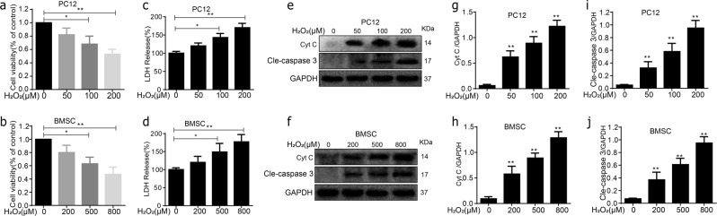
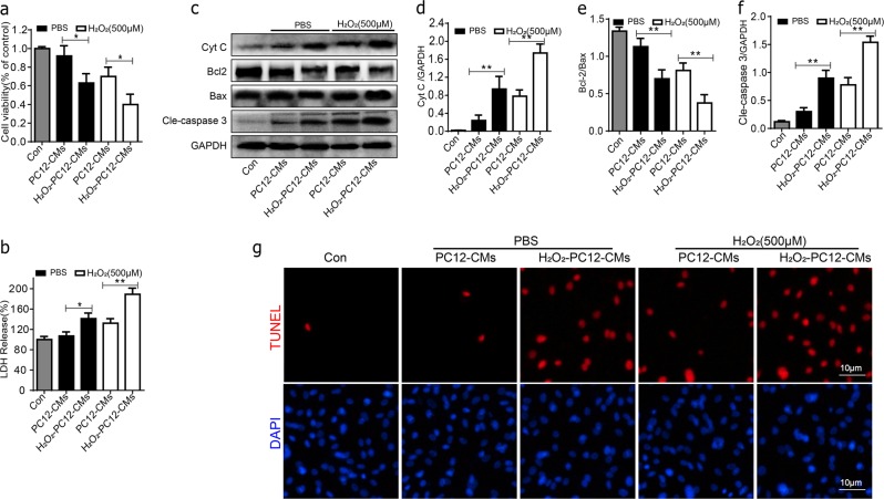
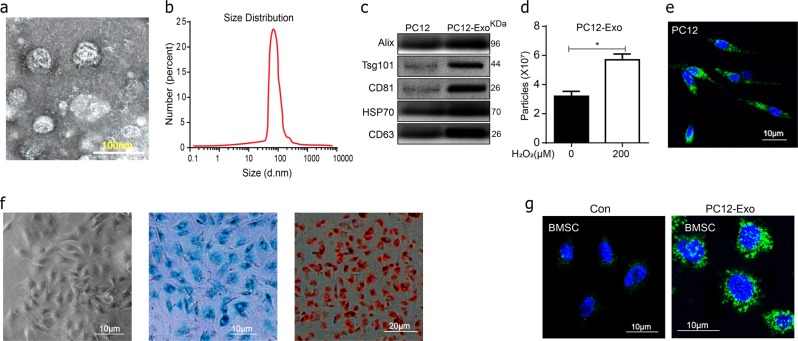
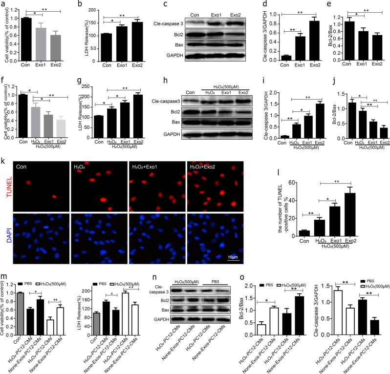
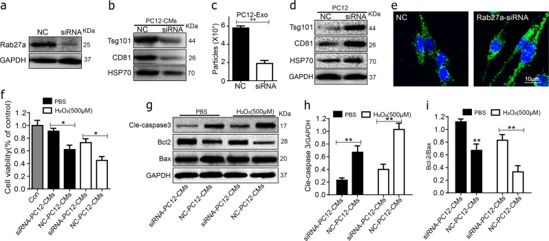
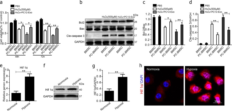
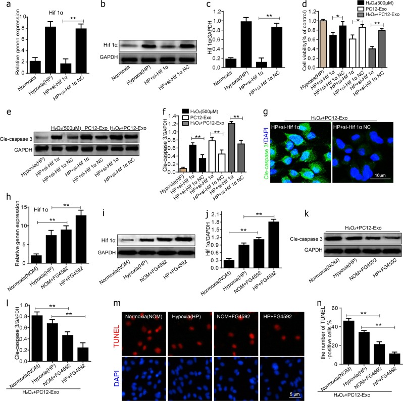
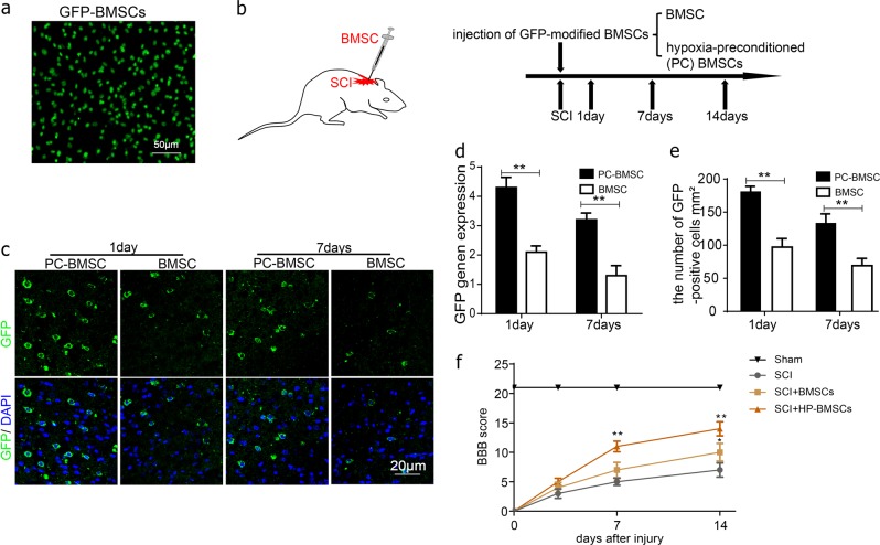
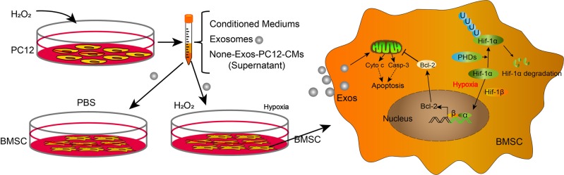
Bone marrow derived stem cells (BMSCs) transplantation are viewed as a promising therapeutic candidate for spinal cord injury (SCI). However, the inflammatory microenvironment in the spinal cord following SCI limits the survival and efficacy of transplanted BMSCs. In this study, we investigate whether injured neuronal cells derived exosomes would influence the survival of transplanted BMSCs after SCI. In order to mimic the microenvironment in SCI that the neuronal cells or transplanted BMSCs suffer in vivo, PC12 cells conditioned medium and PC12 cell's exosomes collected from H2O2-treated PC12 cell's culture medium were cultured with BMSCs under oxidative stress in vitro. PC12 cells conditioned medium and PC12 cell's exosomes significantly accelerated the apoptosis of BMSCs induced by H2O2. Moreover, the cleaved caspase-3, cytochrome (Cyt) C, lactate dehydrogenase (LDH) releases, and apoptotic percentage were increased, and the ratio of Bcl-2/Bax and cell viability were decreased. Inhibition of exosome secretion via Rab27a small interfering RNA prevented BMSCs apoptosis in vitro. In addition, hypoxia-preconditioned promoted the survival of BMSCs under oxidative stress both in vivo after SCI and in vitro. Our results also indicate that HIF-1α plays a central role in the survival of BMSCs in hypoxia pretreatment under oxidative stress conditions. siRNA-HIF-1α increased apoptosis of BMSCs; in contrast, HIF-1α inducer FG-4592 attenuated apoptosis of BMSCs. Taken together, we found that the injured PC12 cells derived exosomes accelerate BMSCs apoptosis after SCI and in vitro, hypoxia pretreatment or activating expression of HIF-1α to be important in the survival of BMSCs after transplantation, which provides a foundation for application of BMSCs in therapeutic potential for SCI.


Conflict of interest statement

The authors declare that they have no conflict of interest.

Figures

Fig. 1
Fig. 1. Oxidative stress caused apoptosis in PC12 cells and BMSCs.
a, b H2O2 induced apoptosis in PC12 cells and BMSCs 24 h posttreatment. c, d PC12 cells and BMSCs exhibited release of LDH in a dose-dependent of H2O2 stimulating concentration. ej The protein expressions of cleaved caspase-3 and cyt C were visualized by western blot. All data in the figures represent the mean ± SD. *P < 0.05; **P < 0.01, compared with each group, n = 5 for each group
Fig. 2
Fig. 2. Apoptosis of BMSCs in PC12 cells conditioned medium under oxidative stress.
BMSCs were cultured with PC12 cells conditioned medium and treated without or with 500 μM H2O2 for 24 h. a Cell viability of BMSCs in each group was detected by MTT assay. b The ratio of LDH release in the group of H2O2-PC12-CMs exhibited a significantly increased compared to PC12-CMs group treated without or with 500 μM H2O2 for 24 h. cf Representative western blots and quantification data of Cyt C, Bcl-2, Bax, cleaved caspase-3 in each group cells. g Double staining for TUNEL (red) and DAPI (blue) of BMSCs in each group cells. All data in the figures represent the mean values ± SD. *P < 0.05; **P < 0.01, compared with each group, n = 5
Fig. 3
Fig. 3. Characterization of PC12 cells derived exosomes.
a Representative transmission electron microscopy (TEM) images of exosomes. Scale bar 100 nm. b Size distributions of exosomes were measured using nanoparticle tracking analysis (NTA). c Western-blotting analysis of exosomal markers including CD63, HSP70, CD81, Tsg101, and Alix isolated from PC12 cells derived exosomes. d Recording of exosome concentrations from conditioned medium from PC12 cells with or without 100 μM H2O2 for 24 h by NTA. e The PKH67-labeled PC12 cells exosomes and stained with DAPI. f Passage three of BMSCs were assessed for their differentiation ability based on Alizarin red staining and alcian blue staining respectively. g Uptake of PC12 exosomes by BMSCs. BMSCs were cultured with PKH67-labeled PC12 exosomes or control medium for 24 h and stained with DAPI. All data represent mean values ± SD. *P < 0.05; **P < 0.01, compared with each group
Fig. 4
Fig. 4. The apoptotic effect of PC12 cells exosomes on BMSCs.
a Cell viability of BMSCs after being cultured with two different concentrations of PC12 cells exosomes was detected by MTT assay. b BMSCs exhibited release of LDH in a dose-dependent of PC12 cells exosomes. ce Representative western blots of cleaved caspase-3, Bcl-2, Bax in BMSCs after 24 h culture with PC12 cells exosomes. Quantitative data of cleaved caspase-3/GAPDH and Bcl-2/Bax ratio were shown. f Cell viability of BMSCs after being co-cultured with two different concentrations of PC12 cells exosomes and 500 μM H2O2 for 24 h was detected by MTT assay. g BMSCs exhibited release of LDH in a dose-dependent of PC12 cells exosomes under oxidative stress. hj Representative western blots and quantification data of cleaved caspase-3, Bcl-2, Bax. k, l Cell apoptosis assessed by TUNEL staining, the percentages of TUNEL + cells were significantly higher in Exo1 and Exo2 group compared with H2O2 alone. All data in the figures represent the Mean values ± SD. *P < 0.05; **P < 0.01, n = 4, compared with each group
Fig. 5
Fig. 5. Effect of Rab27a inhibition on the PC12 cells exosomes secretion and apoptosis of BMSCs.
a, b The Rab27a was significantly decreased and presence of exosomal marker HSP70, CD81, and Tsg101 were lower in Rab27a-siRNA group culture mediums as compared with the non-specific control (NC) group assessed by western blot. c NTA showed that the number of the secretory exosomes greatly decreased in Rab27a-SiRNA group compared to NC. d The expression of exosomal marker HSP70, CD81, and Tsg101 in PC12 cells assessed by western blot. e Immunofluorescence of PKH67-labeled the exosomes in PC12 cells. f Cell viability of BMSCs in Rab27a-siRNA group and NC-siRNA group mediums both in PBS or H2O2 conditions were detected by MTT assay. gi Representative western blots and quantification data of cleaved caspase-3, Bcl-2, Bax in BMSCs in each group. All data represent Mean values ± SD. *P < 0.05; **P < 0.01, compared with each group
Fig. 6
Fig. 6. Hypoxia-preconditioned promoted the survival of BMSCs under oxidative stress.
a Cell viability in the BMSC and PC-BMSC groups cultured in control PBS, H2O2 (500 μM) and H2O2 combine with PC12 cell’s exosomes (H2O2-PC12-Exo) after 48 and 72 h were assessed by MTT. bd) Representative western blots and quantification data of cleaved caspase-3, Bcl-2, Bax in BMSCs in each group. e The gene expression of HIF-1α in normoxia and hypoxia groups. f, g Representative Western blots and quantification data of HIF-1α in normoxia and hypoxia groups. h Immunofluorescence staining of HIF-1α (red) in normoxia and hypoxia groups, and DAPI - labeled nuclei (blue). *P < 0.05; **P < 0.01, compared with each group, n = 5, All data represent Mean values ± SD
Fig. 7
Fig. 7. HIF-1α played an important role in the survival of BMSCs.
ac The gene expression and western blots and quantification data of HIF-1α in the siRNA-HIF-1α group and siRNA-NC groups. d BMSCs viability in each group was assessed by MTT. e, f Representative western blots and quantification data of cleaved caspase-3 in each group. g Representative micrographs showing immunofluorescence of cleaved caspase-3 (green) and DAPI - labeled nuclei (blue) in each group. hj The gene expression, representative western blots and quantification data of HIF-1α in each group, HIF-1α was up-regulated by using FG-4592. k, l Representative western blots and quantification data of cleaved caspase-3 in each group. m, n Cell apoptosis assessed by TUNEL staining in each group. All data in the figures represent the Mean ± SD. *P < 0.05; **P < 0.01, compared with each group
Fig. 8
Fig. 8. Hypoxia-preconditioned promoted the survival of BMSCs in vivo.
a All cultured GFP-BMSC emitted sufficient fluorescence for microscopic observations. b The GFP-modified BMSCs were transplanted into the spine cord of SCI rats. c, e Representative micrographs showing immunofluorescence of GFP (green) and DAPI - labeled nuclei (blue) in each group at 1 and 7 day respectively after transplantation. d GFP gene expression in the PC-BMSCs groups was significantly increased than in the control groups. All data in the figures represent the averages ± SD. *P < 0.05; **P < 0.01, compared with each group
Fig. 9
Fig. 9. Schematic illustration of the survival of hypoxia-preconditioned BMSCs.
Technical and potential molecular mechanism involved in the survival of hypoxia-preconditioned BMSCs

Similar articles

Cited by

References

    1. S. R. Andresen F, et al. Finnerup, pain, spasticity and quality of life in individuals with traumatic spinal cord injury in Denmark. Spinal Cord. 2016;54:973–979. - PubMed
    1. C. S. Rivers N, et al. Health conditions: effect on function, health-related quality of life, and life satisfaction after traumatic spinal cord injury. a prospective observational registry cohort study. Arch. Phys. Med. Rehabil. 2018;99:443–451. - PubMed
    1. S. V. Hiremath NS, et al. Longitudinal prediction of quality-of-life scores and locomotion in individuals with traumatic spinal cord injury. Arch. Phys. Med. Rehabil. 2017;98:2385–2392. - PubMed
    1. Kwon BK, Tetzlaff W, Grauer JN, Beiner J, Vaccaro AR. Pathophysiology and pharmacologic treatment of acute spinal cord injury. Spine J. 2004;4:451–464. - PubMed
    1. Tykocki T, Poniatowski L, Czyz M, Koziara M, Wynne-Jones G. Intraspinal pressure monitoring and extensive duroplasty in the acute phase of traumatic spinal cord injury: a systematic review. World Neurosurg. 2017;105:145–152. - PubMed

Publication types

MeSH terms