. 2019 Feb 12;10(2):130.
doi: 10.1038/s41419-019-1337-3.

LncRNA H19 promotes the committed differentiation of stem cells from apical papilla via miR-141/SPAG9 pathway

Affiliations

LncRNA H19 promotes the committed differentiation of stem cells from apical papilla via miR-141/SPAG9 pathway

Zehan Li et al. Cell Death Dis. .

Abstract

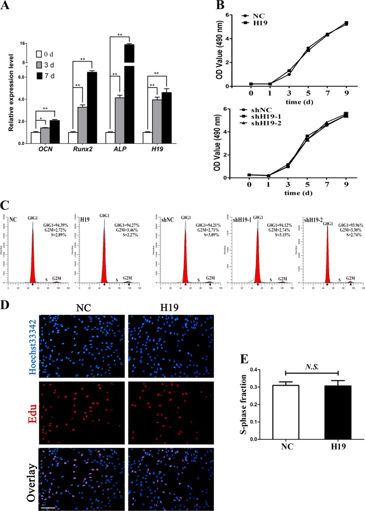
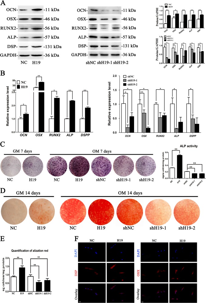
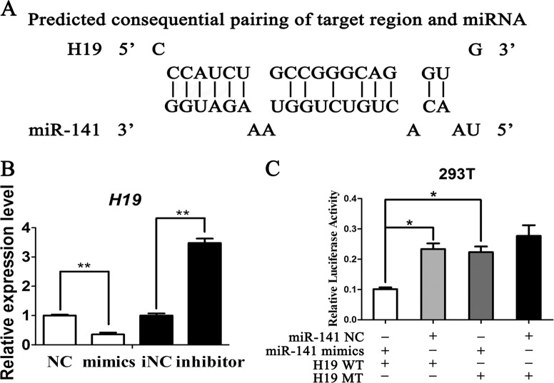
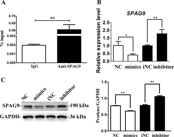
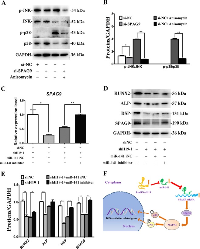
Long noncoding RNAs (lncRNAs) exert significant roles at transcriptional and post-transcriptional levels. Stem cells from apical papilla (SCAPs) differentiate into dentin/bone-like tissues under certain conditions. So far, whether lncRNA-H19 can affect the proliferative behaviors and osteo/odontogenesis of SCAPs, as well as its specific mechanism remain to be elucidated. Here, SCAPs were isolated and transfected with the lentiviruses or packaging vectors. Our results showed that lncRNA-H19 had no significant effect on the proliferative behaviors of SCAPs, as presented by CCK-8 assay, EdU assay and flow cytometry (FCM). Furthermore, alkaline phosphatase (ALP) activity, alizarin red staining, Western blot assay (WB), quantitative real-time polymerase chain reaction (qRT-PCR) and in vivo bone formation assay were conducted to verify the biological influences of H19 on SCAPs. Overexpression of H19 led to the enhanced osteo/odontogenesis of SCAPs, whereas knockdown of H19 inhibited these effects. Mechanistically, H19 competitively bound to miR-141 and prevented SPAG9 from miRNA-mediated degradation, thus significantly elevating phosphorylated levels of p38 and JNK and facilitating the committed differentiation of SCAPs. Taken together, the osteo/odontogenesis of SCAPs was upregulated by overexpression of H19 via miR-141/SPAG9 pathway.


Conflict of interest statement

The authors declare that they have no conflict of interest.

Figures

Fig. 1
Fig. 1. LncRNA-H19 expression during osteoblast differentiation of stem cells from apical papilla (SCAPs) and effects of lncRNA-H19 on proliferation of SCAPs.
a Relative expression of H19 and osteoblastic markers of OCN, Runx2, and ALP were determined by qRT-PCR analysis during osteoblast differentiation of SCAPs at day 0, 3 and 7. The relative expression leves at indicated time points were normalized to day 0. GAPDH was used as an internal control. Results were presented as the mean ± SD (*P < 0.05, **P < 0.01). b CCK-8 assay showed no significant difference in cell proliferation when H19 was overexpression or knockdown from day 0 to day 9. c, d Flow cytometry and EdU assay demonstrated that H19 had no significant difference on cell proliferation of SCAPs at day 7 (Scale Bar = 200 μm; N.S., P > 0.05)
Fig. 2
Fig. 2. LncRNA-H19 promotes osteo/odontogenesis of SCAPs.
a Western blot results revealed that the protein levels of OCN, OSX, RUNX2, ALP and DSP significantly increased in H19 group while decreased in shH19-1 and shH19-2 groups after osteoblast induction for 7 days. GAPDH served as an internal control. Histograms showed the quantification of band intensities (*P < 0.05, **P < 0.01). b Relative mRNA expressions of OCN, OSX, RUNX2, ALP and DSPP measured by qRT-PCR after OB induction for 7 days. GAPDH was used for normalization. Results were presented as the mean ± SD (*P < 0.05, **P < 0.01). c Images of ALP staining in the NC, H19, shNC, shH19-1, and shH19-2 groups. SCAPs were cultured in growth medium (GM) or osteogenic medium (OM) for 7 days. Histograms show the activity of ALP were increased by H19 overexpression and decreased by H19 knockdown after OB induction for 7 days (**P < 0.01). d After cell culture in GM or OM for 14 days, alizarin red staining showed that H19 group generated more calcified nodules than control group. shNC group generated more calcified nodules than shH19-1 and shH19-2 groups. e Histograms showed quantification of Alizarin red staining by spectrophotometry. Values were expressed as means ± SD, **P < 0.01. f Immunofluorescence assay revealed that the expressions of DSP and OSX in Lenti-H19 treated SCAPs were significantly up-regulated (Scale Bar = 50 μm)
Fig. 3
Fig. 3. H19 enhanced the osteo/dentinogenesis of SCAPs in vivo.
a SCAPs in NC and H19 group were transplanted subcutaneously into 5-week-old BALB/c homozygous nude mice for 8 weeks. b Upper: reconstructed three-dimensional micro-CT images of the tissue-engineered bone constructs from NC and H19 groups. Lower: percentages of new BV/TV of cultured bone constructs. Data are shown as the mean ± SD (*P < 0.05). c H&E staining, Masson staining and immunohistochemical staining of osteocalcin in NC and H19 groups. B bone/dentin-like tissues, S around the scaffold, BV/TV bone volume to tissue volume, NC negative control. Scale Bar = 100 μm
Fig. 4
Fig. 4. LncRNA-H19 functions as an endogenous sponge of miR-141.
a The potential binding sites between lncRNA-H19 and miR-141 predicted by biological software. b Relative lncRNA-H19 expression level in SCAPs transfected with with miR-141 mimics or miR-141 inhibitor (**P < 0.01). c Luciferase reporter assay was used to validate the target in 293T cells. The relative luciferase activities of luciferase reporters containing WT or Mut lncRNA-H19 were assayed 48 h after co-transfection with miR-141 mimics or mimics NC. Relative Renilla luciferase activity was normalized to that of firefly luciferase (*P< 0.05, **P < 0.01)
Fig. 5
Fig. 5. MiR-141 inhibits osteo/odontogenic differentiation of SCAPs.
ac Western blot results revealed that the protein levels of OCN, OSX, RUNX2, ALP and DSP significantly decreased in miR-141 mimics group while increased in miR-141 inhibitor group (**P < 0.01). d, e Relative mRNA expressions of OCN, OSX, RUNX2, ALP, and DSPP were measured by qRT-PCR. GAPDH was used for normalization. Results were presented as the mean ± SD (*P < 0.05; **P < 0.01). f, g After cell culturing in OM for 14 days, alizarin red staining showed that miR-141 mimics group generated more calcified nodules than control group. MiR-141 inhibitor group generated less calcified nodules than iNC group (Scale Bar = 200 μm). Histograms showed quantification of Alizarin red staining by spectrophotometry. Values were expressed as means ± SD, **P < 0.01. h, i Images of ALP staining in the NC, mimics, iNC and inhibitor groups. SCAPs were cultured in OM for 7 days. Histograms showed the activity of ALP increased by miR-141 overexpression and decreased by miR-141 knockdown (**P < 0.01). j Immunofluorescence assay revealed that the expressions of DSP and OSX were significantly up-regulated in miR-141 mimics group compared with NC group (Scale Bar = 50 μm)
Fig. 6
Fig. 6. MiR-141 downregulates the expression of SPAG9.
a Relative expression level of miR-141 in SCAPs was accessed by RIP assay (**P < 0.01). b Relative mRNA expression of SPAG9 in SCAPs transfected with with miR-141 mimics or miR-141 inhibitor (*P < 0.05, **P < 0.01). c Western blot analysis of protein expression of SPAG9 in SCAPs transfected with miR-141 mimics or inhibitor. d Results of western blotting was analyzed with ImageJ software and data were presented as ratio of target protein to GAPDH (**P < 0.01)
Fig. 7
Fig. 7. SPAG9 regulates p38 and JNK signaling pathways in SCAPs and the miR-141 inhibitor could rescue the shH19-1 mediated inhibitory effects of osteo/odontogenic differentiation in SCAPs.
a Western blot assay for the expressions of genes relative to p38 and JNK signaling pathways at 72 h. b The ratio changes of p-p38/p38 and p-JNK/JNK at 72 h in different groups. Values were described as the means ± SD, n = 3. *P < 0.05, **P < 0.01. c Results of qRT-PCR analysis revealed the miR-141 inhibitor rescued the shH19-1 mediated downregulation of SPAG9 expression (*P < 0.05, **P < 0.01). d Results of western blot analysis indicated that the miR-141 inhibitor rescued the shH19-1 mediated downregulation of RUNX2, ALP, DSP, SPAG9. e Results of western blotting was analyzed with ImageJ software and data were presented as ratio of target protein to GAPDH in the form of grayscale value. (*P < 0.05, **P < 0.01). f Schematic diagram for lncRNA-H19/miR-141/SPAG9/MAPK axis

Similar articles

Cited by

References

    1. Bianco P, et al. The meaning, the sense and the significance: translating the science of mesenchymal stem cells into medicine. Nat. Med. 2013;19:35–42. doi: 10.1038/nm.3028. - DOI - PMC - PubMed
    1. Kim HJ, et al. Transfection of gene regulation nanoparticles complexed with pDNA and shRNA controls multilineage differentiation of hMSCs. Biomaterials. 2018;177:1–13. doi: 10.1016/j.biomaterials.2018.05.035. - DOI - PubMed
    1. Chen Q, et al. Fate decision of mesenchymal stem cells: adipocytes or osteoblasts? Cell Death Differ. 2016;23:1128–1139. doi: 10.1038/cdd.2015.168. - DOI - PMC - PubMed
    1. Lv FJ, Tuan RS, Cheung KM, Leung VY. Concise review: the surface markers and identity of human mesenchymal stem cells. Stem Cells. 2014;32:1408–1419. doi: 10.1002/stem.1681. - DOI - PubMed
    1. Zhang C, et al. Therapeutic effect of dental pulp stem cell transplantation on a rat model of radioactivity-induced esophageal injury. Cell Death Dis. 2018;9:738. doi: 10.1038/s41419-018-0753-0. - DOI - PMC - PubMed

Publication types