. 2019 Feb 12;10(2):126.
doi: 10.1038/s41419-018-1202-9.

KIAA1199 is a secreted molecule that enhances osteoblastic stem cell migration and recruitment

Affiliations

KIAA1199 is a secreted molecule that enhances osteoblastic stem cell migration and recruitment

Li Chen et al. Cell Death Dis. .

Abstract

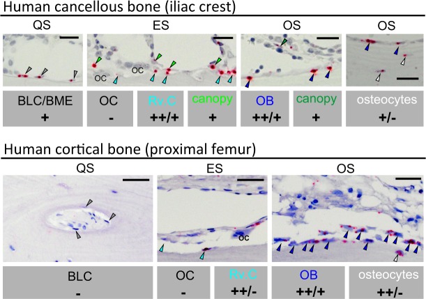
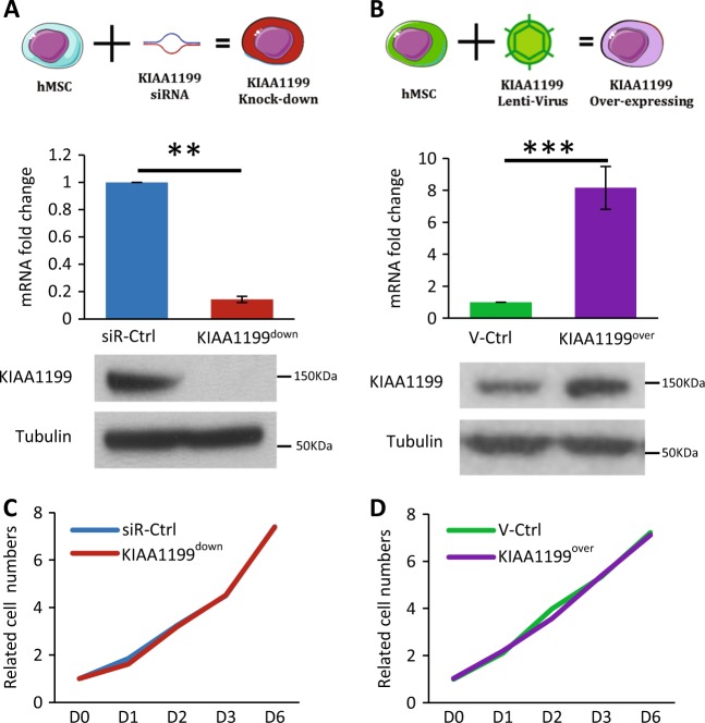
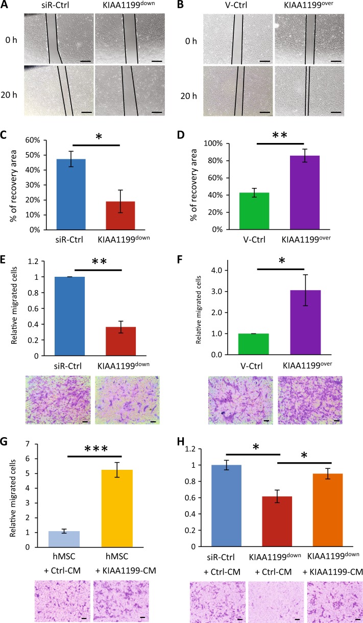
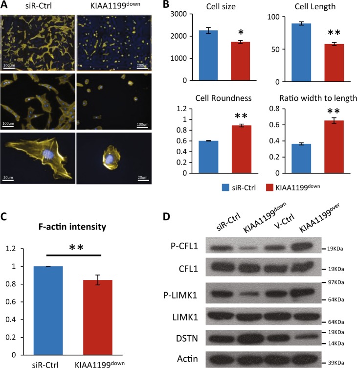
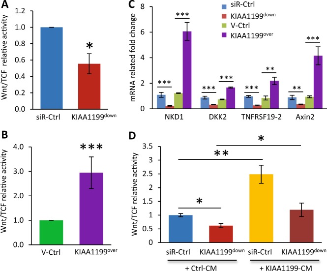
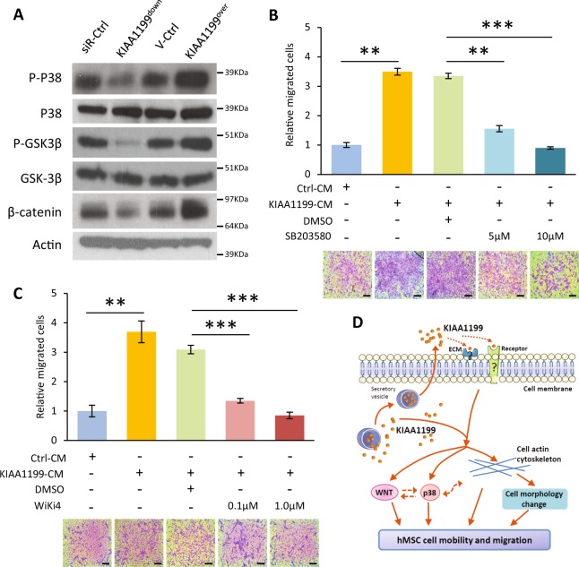
Factors mediating mobilization of osteoblastic stem and progenitor cells from their bone marrow niche to be recruited to bone formation sites during bone remodeling are poorly known. We have studied secreted factors present in the bone marrow microenvironment and identified KIAA1199 (also known as CEMIP, cell migration inducing hyaluronan binding protein) in human bone biopsies as highly expressed in osteoprogenitor reversal cells (Rv.C) recruited to the eroded surfaces (ES), which are the future bone formation sites. In vitro, KIAA1199 did not affect the proliferation of human osteoblastic stem cells (also known as human bone marrow skeletal or stromal stem cells, hMSCs); but it enhanced cell migration as determined by scratch assay and trans-well migration assay. KIAA1199 deficient hMSCs (KIAA1199down) exhibited significant changes in cell size, cell length, ratio of cell width to length and cell roundness, together with reduction of polymerization actin (F-actin) and changes in phos-CFL1 (cofflin1), phos-LIMK1 (LIM domain kinase 1) and DSTN (destrin), key factors regulating actin cytoskeletal dynamics and cell motility. Moreover, KIAA1199down hMSC exhibited impaired Wnt signaling in TCF-reporter assay and decreased expression of Wnt target genes and these effects were rescued by KIAA1199 treatment. Finally, KIAA1199 regulated the activation of P38 kinase and its associated changes in Wnt-signaling. Thus, KIAA1199 is a mobilizing factor that interacts with P38 and Wnt signaling, and induces changes in actin cytoskeleton, as a mechanism mediating recruitment of hMSC to bone formation sites.


Conflict of interest statement

All the authors declare that they have no conflict of interest.

Figures

Fig. 1
Fig. 1. KIAA1199 is expressed in osteoprogenitor cells colonizing future bone formation sites in human bone.
Four human iliac crest bone specimens were in situ-hybridized for KIAA1199. KIAA199 expresses in reversal cells (Rv.C–light blue arrowheads), i.e., progenitor cells, colonizing the eroded surfaces (ES-future bone formation sites), the bone lining cells (BLC), bone marrow envelope (BME) (gray arrowheads) above quiescent surfaces (QS), as well as canopy cells (green arrowheads) above eroded surfaces (ES) that reported to be a local source of osteoprogenitor cells. KIAA1199 expression was also clear present in mature osteoblasts (OB–dark blue arrowheads) on osteoid surfaces (OS) and in some osteocytes, and absent in osteoclasts (OC). The overall expression level in each cell type is indicated below their illustrations: - no expression; + low expression, + + medium expression. Scale bar: 40 µm
Fig. 2
Fig. 2. Establishing of KIAA1199 deficient or overexpressing human bone marrow skeletal (stromal) stem cells (hMSCs).
a KIAA1199 deficient hMSCs (KIAA1199down) were obtained by transfecting the cells with a specific siRNA for KIAA1199 (siR-KIAA1199) or with non-target siRNA control (siR-Ctrl); (b) KIAA1199-overexpression hMSCs (KIAA1199over) were established using a lentiviral transduction and, corresponding control was established by parallel infected by lentiviral vehicle (V-Ctrl). Expression levels of KIAA1199 in cells were determined using qRT-PCR and Western blot analysis (a, b). c–d Cell proliferation was determined by cell counting and cell viability assay
Fig. 3
Fig. 3. KIAA1199 regulates human bone marrow skeletal (stromal) stem cells (hMSCs) cell motility and migration.
a–b In vitro scratch assay was performed in hMSC-KIAA1199down or hMSC-KIAA1199over cells. Photomicrographs were obtained at the same position of culture dish after 20 h incubation in 0.2% fetal bovine serum (FBS) cultured medium, Scale bar: 500 µm. c–d The ability of the cells to ‘heal the wound’ (migrated area) was determined as a ratio of cell covered area per total area by Image-J® program (n ≥ 3). e–f Boyden Chamber trans-well cell migration assay were performed in hMSC-KIAA1199down or hMSC-KIAA1199over and cell migration was measured after 16 h. The migrated cells were calculated by Image-J program (n ≥ 3). g hMSCs were placed in the upper chamber of Boyden chamber system and Lower chambers were filled with conditional mediums (CM) from KIAA1199 overexpression cells (KIAA1199-CM) or its corresponding vehicle control cells (Ctrl-CM). Cell migration was measured after 16 h. h KIAA1199 deficient hMSCs (KIAA1199down) and control cells (siR-Ctrl) were placed in upper chamber of Boyden chamber system, Ctrl-CM or KIAA1199-CM were added into lower chamber. Cell migration was measured after 16 h. The migrated cells were calculated by Image-J program (n ≥ 3). Data represent mean ± SD. *P < 0.05, **P < 0.01, ***P < 0.001. Scale bar for pictures: 500 µm
Fig. 4
Fig. 4. KIAA1199 regulates cell morphology and actin cytoskeleton organization.
Human bone marrow skeletal (stromal) stem cells (hMSCs) were cultured and transfected with siRNA specific for KIAA1199 or non-target control siRNAs to obtain KIAA1199 deficient hMSCs (KIAA1199down) and the corresponding control cells (siR-Ctrl). a The cells were transferred and seeded for 16 h, stained with TRITC-phalloidin and DAPI. Images were acquired on the Operetta® high content image system using ×10 or ×40 objective. b Parameters of cell morphology, including cell size (area) (µm2), cell length (µm), cell width (µm), cell roundness, and cell ratio (width to length) were analyzed by the Harmony® software. c The staining intensity of F-actin was analyzed by the Harmony® software for phalloidin stained F-actin. N ≥ 3, *P < 0.05, **P < 0.01. d Actin depolymerizing factors involved in actin cytoskeletal dynamics: CFL1, LIMK1 and DSTN, were measured by Western-blot analysis in KIAA1199down or KIAA1199over hMSCs
Fig. 5
Fig. 5. KIAA1199 regulates canonical Wnt signaling in human skeletal (stromal) stem cells (hMSCs).
a–b Wnt/TCF-luciferase-reporter was transduced into hMSCs deficient in KIAA1199 (KIAA1199down) that were created by siRNA-mediated knock-down and control cells transfected with non-target siRNA (siR-Ctrl) (a); or transduced into hMSCs overexpressing KIAA1199 (KIAA1199over) that was created by lentiviral transduction and corresponding control (V-Ctrl) was established by parallel infected by lentiviral vehicle (b). In presence of 50% Wnt3a condition medium induction, TCF-Luciferase activity was measured after 24 h incubation, firefly luciferase was normalized to parallel renilla luciferase activities in the same sample using the Dual-Glo Luciferase Assay System. c The mRNA expression levels of canonical Wnt signaling target genes NKD1, DKK2, TNFRSF19-2, and Axin2 were determined by qRT-PCR and normalized to internal controls. d KIAA1199down and control cells were cultured in control conditional medium (Ctrl-CM) or KIAA1199 conditioned medium (KIAA1199-CM), with Wnt3a stimulation, TCF-Luciferase activity was measured after 24 h incubation, firefly luciferase was normalized to parallel renilla luciferase activities in the same sample using the Dual-Glo Luciferase Assay System. At least three independent experiments were preformed, data presented as mean ± SD, *P < 0.05, **P < 0.01, ***P < 0.001
Fig. 6
Fig. 6. Wnt and p38 signaling are involved in the regulation of KIAA1199 at cell migration in hMSCs.
a KIAA1199down or KIAA1199over were starved for 6 h, and stimulated with 5% FBS medium for 15 min. Signaling pathways were analyzed by Western-blot analysis. b hMSCs were pretreated with P38 inhibitor (SB203580, 5 µM or 10 µM) for 2 h and trans-well migration assay was performed in the absence or presence of KIAA1199 conditioned medium (CM). c hMSCs were pretreated with Wnt signaling inhibitor (Wiki4, 0.1 µM or 1 µM) for 2 h and trans-well migration assay was performed in the absence or presence of KIAA1199 conditioned medium (CM). At least three independent experiments were preformed, data presented as mean ± SD, **P < 0.01, ***P < 0.001. Scale bar for pictures: 200 µm. d Mechanisms model for the regulation of KIAA1199 in hMSCs mobility and migration

Similar articles

Cited by

References

    1. Lassen NE, et al. Coupling of bone resorption and formation in real time: new knowledge gained from human haversian BMUs. J. Bone Miner. Res. 2017;32:1395–1405. doi: 10.1002/jbmr.3091. - DOI - PubMed
    1. Zaher W, Harkness L, Jafari A, Kassem M. An update of human mesenchymal stem cell biology and their clinical uses. Arch. Toxicol. 2014;88:1069–1082. doi: 10.1007/s00204-014-1232-8. - DOI - PubMed
    1. Parfitt AM, Villanueva AR, Foldes J, Rao DS. Relations between histologic indices of bone formation: implications for the pathogenesis of spinal osteoporosis. J. Bone Miner. Res. 1995;10:466–473. doi: 10.1002/jbmr.5650100319. - DOI - PubMed
    1. Sims NA, Martin TJ. Coupling the activities of bone formation and resorption: a multitude of signals within the basic multicellular unit. Bone. Rep. 2014;3:481. - PMC - PubMed
    1. Devine SM, et al. Mesenchymal stem cells are capable of homing to the bone marrow of non-human primates following systemic infusion. Exp. Hematol. 2001;29:244–255. doi: 10.1016/S0301-472X(00)00635-4. - DOI - PubMed

Publication types