. 2019 Feb 12;10(2):131.
doi: 10.1038/s41419-018-1188-3.

P53-induced miR-1249 inhibits tumor growth, metastasis, and angiogenesis by targeting VEGFA and HMGA2

Affiliations

P53-induced miR-1249 inhibits tumor growth, metastasis, and angiogenesis by targeting VEGFA and HMGA2

Xiaoxiang Chen et al. Cell Death Dis. .

Abstract

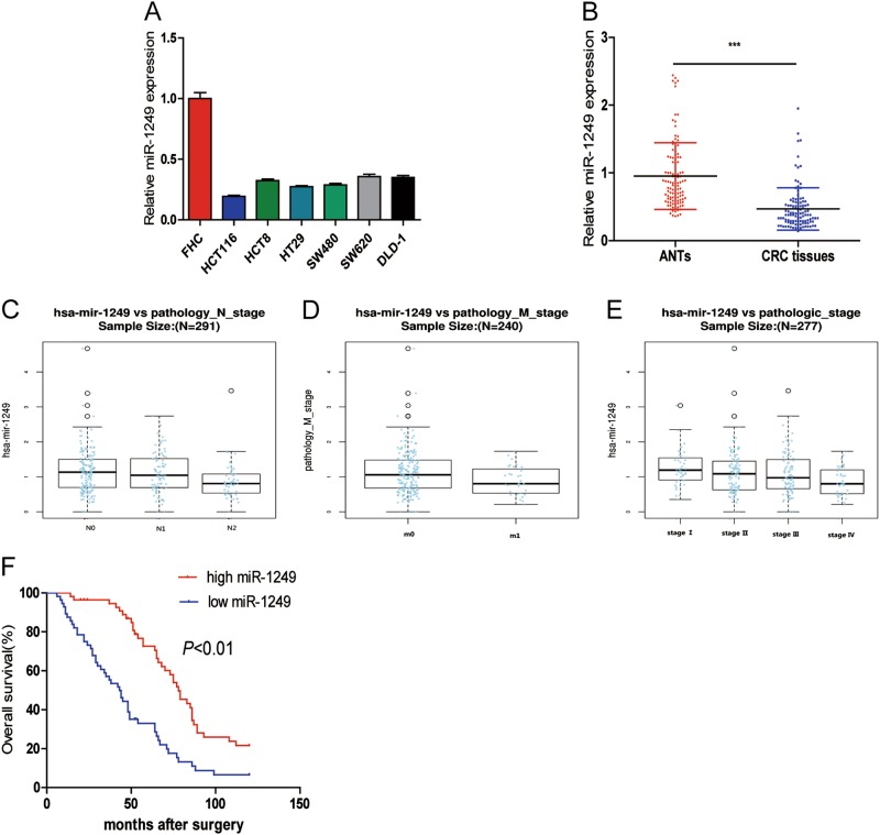
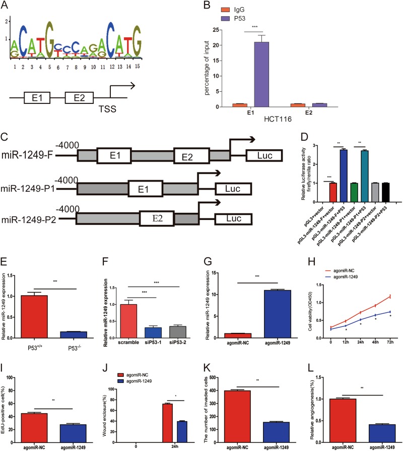
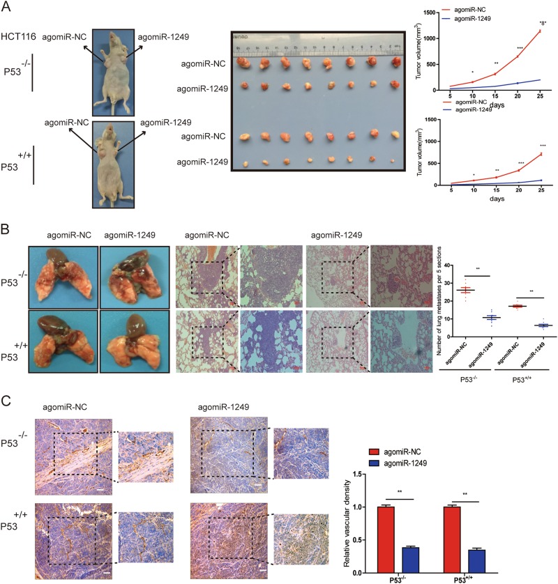
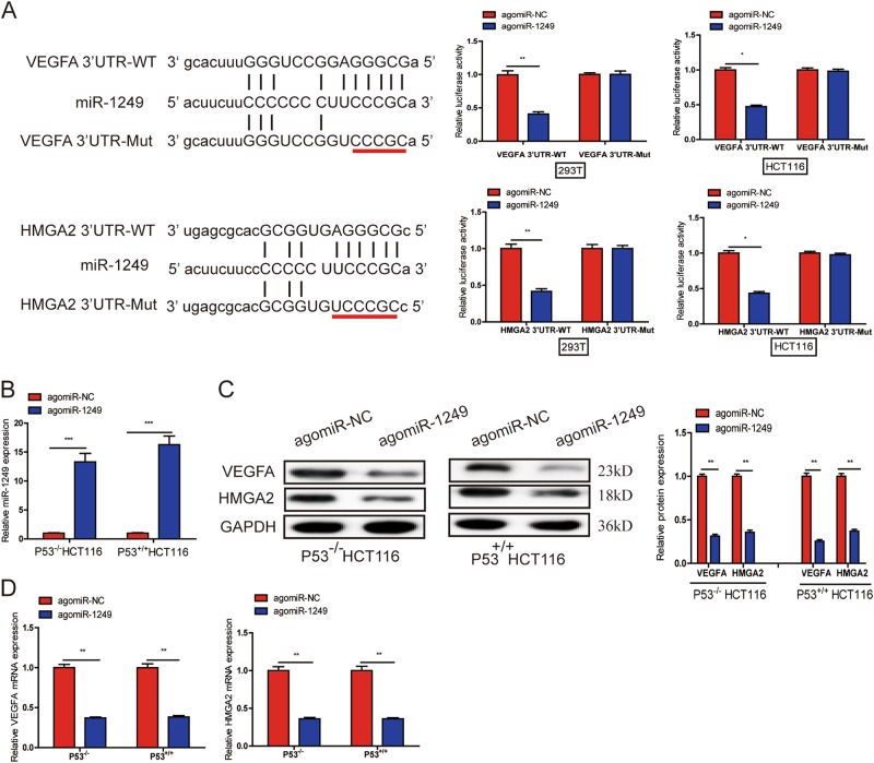
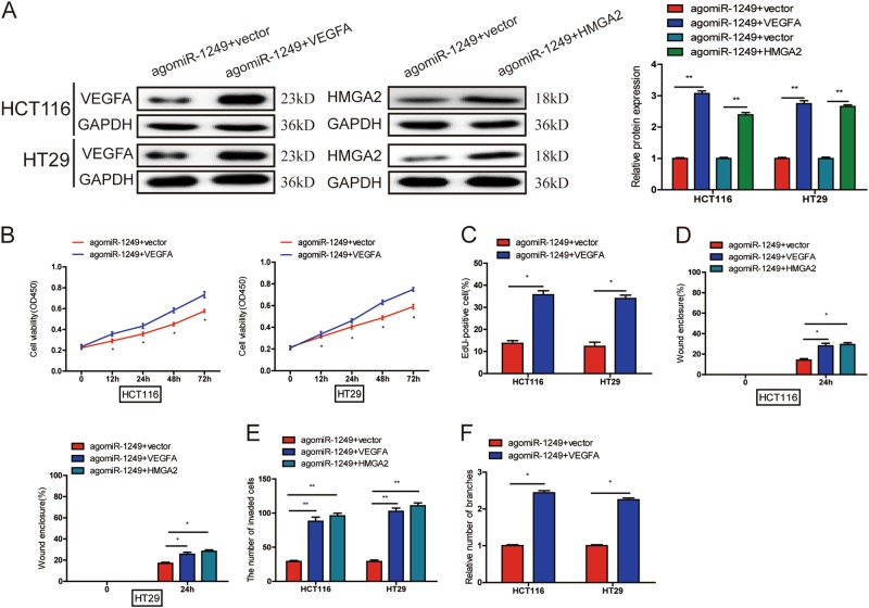
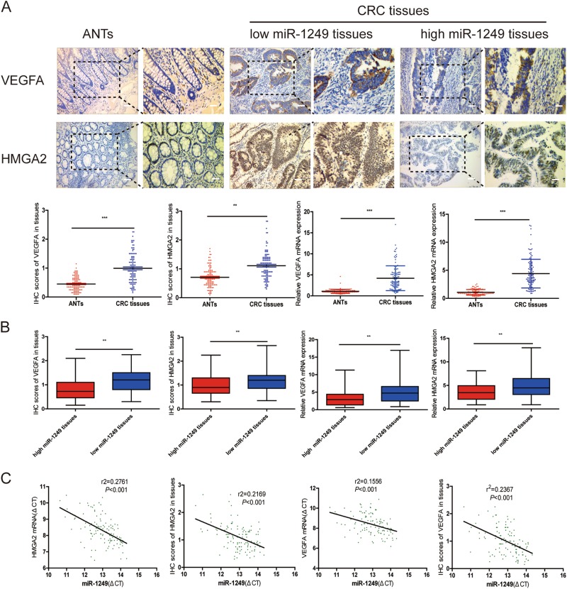
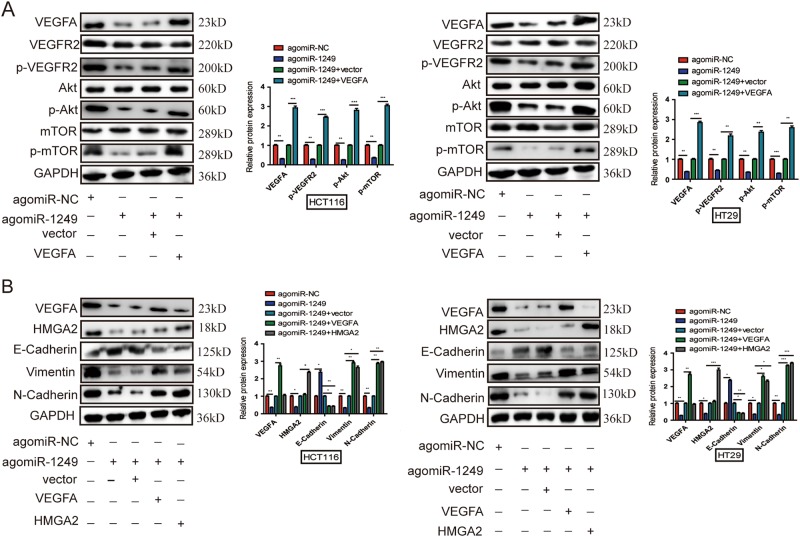
MicroRNAs (miRNAs) are important class of functional regulators involved in human cancers development, including colorectal cancer (CRC). Exploring aberrantly expressed miRNAs may provide us with new insights into the initiation and development of CRC by functioning as oncogenes or tumor suppressors. The aim of our study is to discover the expression pattern of miR-1249 in CRC and investigate its clinical significance as well as biological role in CRC progression. In our study, we found that miR-1249 was markedly downregulated in CRC tissues and cell lines, and negatively related to pN stage, pM stage, TNM stage, and overall survival (OS). Moreover, we demonstrated that miR-1249 was a direct transcriptional target of P53 and revealed that P53-induced miR-1249 inhibited tumor growth, metastasis and angiogenesis in vitro and vivo. Additionally, we verified that miR-1249 suppressed CRC proliferation and angiogenesis by targeting VEGFA as well as inhibited CRC metastasis by targeting both VEGFA and HMGA2. Further studying showed that miR-1249 suppressed CRC cell proliferation, migration, invasion, and angiogenesis via VEGFA-mediated Akt/mTOR pathway as well as inhibited EMT process of CRC cells by targeting both VEGFA and HMGA2. Our study indicated that P53-induced miR-1249 may suppress CRC growth, metastasis and angiogenesis by targeting VEGFA and HMGA2, as well as regulate Akt/mTOR pathway and EMT process in the initiation and development of CRC. miR-1249 might be a novel the therapeutic candidate target in CRC treatment.


Conflict of interest statement

The authors declare that they have no conflict of interest.

Figures

Fig. 1
Fig. 1. miR-1249 was downregulated in CRC cell lines and tissues.
a Decreased miR-1249 expression was observed in all six CRC cell lines compared with the normal colonial epithelial cell (FHC). b qRT-PCR analysis of miR-1249 in 112 pairs of human CRC tissues and their adjacent normal tissues (ANTs). ce miR-1249 was adverse correlated with pN stage (c), pM stage (d), and TNM stage (e). f Kaplan–Meier analysis of the correlation between miR-1249 expression levels and overall survival (OS) of 112 patients. ***P < 0.001
Fig. 2
Fig. 2. miR-1249 inhibited CRC cell proliferation, migration, invasion and HUVECs tube formation.
a HCT116 and HT29 cells were transfected with antagomiR-1249, agomiR-1249 and corresponding negative control (antagomiR-NC, agomiR-NC). GAPDH was used as a internal control. b, c CCK-8 (b) and EdU (c) assays were used to evaluate the proliferation of transfected CRC cells(HCT116 and HT29). d, e wound-healing (d) and transwell assays (e) were performed to detect the ability of migration and invasion of transfected CRC cells. f HUVECs were cultured in TCM from CRC cells transfected with antagomiR-NC, antagomiR-1249, agomiR-NC and agomiR-1249. The number of tube branches were measured in 10 photographic fields randomly. Data were shown as mean ± SD of three independent experiments. *P < 0.05, ** P < 0.01, ***P < 0.001
Fig. 3
Fig. 3. P53 induced miR-1249 expression as a transcription factor.
a The predicted positions of puative P53 binding motif in −4000 bp human miR-1249 promoter. b Quantitative ChIP assays were performed to show direct binding of P53 to endogenous miR-1249 promoter regions. The primers designed for ChIP were provided in supplementary materials and methods. c, d A dual-luciferase reporter assay was used by cotransfecting with full length miR-1249 promoter(miR-1249-F) or deleted miR-1249 E1 or E2 fragment (miR-1249-P2 and miR-1249-P1) with P53 expression plasmid or blank vector in 293T cells. Results are presented as mean ± SD. e The expression of miR-1249 in P53-/- and P53+/+ HCT116 cells. f The expression of miR-1249 was decreased after P53 knockdown in P53+/+ HCT116 cells. g P53-/- HCT116 cells were transfected with agomiR-1249 or agomiR-NC. hl. The ability of proliferation (h, i), migration (j), and invasion (k) was markedly increased in P53-/-HCT116 cells after after agomiR-1249 transfection. l HUVECs were cultured in TCM from P53-/- HCT116 cells transfecting with agomiR-1249 or agomiR-NC. Data were shown as mean ± SD. All the experiments were performed more than three times. *P < 0.05, **P < 0.01, ***P < 0.001
Fig. 4
Fig. 4. P53 induced miR-1249 inhibited tumor growth and distant metastasis in vivo.
a The nude mice were injected with P53-/- or P53+/+ HCT116 cells transfected with agomiR-1249 or agomiR-NC. The diameter of tumors were measured every 5 days. b Representative lungs and representative HE of lungs from mice. AgomiR-1249 group developed lower and smaller lung metastatic foci than agomiR-NC group. c CD31 expression were analyzed in xenografts tissues by IHC. Data were shown as mean ± SD. *P < 0.05, ** P < 0.01, ***P < 0.001
Fig. 5
Fig. 5. VEGFA and HMGA2 are both direct target genes of miR-1249.
a miR-1249 and its putative binding sequence in the wild-type (WT) and mutant (Mut) 3’-UTR of VEGFA or HMGA2. Overexpression of miR-1249 obviously decreased the luciferase activity that carried WT but not Mut 3′-UTR of VEGFA and HMGA2 in 293T and HCT116 cells. b, c Upregulation of miR-1249 could significantly decreased the expression of VEGFA and HMGA2 both on protein (b) and mRNA (c) levels in P53-/- and P53+/+ HCT116 cells. d Both VEGFA and HMGA2 mRNA were downregulated in xenografts tissues with high miR-1249. Data were presented as mean ± SD of three independent experiments. *P < 0.05, **P < 0.01, ***P < 0.001
Fig. 6
Fig. 6. miR-1249 inhibited CRC growth and angiogenesis by VEGFA, and suppressed migration and invasion by both VEGFA and HMGA2.
a miR-1249-overexpressioning HCT116 and HT29 cells that transfected with blank vector or pcDNA3.1-VEGFA and pcDNA3.1-HMGA2 were subjected to western blot for VEGFA and HMGA2. b, c. VEGFA upregulation could promote proliferation of miR-1249-overexpressing CRC cells using CCK-8 (b) and EdU (c). d, e VEGFA or HMGA2 upregulation could abrogate the effects of miR-1249-overexpressing on migration (d) and invasion (e) of CRC cells. f The ability of HUVECs cultured in TCM from VEGFA-upregulating CRC cells was obviously increased. Results were presented as mean ± SD. All the experiments were performed more than three times. *P < 0.05, **P < 0.01
Fig. 7
Fig. 7. The expression of VEGFA and HMGA2 were negative correlated with miR-1249 in CRC tissues.
a The statistical graph showed that IHC scores of VEGFA and HMGA2 in CRC tissues were significantly higher than those in matched adjacent normal tissues (ANTs). qRT-PCR analysis showed that both VEGFA and HMGA2 mRNA levels were higher than those in ANTs. b The statistical graph showed that IHC scores and mRNA of VEGFA and HMGA2 in low-miR-1249 CRC tissues were significantly higher than that of high-miR-1249 CRC tissues, respectively. c The relationship between miR-1249 and VEGFA and HMGA2 expression on protein and mRNA levels in CRC tissues. Data were shown as mean ± SD. **P < 0.01, ***P < 0.001
Fig. 8
Fig. 8. miR-1249 downregulated VEGFA and HMGA2 expression, and inhibited Akt/mTOR signaling pathway and EMT process.
a The downregulation of VEGFA, p-VEGFR2, p-Akt and p-mTOR were countacted by VEGFA. b The downregulation of Vimentin and N-Cadherin and the upregulation fo E-Cadherin were countacted by both VEGFA and HMGA2. GAPDH was performed as a loading control. Data indicate mean ± SD of three independent experiments, *P < 0.05, **P < 0.01, P < 0.001

Similar articles

Cited by

References

    1. Torre LA, et al. Global cancer statistics, 2012. Cancer J. Clin. 2015;65:87–108. doi: 10.3322/caac.21262. - DOI - PubMed
    1. Gill S, Dowden S, Colwell B, Collins LL, Berry S. Navigating later lines of treatment for advanced colorectal cancer - optimizing targeted biological therapies to improve outcomes. Cancer Treat. Rev. 2014;40:1171–1181. doi: 10.1016/j.ctrv.2014.10.002. - DOI - PubMed
    1. Bagga S, et al. Regulation by let-7 and lin-4 miRNAs results in target mRNA degradation. Cell. 2005;122:553–563. doi: 10.1016/j.cell.2005.07.031. - DOI - PubMed
    1. Bartel DP. MicroRNAs: target recognition and regulatory functions. Cell. 2009;136:215–233. doi: 10.1016/j.cell.2009.01.002. - DOI - PMC - PubMed
    1. Peng Y, Croce CM. The role of MicroRNAs in human cancer. Signal Transduct. Target Ther. 2016;1:15004. doi: 10.1038/sigtrans.2015.4. - DOI - PMC - PubMed

Publication types

MeSH terms