. 2019 Feb 12;10(1):710.
doi: 10.1038/s41467-019-08443-3.

Structural basis of ECF-σ-factor-dependent transcription initiation

Affiliations

Structural basis of ECF-σ-factor-dependent transcription initiation

Wei Lin et al. Nat Commun. .

Abstract

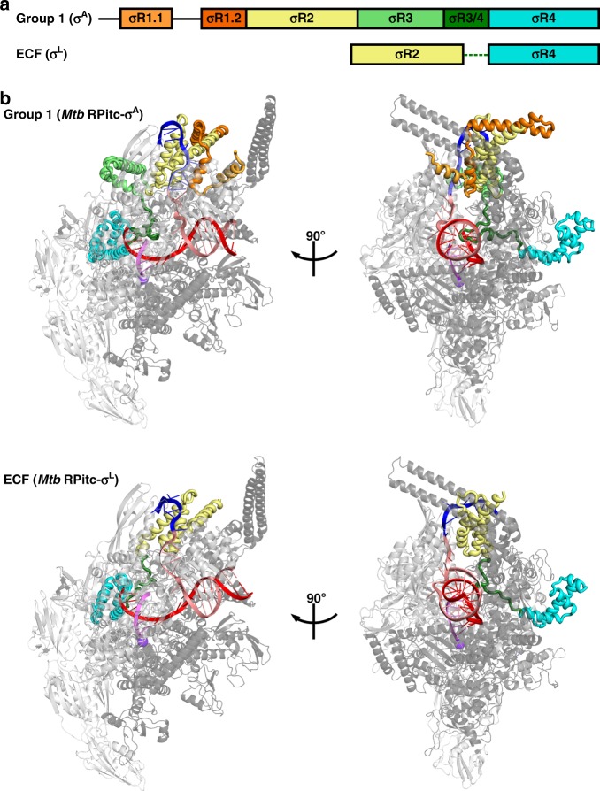
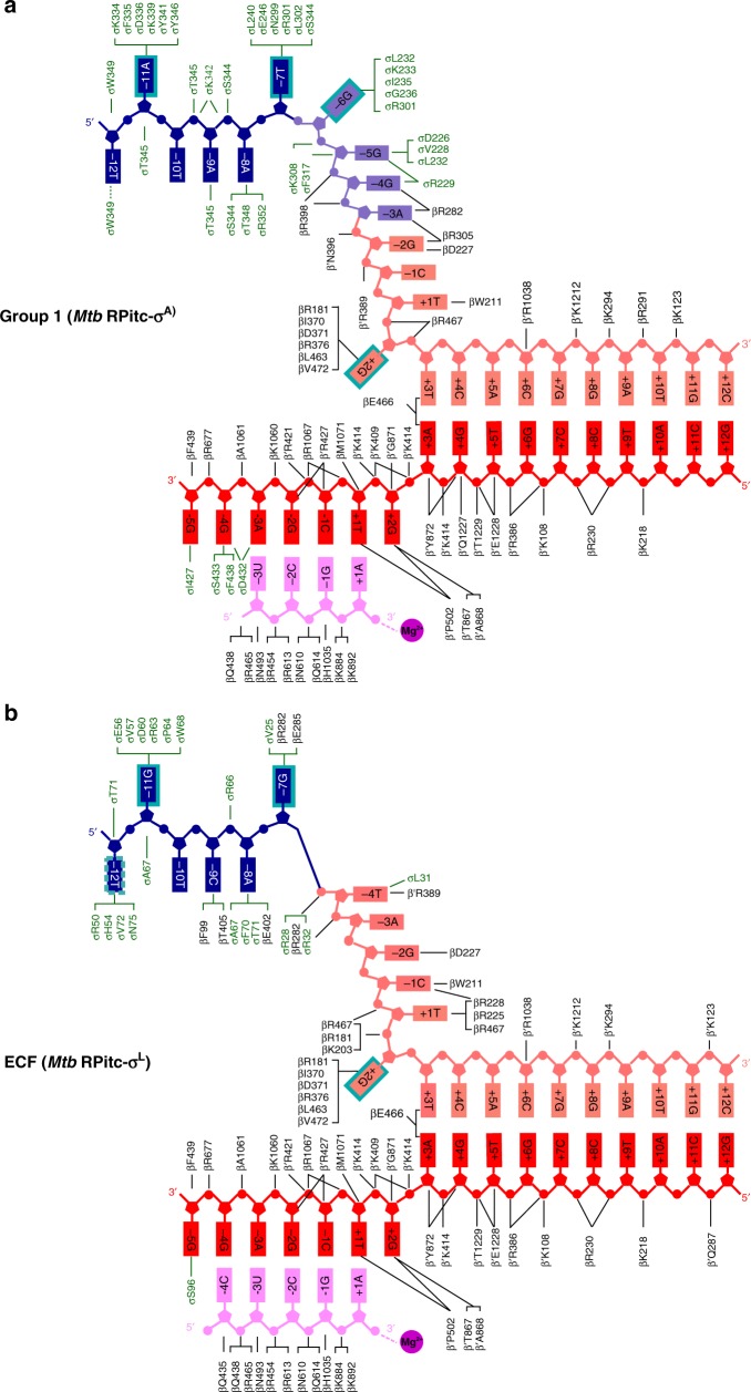
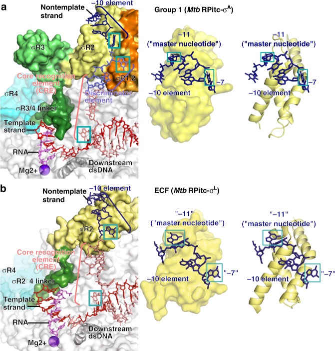
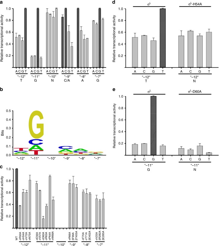
Extracytoplasmic (ECF) σ factors, the largest class of alternative σ factors, are related to primary σ factors, but have simpler structures, comprising only two of six conserved functional modules in primary σ factors: region 2 (σR2) and region 4 (σR4). Here, we report crystal structures of transcription initiation complexes containing Mycobacterium tuberculosis RNA polymerase (RNAP), M. tuberculosis ECF σ factor σL, and promoter DNA. The structures show that σR2 and σR4 of the ECF σ factor occupy the same sites on RNAP as in primary σ factors, show that the connector between σR2 and σR4 of the ECF σ factor-although shorter and unrelated in sequence-follows the same path through RNAP as in primary σ factors, and show that the ECF σ factor uses the same strategy to bind and unwind promoter DNA as primary σ factors. The results define protein-protein and protein-DNA interactions involved in ECF-σ-factor-dependent transcription initiation.


Conflict of interest statement

The authors declare no competing interests.

Figures

Fig. 1
Fig. 1
Structures of group-1 and ECF σ factors. a Structural organization of group-1 (σA) and ECF (σL) σ factors. Conserved regions σR1.1, σR1.2, σR2, σR3, σR3/4 linker, and σR4 are in light orange, dark orange, yellow, green, dark green, and cyan, respectively. Dashed line, non-conserved σR2/4 linker present in ECF σ factors. b Crystal structures of group-1 (Mtb RPitc-σA; PDB 5UH8) and ECF (Mtb RPitc-σL; PDB 6DVC) transcription initiation complexes (two orthogonal views of each). σ factors are shown in tube representations with conserved regions colored as in (a). Gray ribbon, RNAP core enzyme; blue, pink, red, and magenta ribbons, -10 element of DNA nontemplate strand, rest of DNA nontemplate strand, DNA template strand, and RNA product; violet sphere, RNAP active-center Mg2+. Other colors are as in (a). See Supplementary Figs. 1, 2, and 7
Fig. 2
Fig. 2
Protein–protein interactions between group-1 and ECF σ factors and RNAP core enzyme. a Protein–protein interactions by group-1 (σA) σ factor. b Protein–protein interactions by ECF (σL) σ factor. Colors are as in Fig. 1. See Supplementary Fig. 7
Fig. 3
Fig. 3
Protein–nucleic acid interactions by group-1 and ECF σ factors: summary. a Summary of protein–nucleic acid interactions in Mtb RPitc-σA. Black residue numbers and lines, interactions by Mtb RNAP; green residue numbers and lines, interactions by Mtb σA; blue, -10 element of DNA nontemplate strand; light blue, discriminator element of DNA nontemplate strand; pink, rest of DNA nontemplate strand; red, DNA template strand; magenta, RNA product; violet circle, RNAP active-center Mg2+; cyan boxes, bases unstacked and inserted into protein pockets. Residues are numbered as in Mtb RNAP. b Summary of protein–nucleic acid interactions in Mtb RPitc-σL. Green residue numbers and lines, interactions by Mtb σL. Other colors are as in (a). See Supplementary Figs. 3–5 and 7
Fig. 4
Fig. 4
Protein–nucleic acid interactions by group-1 and ECF σ factors: interactions with transcription bubble. a Left: interactions of Mtb RNAP and σA with transcription-bubble nontemplate strand, transcription-bubble template strand, and downstream dsDNA. Right: interactions of σA σR2 with σA-dependent promoter -10 element. For promoter positions -11 (“master nucleotide”) and -7, bases are unstacked and inserted into pockets (cyan boxes). Colors are as in Figs. 1–3. b Left: interactions of Mtb RNAP and σL with transcription-bubble nontemplate strand, transcription-bubble template strand, and downstream dsDNA. Right: interactions of σL σR2 with σL-dependent promoter -10 element. For two promoter positions, here designated “-11” (“master nucleotide”) and “-7”, by analogy to corresponding nucleotides in group-1-σ-factor complex (panel a), bases are unstacked and inserted into pockets (cyan boxes). For one additional nucleotide, here designated “-12”, the base also appears to be unstacked and inserted into a pocket (dashed cyan box). See Supplementary Figs. 4, 5 and 7
Fig. 5
Fig. 5
Recognition by Mtb σL of σL-promoter -10 element: interactions with nontemplate strand positions “-12” through “-7”. For each promoter position, left subpanel shows DNA nucleotides in stick representation to highlight individual protein-nucleotide interactions, and right subpanel shows DNA nucleotide base moieties in space-filling representation to highlight protein-base steric complementarity. Yellow surfaces, solvent-accessible surfaces of Mtb σL σR2; gray surfaces, solvent-accessible surfaces of Mtb RNAP β subunit; dark blue surfaces, van der Waals surfaces of bases of σL-promoter -10 element; yellow ribbons, Mtb σL σR2 backbone; gray ribbons, Mtb RNAP β subunit backbone; yellow, yellow-blue, and yellow-red stick representations, σL carbon, nitrogen, and oxygen atoms, respectively; white, blue, red, and orange stick representations, DNA carbon, nitrogen, oxygen, and phosphorous atoms, respectively; blue dashed lines, H-bonds. Residues are numbered as in Mtb RNAP and σL, and, in parentheses, as in E. coli RNAP and σ70. See Supplementary Fig. 4
Fig. 6
Fig. 6
Recognition by Mtb σL of σL-promoter -10 element: experimental data. a Systematic-substitution experiments defining σL-dependent promoter -10-element consensus sequence. Relative transcriptional activities of derivatives of σL-dependent promoter P-sigL having all possible single base-pair substitutions at each position of promoter -10 element, “-12” through “-7”. Inferred consensus nucleotides are shown at the bottom, and data for inferred consensus nucleotides are hatched. Error bars, SE (N = 3). b Sequence logo for σL-promoter -10-element consensus sequence [generated using transcription data from (a) and enoLOGOS (http://biodev.hgen.pitt.edu/enologos/); input setting “energy (2)” and weight-type setting “probabilities”]. c Alanine-scanning experiments demonstrating functional importance of observed amino acid-base interactions in recognition of σL-promoter -10 element. Effects on transcription of alanine substitutions of σL amino acids that contact σL-dependent promoter -10 element, positions “-12” through “-7” (identities of contacting amino acids from Figs. 3 and 5). d, e Loss-of-contact experiments indicating that σL residues His54 and Asp60 determine specificity at position “-12” and “-11”, respectively. Left: transcriptional activity with wild-type σL for all possible single base-pair-substitutions at indicated position (strong specificity for consensus base pair). Right: transcriptional activity of σL derivatives having alanine substitutions (no specificity for consensus base pair). Error bars, SE (N = 3). See Supplementary Fig. 6. Source data are provided as a Source Data file

References

    1. Feklistov A, Sharon BD, Darst SA, Gross CA. Bacterial sigma factors: a historical, structural, and genomic perspective. Annu. Rev. Microbiol. 2014;68:357–376. doi: 10.1146/annurev-micro-092412-155737. - DOI - PubMed
    1. Murakami K, Masuda S, Darst S. Structural basis of transcription initiation: RNA polymerase holoenzyme at 4 Å resolution. Science. 2002;296:1280–1284. doi: 10.1126/science.1069594. - DOI - PubMed
    1. Vassylyev D, et al. Crystal structure of a bacterial RNA polymerase holoenzyme at 2.6 Å resolution. Nature. 2002;417:712–719. doi: 10.1038/nature752. - DOI - PubMed
    1. Mekler V, et al. Structural organization of bacterial RNA polymerase holoenzyme and the RNA polymerase-promoter open complex. Cell. 2002;108:599–614. doi: 10.1016/S0092-8674(02)00667-0. - DOI - PubMed
    1. Kulbachinskiy A, Mustaev A. Region 3.2 of the sigma subunit contributes to the binding of the 3′-initiating nucleotide in the RNA polymerase active center and facilitates promoter clearance during initiation. J. Biol. Chem. 2006;281:18273–18276. doi: 10.1074/jbc.C600060200. - DOI - PubMed

Publication types

MeSH terms