. 2019 Feb 12;9(1):1852.
doi: 10.1038/s41598-018-38156-4.

IL-24 contributes to skin inflammation in Para-Phenylenediamine-induced contact hypersensitivity

Affiliations

IL-24 contributes to skin inflammation in Para-Phenylenediamine-induced contact hypersensitivity

Astrid B Van Belle et al. Sci Rep. .

Abstract

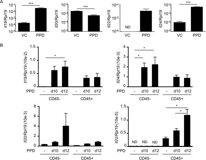
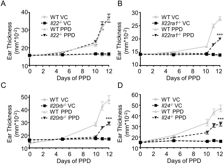
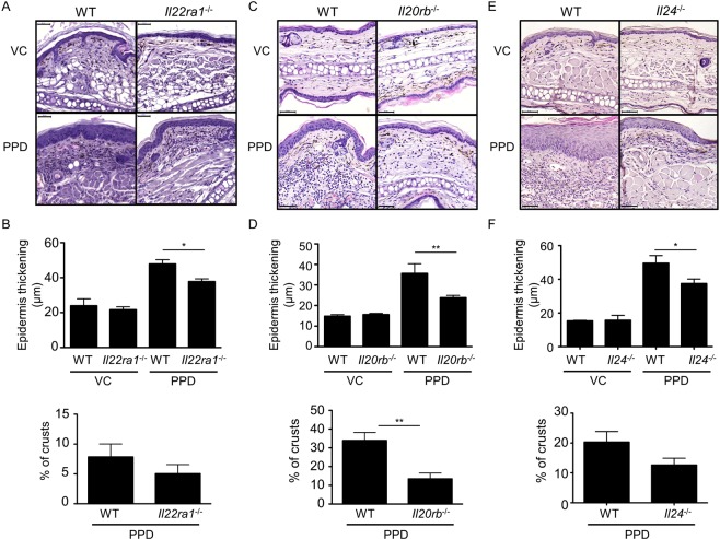
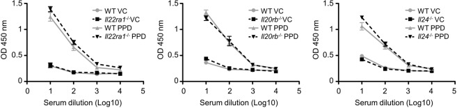
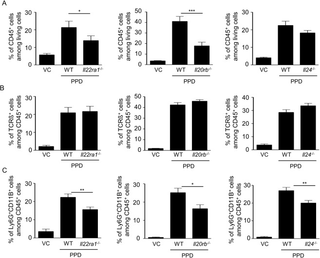
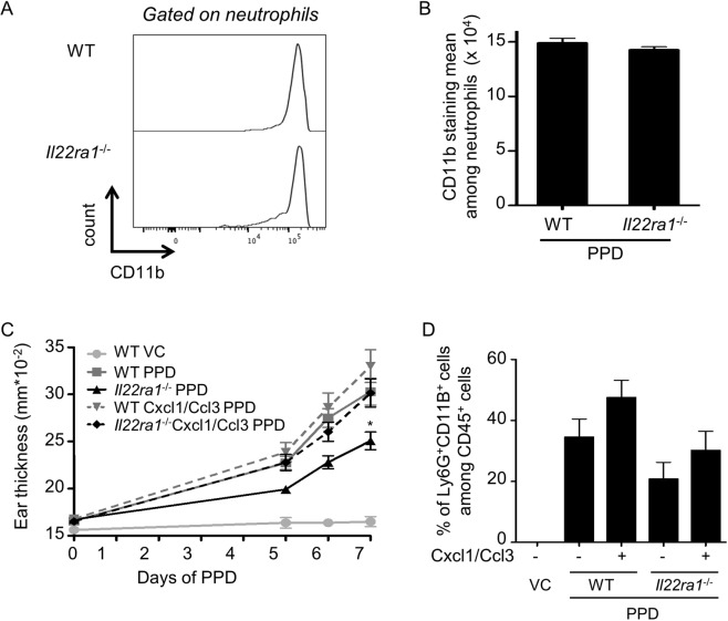
Para-Phenylenediamine (PPD) is an aromatic amine used in hair dyes and in temporary black henna tattoos, which is a frequent cause of allergic contact dermatitis (ACD). ACD is a skin inflammatory reaction characterized by modifications such as spongiosis, exocytosis and acanthosis. The aim of this study is to characterize the expression and the role of IL-20-related cytokines, including IL-19, IL-20, IL-22 and IL-24, in ACD. The expression of IL19, IL20, IL22 and IL24 is increased in affected skin from PPD allergic patients compared with uninvolved skin. In addition, the expression of these cytokines positively correlates with clinical symptoms. To assess their role in ACD, we set up a mouse model of PPD-induced allergic contact dermatitis and we showed that, in contrast to Il22-deficient mice, Il22ra1-, Il20rb- and Il24-deficient mice are partially protected against development of PPD-induced contact hypersensitivity. These mice have decreased ear thickening and less acanthosis compared with WT mice after PPD treatment. In addition, the absence of IL-22R, IL-20R2 or IL-24 affects the recruitment of neutrophils into the skin but not the total IgE production. Taken together, these results demonstrate the implication of IL-24 via the IL-20R type II receptor in the inflammatory process of ACD.


Conflict of interest statement

The authors declare no competing interests.

Figures

Figure 1
Figure 1
Expression of IL-20-related cytokines is increased in skin of PPD-allergic patients. (A) RNA was isolated from healthy skin and patch test biopsies (at indicated period of time) of allergic patients (N = 11). B. RNA was isolated from PBMCs of healthy control (HC, N = 16) and PPD-allergic patients (PPD, N = 24). PBMCs were stimulated with anti-CD3, anti-CD28 and PPD for 48 hours. Next, qPCR for IL19, IL20, IL22, IL24 and EF1 mRNA expression were performed. (A) Black curves represent patients with at least a positive patch test reaction at 24 hours. Grey curves represent patients with a negative patch test reaction at 24 hours. (B) The induction is calculated by comparing the expression of cytokines in stimulated PBMCs vs unstimulated PBMCs. *p < 0.05, **p < 0.01 and ***p < 0.001 (A: Friedman test, Dunn’s multiple comparison and B: Mann-Whitney).
Figure 2
Figure 2
Expression of IL-20-related cytokines is increased in a mouse model of PPD-induced allergic contact dermatitis. 129/Sv mice were treated with PPD solutions. Quantitative RT-PCR analysis was performed for each indicated gene. (A) RNA was isolated from the total ear 24 hours after the second application. (B) 24 hours after the third (day 10) and the fifth (day 12) application, CD45+ cells were purified from the epidermis by MACS. RNA was isolated from both the CD45-positive and CD45-negative fraction. Data correspond to the mean ± SEM (N = 4 mice per group). Data are representative of three independent experiments. *p < 0.05 and ***p < 0.001 (A: Mann-Whitney and B: one-way ANOVA, Bonferroni multiple comparison). ND: not detected, VC: vehicle control.
Figure 3
Figure 3
Il24−/−, Il22ra1−/− and Il20rb−/− mice, but not Il22−/− mice, are partially protected against CHS induced by PPD. Ear thickness was measured before each PPD treatment and 24 hours after the last application with a micrometer screw to evaluate the development of CHS. (A) Ear thickness in Il22+/+ and Il22−/− 129/Sv mice. (B) Ear thickness in Il22ra1+/+ and Il22ra1−/− 129/Sv mice. (C) Ear thickness in Il20rb+/+ and Il20rb−/− C57BL/6 mice. (D) Ear thickness in Il24+/+ and Il24−/− C57BL/6 mice. Data are means ± SEM (N = at least 4 for VC groups and N = at least 5 for PPD-treated groups) and representative of at least three independent experiments ***p < 0.001 Comparison of PPD treated mice (two-way Anova, Bonferroni multiple comparison). VC: vehicle control.
Figure 4
Figure 4
Acanthosis is less prominent in Il24−/−, Il22ra1−/− and Il20rb−/− mice compared with WT mice after PPD treatment. (A,C,E) HE staining of ear skin sections from mice, treated or not with PPD, 24 hours after the sixth application. One representative picture is shown for each treatment regimen (original magnification x30, scale bar = 50 μm). (B,D,F) Acanthosis was evaluated by measuring the epidermal thickness at six different places by using Pannoramic viewer measuring tool. The percentages of crusts are calculated by dividing the length of crusts by the length of the section. These analyses were performed in Il22ra1+/+ and Il22ra1−/− 129/Sv mice (A,B), in Il20rb+/+ and Il20rb−/− C57BL/6 mice (C,D) and Il24+/+ and Il24−/− C57BL/6 mice (E,F). Data are means ± SEM (N = 5 for VC groups and N = 8 for PPD-treated groups) and representative of four independent experiments. *p < 0.05 and **p < 0.01 (Mann-Whitney to compare treated mice). Histological analysis was performed by two evaluators. VC: vehicle control.
Figure 5
Figure 5
IgE-dependent allergy is similar in WT and Il22ra1−/−, Il20rb−/− and Il24−/− mice after PPD treatment. Mice were treated or not with PPD and 24 hours after the third application, IgE levels in the sera were assessed by ELISA. This analysis was performed in Il22ra1+/+ vs Il22ra1−/− 129/Sv mice (left panel), in Il20rb+/+ vs Il20rb−/− C57BL/6 mice (central panel) and in Il24+/+ and Il24−/− C57BL/6 mice (right panel). Data are means ± SEM (N = 3 for VC groups and N = 6 for PPD-treated groups). VC: vehicle control.
Figure 6
Figure 6
Epidermal infiltration of CD45+ cells decreases in Il22ra1−/− and Il20rb−/− mice compared with WT mice. Flow cytometry on epidermal cells from mice, treated or not with PPD (VC), 24 hours after the second application. (A) The percentage of CD45+ cells among living cells was analyzed. (B) The proportion of TCRß+ cells among CD45+ living cells was analyzed. (C) The proportion of Ly6G+CD11B+ cells among CD45+ living cells was analyzed. These analyses were performed in Il22ra1+/+ and Il22ra1−/− 129/Sv mice (left panels), in Il20rb+/+ and Il20rb−/− C57BL/6 mice (central panels) and Il24+/+ and Il24−/− C57BL/6 mice (right panels). Data are means ± SEM (N = 5 for VC groups and PPD-treated groups) and representative of three independent experiments. *p < 0.05 and ***p < 0.001 (Mann-Whitney to compare treated mice). VC: vehicle control.
Figure 7
Figure 7
Neutrophil activity is similar in WT and Il22ra1−/− mice but induction of neutrophil recruitment increases ear thickness. (A,B) After two PPD applications, ears from Il22ra1+/+ and Il22ra1−/− C57BL/6 mice were collected to analyze neutrophil population by flow cytometry. (A) CD11B staining in neutrophil population (gated on Ly6G+ CD11B+ cells among CD45+ living cells) from Il22ra1+/+ and Il22ra1−/− mice after PPD treatment (one representative mouse per group). (B) Mean of CD11B staining among neutrophil populations. Data are means ± SEM (N = at least 5 mice per group) and representative of two independent experiments. (C,D) Cxcl1 and Ccl3 chemokines were injected in ears during first and second applications to induce neutrophil recruitment and flow cytometry analysis was performed on epidermal cells from mice, treated or not with PPD (VC), 48 hours after the second application. PBS was injected in mice without chemokine injection. This analysis was performed in Il22ra1+/+ vs Il22ra1−/− C57BL/6 mice. (C) Ear thickness was measured before each PPD application and each day after the second application. (B) The proportion of Ly6G+CD11B+ cells among CD45+ living cells was analyzed. Data are means ± SEM (N = 4 for VC group and at least 5 for PPD-treated groups) and representative of three independent experiments. *p < 0.05 (C: two-way Anova, Bonferroni multiple comparison). VC: vehicle control.

References

    1. Calogiuri G, et al. Allergic reactions to henna-based temporary tattoos and their components. Immunopharmacol Immunotoxicol. 2010;32:700–704. doi: 10.3109/08923971003685942. - DOI - PubMed
    1. Armstrong DK, et al. Occupational sensitization to p-phenylenediamine: a 17-year review. Contact Dermatitis. 1999;41:348–349. doi: 10.1111/j.1600-0536.1999.tb06191.x. - DOI - PubMed
    1. Thyssen JP, White JM. & European Society of Contact, D. Epidemiological data on consumer allergy to p-phenylenediamine. Contact Dermatitis. 2008;59:327–343. doi: 10.1111/j.1600-0536.2008.01427.x. - DOI - PubMed
    1. Diepgen TL, et al. Prevalence of Contact Allergy to p-Phenylenediamine in the European General Population. J Invest Dermatol. 2016;136:409–415. doi: 10.1016/j.jid.2015.10.064. - DOI - PubMed
    1. Phelps RG, Miller MK, Singh F. The varieties of “eczema”: clinicopathologic correlation. Clin Dermatol. 2003;21:95–100. doi: 10.1016/S0738-081X(02)00357-7. - DOI - PubMed

Publication types

MeSH terms

LinkOut - more resources