. 2019 Feb 12;9(1):1829.
doi: 10.1038/s41598-019-38646-z.

Three Dimensional Glomerular Reconstruction: A Novel Approach to Evaluate Renal Microanatomy in Diabetic Kidney Disease

Affiliations

Three Dimensional Glomerular Reconstruction: A Novel Approach to Evaluate Renal Microanatomy in Diabetic Kidney Disease

Niloufar Torkamani et al. Sci Rep. .

Abstract

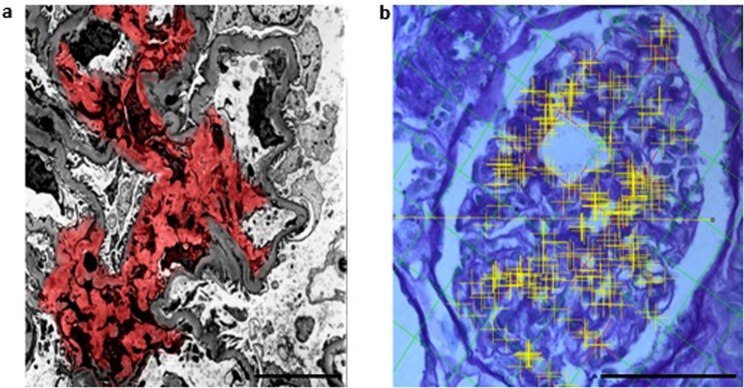
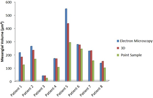
Mesangial metrics reflect glomerular filtration surface area in diabetes. The point-sampled intercept (PSI) method is the conventional method to calculate these parameters. However, this is time consuming and subject to underestimation. We introduce a novel three-dimensional (3D) reconstruction method applicable to light microscopy to measure mesangial metrics. Transmission electron microscopy (TEM), PSI and our new 3D imaging methods were used to quantify mesangial metrics from 22 patients with type 2 diabetes, normo-, micro- and macroalbuminuria and an estimated glomerular filtration rate of <60 mL/min/1.73 m2. Repeated-measures ANOVA test was used to test the equality of the measurement means from the three methods and the degree of inter method variability. Repeated-measures and post-estimation ANOVA tests together with correlation coefficient measurements were used to compare the methods with TEM as reference. There was a statistically significant difference in mesangial volume measurements (F(2, 16) = 15.53, p = 0.0002). The PSI method underestimated measurements compared to TEM and 3D methods by 30% (p = 0.001) and 15%, respectively (p < 0.001). 3D and TEM measurements did not differ significantly. 3D reconstruction is a reliable and time efficient method for calculating mesangial metrics. It may prove to be a useful tool in clinical and experimental diabetic kidney disease.


Conflict of interest statement

The authors declare no competing interests.

Figures

Figure 1
Figure 1
Three-dimensional reconstruction of the glomerulus. (a) Glomerular Mesangium (blue) was identified using Reconstruct software based on PAS staining differentiation (scale bar = 50 μm). The eye symbol represents the direction of the view (b) Superior view of glomerulus. 3-dimensional model of the glomerulus was generated using serial horizontal (transverse) sections (c) Close-up view of the 3D model of the glomerular mesangial area. Glomerular mesangial (red) surface area and volume was quantified.
Figure 2
Figure 2
Mesangial area quantification using Transmission Electron Microscopy (TEM) and Point Sample Intercept (PSI) methods. (a) Mesangial area and estimated volume was calculated from TEM images (Scale bar 4 µm). Images were analysed (Digimizer 4.2.2) and mesangial area (red) was identified by an expert operator. Transverse sections through the middle of representative glomeruli were chosen and all identifiable mesangial area per glomerulus was measured. The mean mesangial area in a glomerulus per subject was then calculated. (b) Fractional volumes of glomerular components were calculated using PSI method. Using the Cavalieri’s method of segmentation combined with automated point counting (yellow) the mesangial volume was estimated (Fiji by Image J 1.51e) (Scale bar 50 µm).
Figure 3
Figure 3
Comparison of three methods in measuring the mesangial volume. TEM (blue), 3D method (red) and PSI (green). Correlation coefficient between TEM/3D, TEM/PSI and 3D/PSI methods was 0.98, 0.93, and 0.96 respectively.

References

    1. MacIsaac RJ, Jerums G, Ekinci EI. Effects of glycaemic management on diabetic kidney disease. World J Diabetes. 2017;8:172–186. doi: 10.4239/wjd.v8.i5.172. - DOI - PMC - PubMed
    1. Mauer SM, Steffes MW, Brown DM. The kidney in diabetes. Am J Med. 1981;70:603–612. doi: 10.1016/0002-9343(81)90582-9. - DOI - PubMed
    1. Mauer SM, et al. Structural-functional relationships in diabetic nephropathy. J Clin Invest. 1984;74:1143–1155. doi: 10.1172/JCI111523. - DOI - PMC - PubMed
    1. Tandrup T, Gundersen HJ, Jensen EB. The optical rotator. J Microsc. 1997;186:108–120. doi: 10.1046/j.1365-2818.1997.2070765.x. - DOI - PubMed
    1. Ekinci EI, et al. Renal structure in normoalbuminuric and albuminuric patients with type 2 diabetes and impaired renal function. Diabetes Care. 2013;36:3620–3626. doi: 10.2337/dc12-2572. - DOI - PMC - PubMed

MeSH terms

Substances