. 2019 Feb 12;9(1):1864.
doi: 10.1038/s41598-018-38205-y.

Epithelial Cells and Fibroblasts from the Human Female Reproductive Tract Accumulate and Release TFV and TAF to Sustain Inhibition of HIV Infection of CD4+ T cells

Affiliations

Epithelial Cells and Fibroblasts from the Human Female Reproductive Tract Accumulate and Release TFV and TAF to Sustain Inhibition of HIV Infection of CD4+ T cells

Zheng Shen et al. Sci Rep. .

Abstract

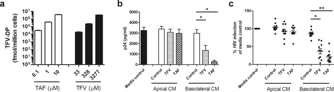
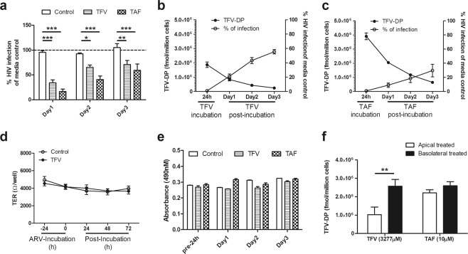
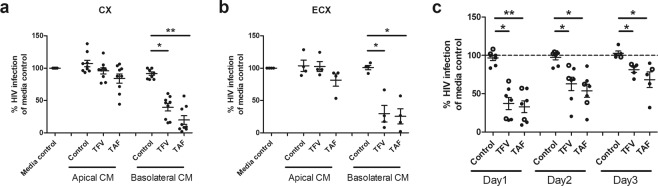
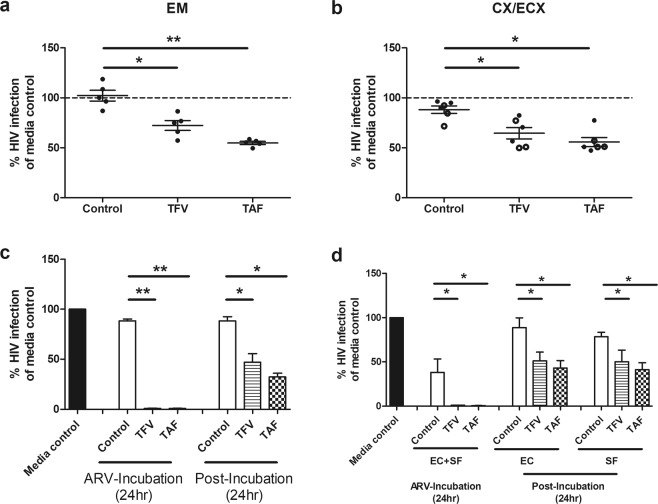
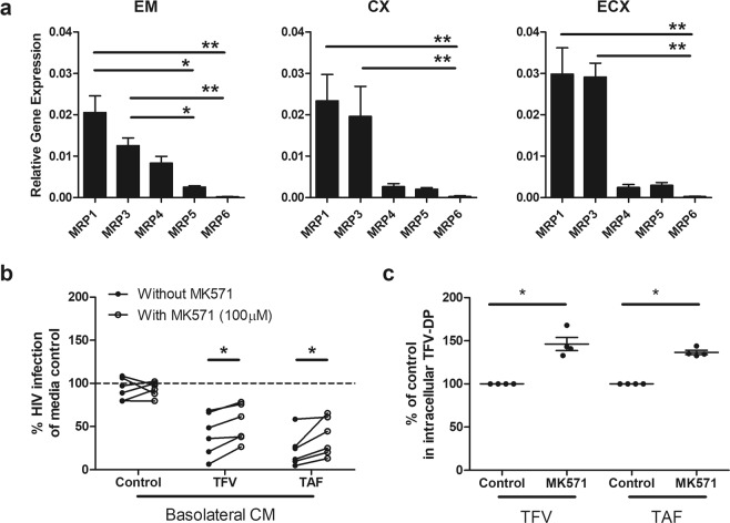
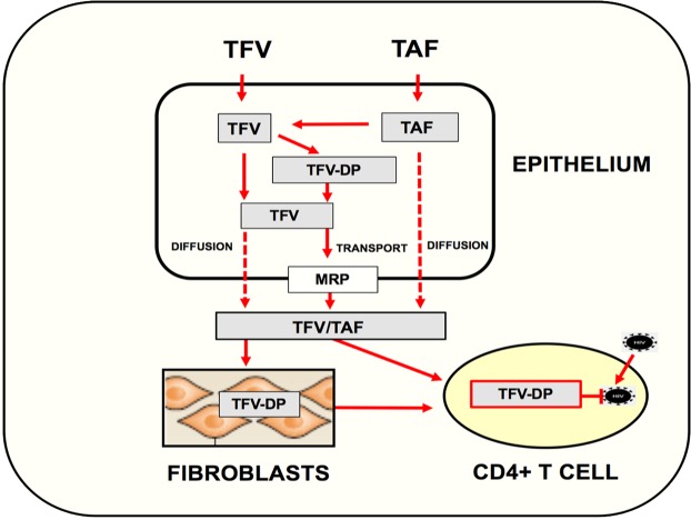
Tenofovir (TFV) treatment of female reproductive tract (FRT) cells results in differential accumulation of intracellular Tenofovir diphosphate (TFV-DP) in different cell types, with greater concentrations in epithelial cells (100-fold) and fibroblasts (10-fold) than in CD4+ T cells. The possibility that TFV-DP accumulation and retention in epithelial cells and fibroblasts may alter TFV availability and protection of CD4+ T cells against HIV infection, prompted us to evaluate TFV and/or Tenofovir alafenamide (TAF) release from FRT cells. Endometrial, endocervical and ectocervical polarized epithelial cells and fibroblasts were pre-loaded with TFV or TAF, and secretions tested for their ability to inhibit HIV infection of activated blood CD4+ T cells. Epithelial cell basolateral secretions (1, 2 and 3 days post-loading), but not apical secretions, suppressed HIV infection of CD4+ T cells, as did secretions from pre-loaded fibroblasts from each site. Intracellular TFV-DP levels in epithelial cells following preloading with TFV or TAF correlated directly with ARV protection of CD4+ T cells from HIV infection. When added apically to epithelial cells, TFV/TAF was released basolaterally, in part through Multidrug Resistant Protein transporters, taken up by fibroblasts and released into secretions to partially protect CD4+ T cells. These findings demonstrate that epithelial cells and fibroblasts release TFV/TAF for use by CD4+ T cells and suggest that the tissue environment plays a major role in the sustained protection against HIV infection.


Conflict of interest statement

The authors declare no competing interests.

Figures

Figure 1
Figure 1
Evaluation of the protective effect of secretions from endometrial epithelial cells following preincubation with TFV or TAF on HIV infection of blood CD4+ T cells. (a) Dose-dependent increase in intracellular TFV-DP levels following treatment of purified polarized endometrial (EM) epithelial cells with TAF (white bars) or TFV (black bars) for 24 hr. Bars and horizontal lines represent mean and SEM respectively from triplicate cultures of cells from a representative patient. Values are expressed as fmol/million cells. (b,c) Levels of HIV infection (released p24) in CD4+ T cells after incubation with apical and basolateral CM from EM epithelial cells pre-treated with TFV (3277 µM) or TAF (10 µM) as described in Methods. (b) Representative example of secreted p24 levels from a single patient run in quadruplicate. Columns and horizontal lines represent the mean and SEM respectively. (c) Secreted p24 values are normalized to the infection of CD4+ T cells in the absence of CM (media control) which is set to 100%. Each circle represents an individual patient (n = 10) and horizontal lines represent the mean and SEM. Blood CD4+ T cells were isolated from 4 donors. *p < 0.05. **p < 0.01.
Figure 2
Figure 2
Protective anti-HIV effect of basolateral secretions from endometrial (EM) epithelial cells following treatment with TFV or TAF correlates with epithelial levels of intracellular TFV-DP. (a) HIV infection levels in CD4+ T cells after incubation with CM from EM epithelial cells pre-treated with TFV or TAF. EM epithelial cells were pre-treated with either TFV (3277 µM) or TAF (10 µM) for 24 hr, after which ARVs were washed out of cell culture. Cells were then incubated with fresh media that was replaced daily for an initial 24 hr (Day 1), a second 24 hr period (Day 2), and a third 24 hr period (Day 3) after which basolateral conditioned media (CM) was collected. Activated CD4+ T cells were treated for 24 hr with CM recovered at Day 1, 2, and 3. Following washout of CM, CD4+ T cells were infected after which secreted p24 levels measured by p24 ELISA after 5 days as described in Methods. Data are normalized to the infection of CD4+ T cells in the absence of CM (media control) which is set to 100% (dashed line). (n = 4 individual patients). Columns and horizontal lines represent the mean and SEM respectively. *p < 0.05. **p < 0.01. ***p < 0.001. (b and c) Intracellular TFV-DP levels in EM epithelial cells are inversely related to HIV infection of CD4+ T cells treated with EM epithelial CM. Intracellular TFV-DP levels in EM epithelial cells were measured by LC-MS/MS following TFV (b) and TAF (c) treatment (24 hr), followed by incubation for 24 hr intervals with fresh media collection on Day 1, Day 2, and Day 3. CM was collected at each time point along with cell recovery to measure HIV infection as described in Methods. Circles and horizontal lines represent the mean and SEM from triplicates in a single representative patient. (d) Time course of the lack of an effect on transepithelial resistance (TER) of polarized epithelial cells treated with TFV (3277 µM) for 24 hr after which ARV was washed out of cell culture. Cells were then incubated with fresh media that was replaced daily for 3 days. (e) Lack of an effect of TFV and TAF on epithelial cell viability. Polarized EM epithelial cells were apically and basolaterally treated with TFV (3277 µM) or TAF (10 µM) for 24 hr prior to washout and measurement of viability on days 1, 2 and 3. Cell viability was tested with CellTiter 96 AQueous One Solution cell proliferation assay. The bars represent the mean and SEM of triplicate cell inserts. (f) Intracellular accumulation of TFV-DP on endometrial epithelial cells following apical or basolateral incubation with TFV or TAF. Intracellular TFV-DP levels were measured by LC-MS/MS in purified polarized endometrial epithelial cells treated with TFV (3277 µM) or TAF (10 µM) apical or basolateral for 24 hr. Values are expressed as fmol/million cells. The bars represent the mean and SEM from 3 patients. **p < 0.01.
Figure 3
Figure 3
Secretions from endocervical and ectocervical epithelial cells treated with TFV or TAF inhibit HIV infection of CD4+ T cells. Apical and basolateral conditioned media (CM) were collected from endocervix (CX) and ectocervix (ECX) epithelial cells pre-treated with TFV (3277 µM) or TAF (10 µM) for 24 hr. CM were collected 24 hr post ARV washout and incubation with fresh media; CM was incubated with activated CD4+ T cells prior to HIV infection. Secreted p24 levels in the culture media after 5 days of infection were measured by p24 ELISA as described in Methods. Data are normalized to the infection of CD4+ T cells in the absence of CM (media control) which is set to 100% using (a) CM from CX epithelial cells from 9 patients, (b) CM from ECX epithelial cells from 4 patients. (c) Time course of HIV protection of CD4+ T cells by basolateral CM from CX (dark circles) and ECX (open circles) epithelial cells pre-treated with either ARVs for 24 hr prior to wash out. Cells were then incubated with fresh media that was replaced each day for 3 days, basolateral CM was collected daily. Activated CD4+ T cells were treated for 24 hr with CM. Following washout of CM, CD4+ T cells were infected after which secreted p24 levels measured by p24 ELISA as described in Methods. Data are normalized to the infection of CD4+ T cells in the absence of CM (media control) which is set to 100% (dashed line). (n = 7 individual patients). Columns and horizontal lines represent the mean and SEM respectively. *p < 0.05. **p < 0.01.
Figure 4
Figure 4
Effect of fibroblasts and endometrial (EM) epithelial cells in decreased HIV infection of CD4+ T cells. Conditioned media (CM) was collected from (a) endometrium (EM), (b) CX/ECX fibroblasts pre-treated with TFV (3277 µM) or TAF (10 µM) for 24 hr. CM were collected 24 hr post ARV washout and incubation with fresh media; CM was incubated with activated CD4+ T cells prior to HIV infection. Secreted p24 levels in the culture media after 5 days of infection were measured by p24 ELISA as described in Methods. CM was collected from EM fibroblasts of 5 patients while CM from CX (dark circle) and ECX (open circle) fibroblasts was from 3 matched patients. Each circle represents an individual patient. Data are normalized in (a,b) to the infection of CD4+ T cells in the absence of CM (media control) and set to 100%. Each circle represents a different patient. Blood CD4+ T cells were isolated from 4 donors. Horizontal lines represent the mean and SEM respectively. *p < 0.05. **p < 0.01. (c) Polarized EM epithelial cells were apically treated with TFV or TAF for 24 hr, after which basolateral CM was immediately collected (ARV-Incubation). Following rinsing, cells were incubated with fresh media (no TFV or TAF) for an additional 24 hr prior to CM collection (Post-Incubation 24 hr). Activated CD4+ T cells from blood were incubated with CM for 24 hr and washed and infected with HIV for 2 hr. Secreted viral p24 levels were measured after 5 days of infection (Methods). Data are normalized to the infection of CD4+ T cells in the absence of CM (media control) which is set to 100%. Bars represent EM tissues from 3 patients. (d) Effect of epithelial cells and fibroblasts on prevention of HIV infection. Polarized EM epithelial cells grown in the upper chamber of cell inserts were treated apically with TFV (3277 µM) or TAF (10 µM) for 24 hr in the presence of confluent fibroblasts (SF) from the same donor grown in the lower chamber (no cell contact). Following incubation, epithelial cells and stromal fibroblasts (EC + SF), basolateral CM was collected for analysis (ARV-Incubation). Following extensive washing to remove extracellular ARVs (Post-Incubation), epithelial cells alone (EC) and stromal fibroblasts alone (SF) were incubated in fresh media for 24 hr, after which CM were collected. CD4+ T cell protection against HIV infection for each CM was measured as described in Methods. Data are normalized to the infection of CD4+ T cells in the absence of CM (media control). Bars represent 4 patients. Blood CD4+ T cells were isolated from 2 donors. Column and horizontal lines represents the mean and SEM respectively. *p < 0.05. **p < 0.01.
Figure 5
Figure 5
Blockade of Multidrug resistance-associated proteins (MRP) leads in part to decreased anti-HIV activity in basolateral epithelial CM via increased intracellular accumulation of TFV-DP. (a) MRP (1, 3, 4, 5 and 6) gene expression in epithelial cells from endometrium (EM), endocervix (CX) and ectocervix (ECX). Gene expression was determined by real time RT-PCR and normalized to β-Actin using epithelial cells from 4 patients with matched EM, CX and ECX. Y axis represents gene expression relative to the house keeping gene β-actin. Columns and horizontal lines represent the mean and SEM respectively. *p < 0.05. **p < 0.01. (b) Blockade of MRP transporter function decreases the release of TFV and TAF into the extracellular environment. EM epithelial cells were incubated with TFV (3277 µM) or TAF (10 µM) for 20 hr, followed by the addition of a human MRP-specific inhibitor MK571 (100 µM) to both the apical and basolateral compartments for 4 hr. Following washout, fresh media +/− MK571 (100 uM) was added to both compartments for an additional 24 hr after which the basolateral CM was collected. Activated CD4+ T cells from blood were incubated with basolateral CM for 24 hr prior to HIV infection (Methods). Data were normalized to % of HIV infection of CD4+ T cells infected in the absence of basolateral CM (media control). Each circle represents a different individual patient (n = 6) with dark circles representing basolateral CM collected from cells not treated with MK571, and open circles basolateral CM from cells treated with MK571 Data are normalized to the infection of CD4+ T cells in the absence of CM (media control) and set to 100%. *p < 0.05. (c) Intracellular TFV-DP levels in purified polarized EM epithelial cells increase following incubation with TFV (3277 µM) or TAF (10 µM) for 24 hr in the presence of MK571 (100 µM). Data were normalized to % of intracellular TFV-DP without MK571 in experiments using EM epithelial cells from 4 individual patients. Each circle represents a different patient. Horizontal lines represent the mean and SEM respectively. *p < 0.05.
Figure 6
Figure 6
Proposed pathway of TFV and TAF mediated protection of CD4+ T cells by epithelial cells and fibroblasts in the female reproductive tract. TFV and TAF readily enter epithelial cells throughout the female reproductive tract (FRT) where they are converted to TFV-DP, the active metabolite that inhibits HIV replication. Intracellular TFV-DP can be converted back into TFV. While movement of TFV-DP out of the cell is restricted, TFV and TAF are released basolaterally into the extracellular environment both by diffusion across the plasma membrane and/or active transport out of cells by MRPs expressed on the cell surface. Once released to the underlying stroma, TFV/TAF either act directly on CD4+ T cells to prevent HIV infection or are taken up by stromal fibroblasts, which similar to epithelial cells, gradually release TFV/TAF to the local environment. In doing so, both epithelial cells and stromal fibroblasts function as reservoirs that extend the time interval of protection beyond the initial uptake of TFV/TAF by mucosal CD4+ T cells, the primary targets of HIV infection.

Similar articles

Cited by

References

    1. WHO. Number of people (all ages) living with HIV: Estimates by WHO region, http://apps.who.int/gho/data/view.main.22100WHO?lang=en (2018).
    1. UNAIDS. Women, girls, gender equality and HIV. (2010).
    1. Wira CR, Rodriguez-Garcia M, Patel MV. The role of sex hormones in immune protection of the female reproductive tract. Nat Rev Immunol. 2015;15:217–230. doi: 10.1038/nri3819. - DOI - PMC - PubMed
    1. Rodriguez-Garcia M, Barr FD, Crist SG, Fahey JV, Wira CR. Phenotype and susceptibility to HIV infection of CD4+ Th17 cells in the human female reproductive tract. Mucosal Immunol. 2014;7:1375–1385. doi: 10.1038/mi.2014.26. - DOI - PMC - PubMed
    1. Rodriguez-Garcia M, et al. Estradiol reduces susceptibility of CD4+ T cells and macrophages to HIV-infection. PLoS One. 2013;8:e62069. doi: 10.1371/journal.pone.0062069. - DOI - PMC - PubMed

Publication types

MeSH terms