. 2019 Feb 12;9(1):1853.
doi: 10.1038/s41598-019-38534-6.

Niraparib activates interferon signaling and potentiates anti-PD-1 antibody efficacy in tumor models

Affiliations

Niraparib activates interferon signaling and potentiates anti-PD-1 antibody efficacy in tumor models

Zebin Wang et al. Sci Rep. .

Abstract

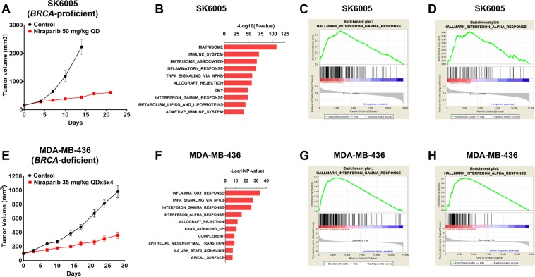
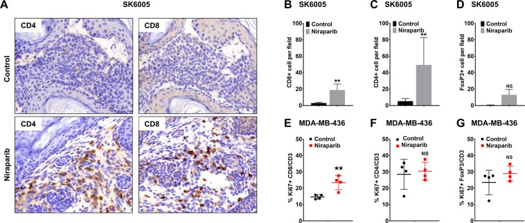
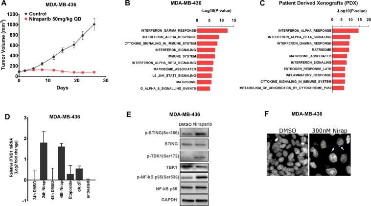
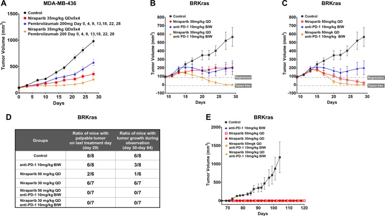
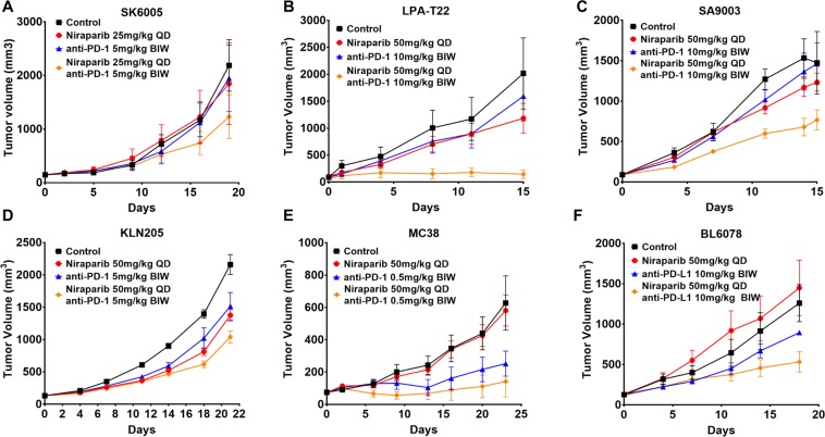
PARP inhibitors have been proven clinically efficacious in platinum-responsive ovarian cancer regardless of BRCA1/2 status and in breast cancers with germline BRCA1/2 mutation. However, resistance to PARP inhibitors may preexist or evolve during treatment in many cancer types and may be overcome by combining PARP inhibitors with other therapies, such as immune checkpoint inhibitors, which confer durable responses and are rapidly becoming the standard of care for multiple tumor types. This study investigated the therapeutic potential of combining niraparib, a highly selective PARP1/2 inhibitor, with anti-PD-1 immune checkpoint inhibitors in preclinical tumor models. Our results indicate that niraparib treatment increases the activity of the type I (alpha) and type II (gamma) interferon pathways and enhances the infiltration of CD8+ cells and CD4+ cells in tumors. When coadministered in immunocompetent models, the combination of niraparib and anti-PD-1 demonstrated synergistic antitumor activities in both BRCA-proficient and BRCA-deficient tumors. Interestingly, mice with tumors cured by niraparib monotherapy completely rejected tumor growth upon rechallenge with the same tumor cell line, suggesting the potential establishment of immune memory in animals treated with niraparib monotherapy. Taken together, our findings uncovered immunomodulatory effects of niraparib that may sensitize tumors to immune checkpoint blockade therapies.


Conflict of interest statement

Z. Wang., K. Sun., Y. Xiao., B. Fen., K. Mikule., J. Hanke., S. Ramaswamy. and J. Wang. are employees and/or shareholders of TESARO, Inc. G. B. Mills receives sponsored research support from TESARO, Inc.

Figures

Figure 1
Figure 1
Interferon response signature genes were significantly enriched in niraparib-treated tumors. (A) Tumor growth curve for the SK6005 syngeneic model with control or 50 mg/kg niraparib (QD) treatment. (B) Genes identified with DEseq as significantly upregulated upon niraparib treatment (p <=0.05, fold change >=1.5) were subjected to enrichment analysis of pathway gene sets. Gene Set Enrichment Analysis (GSEA) demonstrated a significant enrichment of interferon gamma signature (C) and interferon alpha signature (D) genes in niraparib-treated samples. (E) Tumor growth curve for the MDA-MB-436 NOG-EXL humanized model treated with control or 35 mg/kg niraparib daily for 5 days on and 2 days off for 4 weeks (QD × 5 × 4). (F) Significantly upregulated genes identified with a two-sample t test (p <=0.05, fold change >=1.5) were subjected to enrichment analysis of pathway gene sets. Gene Set Enrichment Analysis (GSEA) demonstrated a significant enrichment of interferon gamma signature (G) and interferon alpha signature (H) genes in niraparib-treated samples.
Figure 2
Figure 2
Niraparib promoted tumor immune cell infiltration in both the BRCA-proficient SK6005 syngeneic and BRCA-deficient MDA-MB-436 NOG-EXL humanized tumor models (A) Representative images of CD4 and CD8 immunohistochemical staining in control- and niraparib-treated BRCA-proficient SK6005 tumors. (BD) Quantification of the number of CD4+ cells, CD8+ cells and FoxP3+ cells per field upon niraparib treatment in BRCA-proficient SK6005 tumors. (E-G) Percentage of Ki67-positive CD4+ cells, CD8+ cells and FoxP3+ cells among the total CD3+ population by flow cytometry in humanized NOG-EXL MDA-MB-436 tumors. **p-value is less than 0.05 by Student’s t test.
Figure 3
Figure 3
Niraparib-induced interferon activation is present in xenograft tumors established in immunocompromised mice and in cultured tumor cells (A) Tumor growth curve for MDA-MB-436 tumors in an immunodeficient NOG model treated with control or 50 mg/kg niraparib (QD). (B) Genes identified with a two-sample t-test as significantly upregulated upon niraparib treatment (p <= 0.05, fold change >=1.5) in MDA-MB-436 tumors in immune-deficient NOG mice were subjected to enrichment analysis of pathway gene sets. (C) Genes identified with a paired two-sample t test as significantly upregulated upon niraparib treatment (p <=0.05, fold change >=1.5) in 8 niraparib sensitive PDX models were subjected to enrichment analysis of pathway gene sets. (D) mRNA expression of IFNB1 upon niraparib treatment (300 nM, 24 h and 48 h), etoposide treatment (50 µM, 18 h), or dA:dT transfection (0.5 µg/ml) in MDA-MB-436 cells. (E) Protein expression of p-STING (Ser366), STING, p-TBK1 (Ser172), TBK1, p-NF-κB p65 (Ser536) and NF-κB p65 upon 1 µM niraparib treatment (48 h) by western blotting from the same MDA-MB-436 lysate run on different gels indicated by the divider lines. (F) DAPI staining of nuclear structures following DMSO or 300 nM niraparib treatment in MDA-MB-436 cells cultured in vitro with arrows indicating micronuclei formation in niraparib-treated cells in vitro.
Figure 4
Figure 4
Combination therapy with niraparib and anti-PD-1 augmented antitumor activity and conferred durable responses in BRCA-deficient tumor models (A) Tumor growth in the BRCA-deficient MDA-MB-436 model in huNOG-EXL mice treated with 200 mg anti-PD-1 (pembrolizumab) on days 0, 4, 9, 13, 18, 22, and 28; 35 mg/kg niraparib daily for 5 days on and 2 days off for 4 weeks; and the combination of these agents. (B) The BRCA1-null ovarian cancer mouse syngeneic model was treated with 30 mg/kg niraparib QD, 10 mg/kg anti-PD-1 (BioXCell RMP1-14) BIW, and the combination of these agents for 21 days (day 9–29). Tumor regrowth was monitored post treatment (days 29–64). (C) The BRCA1-null ovarian cancer mouse syngeneic model was treated with niraparib 50 mg/kg QD, anti-PD-1 10 mg/kg BIW, and the combination of these agents for 21 days (days 9–29). Tumor regrowth was monitored post treatment (days 29–64). (D) Table summarizing the ratio of mice with palpable tumors on day 29 (last treatment day) and the ratio of mice with tumor growth observed during the drug-free, posttreatment observation period (days 30–64). (E) Growth curves for rechallenge with BRCA1-null ovarian cancer cells implanted on day 65 in the tumor-free mice from (D) and age-matched treatment-naïve mice.
Figure 5
Figure 5
Combination therapy with niraparib and an anti-PD-(L)1 antibody demonstrated significantly enhanced antitumor activity in a BRCA-proficient mouse syngeneic model (A) Tumor growth in the SK6005 skin syngeneic transplant model treated with anti-PD-1 (BioXCell RMP1–14), niraparib, and the combination of these agents. Niraparib was administered orally at 25 mg/kg daily, and the anti-PD-1 antibody was administered intraperitoneally at 5 mg/kg twice weekly. (B) Tumor growth in the MMTV-LPA1-T22 syngeneic transplant models treated with anti-PD-1 (2C4), niraparib, and the combination of these agents. Niraparib was administered orally at 50 mg/kg daily, and the anti-PD-1 antibody was administered intraperitoneally at 10 mg/kg twice weekly. (C) Tumor growth in the SA9003 sarcoma syngeneic model treated with anti-PD-1 (BioXCell RMP1–14), niraparib, and the combination of these agents. Niraparib was administered orally at 50 mg/kg daily, and the anti-PD-1 antibody was administered intraperitoneally at 10 mg/kg twice weekly. (D) Tumor growth in the KLN205 lung syngeneic model treated with anti-PD-1 (2C4), niraparib, and the combination of these agents. Niraparib was administered orally at 50 mg/kg daily, and the anti-PD-1 antibody was administered intraperitoneally at 5 mg/kg twice weekly. (E) Tumor growth in the MC38 colon syngeneic model treated with anti-PD-1 (BioXCell RMP1–14), niraparib, and the combination of these agents. Niraparib was administered orally at 50 mg/kg daily, and the anti-PD-1 antibody was administered intraperitoneally at 0.5 mg/kg twice weekly. (F) Tumor growth in the BL6078 bladder syngeneic model treated with anti-PD-L1 (BioXCell 10 f.9G2), niraparib, and the combination of these agents. Niraparib was administered orally at 50 mg/kg daily, and the anti-PD-L1 antibody was administered intraperitoneally at 10 mg/kg twice weekly.

References

    1. Vyas S, Chang P. New PARP targets for cancer therapy. Nat Rev Cancer. 2014;14:502–509. doi: 10.1038/nrc3748. - DOI - PMC - PubMed
    1. Pommier Y, O’Connor MJ, de Bono J. Laying a trap to kill cancer cells: PARP inhibitors and their mechanisms of action. Sci Transl Med. 2016;8:362ps317. doi: 10.1126/scitranslmed.aaf9246. - DOI - PubMed
    1. Bryant HE, et al. Specific killing of BRCA2-deficient tumours with inhibitors of poly(ADP-ribose) polymerase. Nature. 2005;434:913–917. doi: 10.1038/nature03443. - DOI - PubMed
    1. Murai J, et al. Trapping of PARP1 and PARP2 by Clinical PARP Inhibitors. Cancer Res. 2012;72:5588–5599. doi: 10.1158/0008-5472CAN-12-2753. - DOI - PMC - PubMed
    1. Mirza MR, et al. Niraparib Maintenance Therapy in Platinum-Sensitive, Recurrent Ovarian Cancer. N Engl J Med. 2016;375:2154–2164. doi: 10.1056/NEJMoa1611310. - DOI - PubMed

Publication types

MeSH terms