. 2019 Feb 6:7:e6154.
doi: 10.7717/peerj.6154. eCollection 2019.

Snake venom NAD glycohydrolases: primary structures, genomic location, and gene structure

Affiliations

Snake venom NAD glycohydrolases: primary structures, genomic location, and gene structure

Ivan Koludarov et al. PeerJ. .

Abstract

NAD glycohydrolase (EC 3.2.2.5) (NADase) sequences have been identified in 10 elapid and crotalid venom gland transcriptomes, eight of which are complete. These sequences show very high homology, but elapid and crotalid sequences also display consistent differences. As in Aplysia kurodai ADP-ribosyl cyclase and vertebrate CD38 genes, snake venom NADase genes comprise eight exons; however, in the Protobothrops mucrosquamatus genome, the sixth exon is sometimes not transcribed, yielding a shortened NADase mRNA that encodes all six disulfide bonds, but an active site that lacks the catalytic glutamate residue. The function of this shortened protein, if expressed, is unknown. While many vertebrate CD38s are multifunctional, liberating both ADP-ribose and small quantities of cyclic ADP-ribose (cADPR), snake venom CD38 homologs are dedicated NADases. They possess the invariant TLEDTL sequence (residues 144-149) that bounds the active site and the catalytic residue, Glu228. In addition, they possess a disulfide bond (Cys121-Cys202) that specifically prevents ADP-ribosyl cyclase activity in combination with Ile224, in lieu of phenylalanine, which is requisite for ADPR cyclases. In concert with venom phosphodiesterase and 5'-nucleotidase and their ecto-enzyme homologs in prey tissues, snake venom NADases comprise part of an envenomation strategy to liberate purine nucleosides, and particularly adenosine, in the prey, promoting prey immobilization via hypotension and paralysis.

Keywords: 5′-nucleotidase; ADP-ribosyl cyclase; Adenosine; CD38; Exons; NAD glycohydrolase; NADase; Phosphodiesterase; Protobothrops mucrosquamatus genome; Snake venom.


Conflict of interest statement

The authors declare there are no competing interests.

Figures
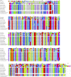
Figure 1
Figure 1. Alignment of 10 snake venom NADases with human, frog, and chicken CD38 sequences.
The snake NADase sequences lack signal peptides. They are readily distinguished from the former three sequences, and are furthermore clearly resolvable as elapid and crotalid sequences, based upon sequence differences at various positions. Like the Homo sapiens CD38, Micrurus sequences have 12 cysteines, presumably arranged in 6 disulfide bonds (Egea et al., 2012). The two complete Protobothrops sequences have 13 cysteines, like the chicken enzyme, raising the possibility of a covalent homodimer. The truncated C-terminus of the P. flavoviridis sequence is almost certainly a misassembly or less likely, a pseudogene, since the last 14 residues (226–239) and the premature stop codon bear no resemblance to the other sequences. Asterisks denote stop codons. Accession numbers for other vertebrate sequences: Homo sapiens, BAA18966.1; Xenopus laevis, BAL72804.1; Gallus gallus, ADQ89191.1; Anolis carolinensis, XP_016848812.1. Amino acid color scheme: Orange, methionine; Yellow, cysteine; Pink, proline; Gray, aliphatic; Purple, aromatic; Royal Blue, basic; Red, acidic; Green, hydroxylated; Sky Blue, amidated.
Figure 2
Figure 2. The predicted 3D structure of a soluble venom NADase is more compact than that of its membrane-bound human homolog, owing to its somewhat condensed, soluble N-terminus.
The enzyme from Micrurus surinamensis venom (left) is compared with human CD38 (right). (A) Venom enzymes lack the membrane-spanning alpha-helix and the intracellular domain (residues 1–44) of human CD38. Venom enzymes possess a soluble random coil instead. The two models are shown as ribbon structures with rainbow coloring to distinguish different regions from the N- (blue) to the C-terminus (red). (B) Superimposed portions of the two structures, corresponding to the soluble domain of human CD38, modeled upon the crystal structure of the soluble domain of human CD38 (3F6Y_A). The two structures superimpose almost perfectly except for the divergent C-termini and the truncated N-termini. Residues 45–300 of the M. surinamensis NADase (red) and residues 45–296 of human CD38 (blue) are shown. Left, frontal view; Right, left-side view. (C) Disulfide bonds are identical in the M. surinamensis NADase and human CD38. The Cys121–Cys202 disulfide bond, in combination with other residues, helps to prevent ADP-ribosyl cyclase activity, forcing the conversion of β-NAD and other suitable substrates to ADP-ribose, which is subsequently hydrolyzed to adenosine, the strategic target. (D) The M. surinamensis NADase and human CD38 are both hydrophilic overall, with Gravy scores of −0.292 and −0.306, respectively. However, while the N-terminal 44 residues of the M. surinamensis NADase are slightly hydrophobic (0.142), the N-terminus of CD38 is strongly hydrophobic (0.710). Gravy scores below 0 are more likely globular, hydrophilic proteins, while scores above 0 are more likely membrane-bound and hydrophobic (Magdeldin et al., 2012). Surfaces are rendered to show Kyte-Doolittle hydrophobicity with blue residues being most hydrophilic and red residues being most hydrophobic. Models were created with GalaxyTBM using human CD38 (3F6Y) as a template (Ko et al., 2012) . Disulfide bond formation, energy minimization, and structural manipulations were performed using Chimera 1.13 (Pettersen et al., 2004).
Figure 3
Figure 3. Structure of the NAD glycohydrolase gene from the genome of Protobothrops mucrosquamatus (Aird et al., 2017a), showing locations of the 8 exons, and the amino acid sequence of the enzyme expressed in the venom glands.
The mature venom protein comprises 304 amino acids linked by six disulfide bonds. Exon 1 actually encodes part of the 5′-untranslated region, as well as the N-terminus of the protein, and Exon 8 extends well beyond the stop codon, but only the mature protein sequence is shown here. Because of split arginine codons that bridge the Exon 6–7 and 7–8 boundaries, splicing out the sixth exon in the short P. mucrosquamatus NADase variants would retain the arginine after the splice. Distances between exons shown above are for purposes of illustration only, and are not proportionally scaled.
Figure 4
Figure 4. In Protobothrops mucrosquamatus, the 8-exon gene is transcribed into a long NADase and a shorter form that lacks exon 6.
(A) Its deletion results in the loss of 31 amino acids from Ser223-Gln253. With split arginine codons at both the exon 6–7 and exon 7–8 boundaries, no amino acid substitution occurs in the shortened (272-residue) structure. (B) The effect of removing these 31 residues is to delete the loose helical region (Ser223-Gln253) from the center of the molecule, including the catalytic residue, Glu229, (Glu232 in Fig. 1) leaving the remainder of the structure essentially intact. If this alternately spliced protein is expressed, it is difficult to imagine what its function might be. Models were created using GalaxyTBM (Ko et al., 2012). Disulfide bond formation, energy minimization, and structural manipulations were performed using Chimera 1.13 (Pettersen et al., 2004). Amino acid classes are colored as in Fig. 1.

Similar articles

Cited by

  • Solenodon genome reveals convergent evolution of venom in eulipotyphlan mammals.
    Casewell NR, Petras D, Card DC, Suranse V, Mychajliw AM, Richards D, Koludarov I, Albulescu LO, Slagboom J, Hempel BF, Ngum NM, Kennerley RJ, Brocca JL, Whiteley G, Harrison RA, Bolton FMS, Debono J, Vonk FJ, Alföldi J, Johnson J, Karlsson EK, Lindblad-Toh K, Mellor IR, Süssmuth RD, Fry BG, Kuruppu S, Hodgson WC, Kool J, Castoe TA, Barnes I, Sunagar K, Undheim EAB, Turvey ST. Casewell NR, et al. Proc Natl Acad Sci U S A. 2019 Dec 17;116(51):25745-25755. doi: 10.1073/pnas.1906117116. Epub 2019 Nov 26. Proc Natl Acad Sci U S A. 2019. PMID: 31772017 Free PMC article.

References

    1. Aird SD. Ophidian envenomation strategies and the role of purines. Toxicon. 2002;40:335–393. doi: 10.1016/S0041-0101(01)00232-X. - DOI - PubMed
    1. Aird SD. The role of purine and pyrimidine nucleosides in snake venoms. In: Mackessy SP, editor. Handbook of venoms and toxins of reptiles. CRC Press; Boca Raton: 2009. pp. 393–419.
    1. Aird SD, Aggarwal S, Villar-Briones A, Tin MM-Y, Terada K, Mikheyev AS. Snake venoms are integrated systems, but abundant venom proteins evolve more rapidly. BMC Genomics. 2015;16:647. doi: 10.1186/s12864-015-1832-6. - DOI - PMC - PubMed
    1. Aird SD, Arora J, Barua A, Qiu L, Terada K, Mikheyev AS. Population genomic analysis of a pitviper reveals microevolutionary forces underlying venom chemistry. Genome Biology and Evolution. 2017a;9:2640–2649. doi: 10.1093/gbe/evx199. - DOI - PMC - PubMed
    1. Aird SD, Da Silva Jr NJ, Qiu L, Villar-Briones A, Saddi VA, Pires de Campos Telles M, Grau ML, Mikheyev AS. Coralsnake venomics: analyses of venom gland transcriptomes and proteomes of six brazilian taxa. Toxins. 2017b;9:1–64. - PMC - PubMed