Spondylodiscitis with Epidural and Psoas Muscle Abscesses as Complications After Transrectal Ultrasound-guided Prostate Biopsy: Report of a Rare Case
- PMID: 30755964
- PMCID: PMC6346856
- DOI: 10.12890/2017_000694
Spondylodiscitis with Epidural and Psoas Muscle Abscesses as Complications After Transrectal Ultrasound-guided Prostate Biopsy: Report of a Rare Case
Abstract
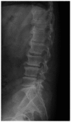
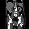
A 71-year-old man presented with spondylodiscitis with epidural and psoas muscle abscesses following transrectal ultrasound (TRUS)-guided prostate biopsy. These rare complications were detected by computed tomography of the abdomen and magnetic resonance imaging of the lumbar spine. The patient was successfully treated with antibiotics and underwent neurosurgery. Awareness of presentations such as backache and weakness of the lower limbs after prostate biopsy is important as these symptoms are usually mistaken for bone or muscle problems and often not recognized as being related to infection.
Learning points: We describe the case of a patient who experienced two major complications (spondylodiscitis with epidural abscess and psoas muscle abscess) following prostate biopsy.Awareness of these potential complications following prostate biopsy is essential to prevent life-threatening consequences.
Keywords: Spondylodiscitis; prostate biopsy; psoas muscle abscess; transrectal ultrasound.
Conflict of interest statement
Conflicts of Interests: The Authors declare that there are no competing interests.
Figures


Similar articles
-
Prostatic abscess with infected aneurysms and spondylodiscitis after transrectal ultrasound-guided prostate biopsy: a case report and literature review.BMC Urol. 2021 Jan 21;21(1):11. doi: 10.1186/s12894-021-00780-0. BMC Urol. 2021. PMID: 33478455 Free PMC article. Review.
-
Spondylodiscitis as a spinal complication of transrectal ultrasound-guided needle biopsy of the prostate.Spine (Phila Pa 1976). 2012 Jun 15;37(14):E870-2. doi: 10.1097/BRS.0b013e318256ed45. Spine (Phila Pa 1976). 2012. PMID: 22472806
-
Degenerative Disc Disease Mimicking Spondylodiscitis with Bilateral Psoas Abscesses.World Neurosurg. 2018 Dec;120:43-46. doi: 10.1016/j.wneu.2018.08.116. Epub 2018 Aug 24. World Neurosurg. 2018. PMID: 30149157
-
[Spondylodiscitis as a complication to ultrasound-guided transrectal prostatic biopsy].Ugeskr Laeger. 2002 Dec 30;165(1):51-2. Ugeskr Laeger. 2002. PMID: 12529951 Danish.
-
Elastic Versus Rigid Image Registration in Magnetic Resonance Imaging-transrectal Ultrasound Fusion Prostate Biopsy: A Systematic Review and Meta-analysis.Eur Urol Focus. 2018 Mar;4(2):219-227. doi: 10.1016/j.euf.2016.07.003. Epub 2016 Jul 29. Eur Urol Focus. 2018. PMID: 28753777
Cited by
-
Spinal epidural abscess post-ureteroscopy: a case report.BMC Urol. 2025 Apr 24;25(1):100. doi: 10.1186/s12894-025-01789-5. BMC Urol. 2025. PMID: 40269870 Free PMC article.
-
Pyogenic Spondylodiscitis Following Nonspinal Cesarean Section.Cureus. 2023 Sep 25;15(9):e45966. doi: 10.7759/cureus.45966. eCollection 2023 Sep. Cureus. 2023. PMID: 37900374 Free PMC article.
-
Prostatic abscess with infected aneurysms and spondylodiscitis after transrectal ultrasound-guided prostate biopsy: a case report and literature review.BMC Urol. 2021 Jan 21;21(1):11. doi: 10.1186/s12894-021-00780-0. BMC Urol. 2021. PMID: 33478455 Free PMC article. Review.
-
Management of Pyogenic Spondylodiscitis Following Nonspinal Surgeries: A Tertiary Care Center Experience.Int J Spine Surg. 2021 Jun;15(3):591-599. doi: 10.14444/8080. Epub 2021 May 13. Int J Spine Surg. 2021. PMID: 33985997 Free PMC article.
-
Discitis following urinary tract infection manifesting as recurrent autonomic dysreflexia related to truncal movements in a person with tetraplegia.BMJ Case Rep. 2020 Dec 17;13(12):e238202. doi: 10.1136/bcr-2020-238202. BMJ Case Rep. 2020. PMID: 33334762 Free PMC article.
References
-
- Nathoo N, Caris EC, Wiener JA, Mendel E. History of the vertebral venous plexus and the significant contributions of Breschet and Batson. Neurosurgery. 2011;69:1007–1014. - PubMed
-
- Shields D, Robinson P, Crowley TP. Iliopsoas abscess—a review and update on the literature. Int J Surg. 2012;10:466–469. - PubMed
LinkOut - more resources
Full Text Sources
