

Klebsiella pneumoniae Invasive Syndrome
- PMID: 30756018
- PMCID: PMC6346957
- DOI: 10.12890/2018_000800
Klebsiella pneumoniae Invasive Syndrome
Abstract
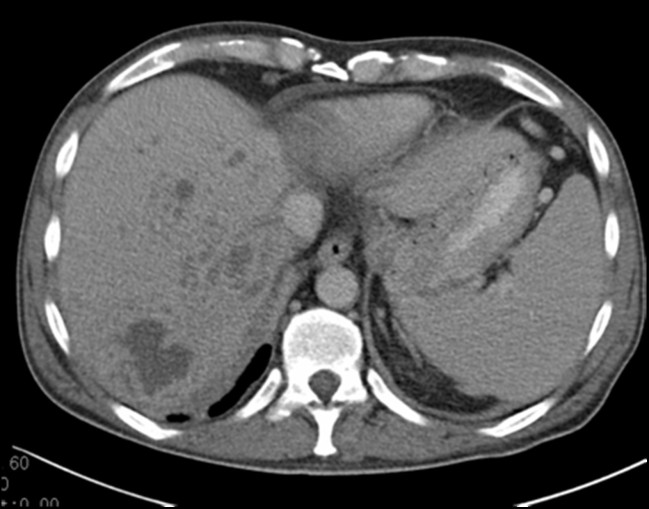
Klebsiella pneumoniae invasive syndrome (KPIS) is a rare clinical condition characterized by primary liver abscess associated with metastatic infection. Most case reports are from Southeast Asia, with only one case described in Portugal. The Authors present the case of a 44-year-old man with a history of fever, dry cough and cervicalgia. A thoracic computed tomography (CT) scan showed multiple pulmonary and hepatic nodules, suggestive of metastatic malignancy. Both blood cultures and bronchoalveolar lavage were positive for Klebsiella pneumoniae. Imaging studies were repeated during his hospital stay, showing a reduction in both number and volume of identified lesions, thus revealing their infectious nature. This case illustrates how much this entity can mimic other illnesses.
Learning points: Klebsiella pneumoniae invasive syndrome is emerging as a global disease.The imaging-led diagnosis of neoplasia was proved incorrect and could have been deleterious for the patient.The lack of diagnostic suspicion can lead to shorter antibiotic treatment regimens, therefore compromising the patient's full recovery.
Keywords: Klebsiella pneumoniae; invasive syndrome; liver abscess.
Conflict of interest statement
Conflicts of Interests: The Authors declare that there are no competing interests.
Figures


References
-
- Liu YC, Cheng DL, Lin CL. Klebsiella pneumoniae liver abscess associated with septic endophthalmitis. Arch Intern Med. 1986;146:1913–16. - PubMed
-
- Chang FY, Chou MY, Fan RL, Shaio MF. A clinical study of Klebsiella liver abscess. Taiwan Yi Xue Hui Za Zhi. 1988;87:282–87. - PubMed
-
- Wang JH, Liu YC, Lee SS, Yen MY, Chen YS, Wang JH, et al. Primary liver abscess due to Klebsiella pneumoniae in Taiwan. Clin Infect Dis. 1998;26:1434–8. - PubMed
-
- Siu LK, Yeh KM, Lin JC, Fung CP, Chang FY. Klebsiella pneumoniae liver abscess: a new invasive syndrome. Lancet Infect Dis. 2012;12:881–87. - PubMed
-
- Moore R, O’Shea D, Geoghegan T, Mallon PW, Sheehan G. Community-acquired Klebsiella pneumoniae liver abscess: an emerging infection in Ireland and Europe. Infection. 2013;41:681–86. - PubMed
LinkOut - more resources
Full Text Sources