

Quantitative Analysis of the Corneal Collagen Distribution after In Vivo Cross-Linking with Second Harmonic Microscopy
- PMID: 30756083
- PMCID: PMC6348900
- DOI: 10.1155/2019/3860498
Quantitative Analysis of the Corneal Collagen Distribution after In Vivo Cross-Linking with Second Harmonic Microscopy
Abstract
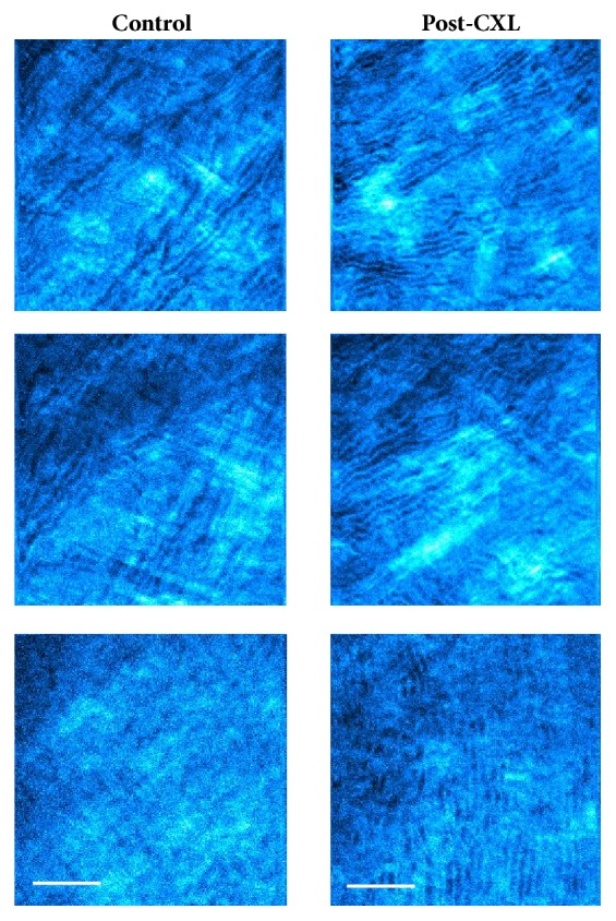
Corneal cross-linking (CXL) is a surgical procedure able to modify corneal biomechanics and stabilize keratoconus progression. Although it is known that CXL produces changes in corneal collagen distribution, these are still a topic of discussion. Here we quantitatively compare the corneal stroma architecture between two animal models four weeks after in vivo conventional CXL treatment, with second harmonic generation (SHG) imaging microscopy and the structure tensor (ST). The healing stage and the stroma recovery were also analyzed by means of histological sections. Results show that the CXL effects depend on the initial arrangement of the corneal collagen. While the treatment increases the order in corneas with a low level of initial organization, corneas presenting a fairly regular pattern are hardly affected. Histological samples showed active keratocytes in anterior and middle stroma, what means that the recovery is still in progress. The combination of SHG imaging and the ST is able to objectively discriminate the changes suffered by the collagen arrangement after the CXL treatment, whose effectiveness depends on the initial organization of the collagen fibers within the corneal stroma.
Figures


Similar articles
-
Morphological and immunohistochemical changes after corneal cross-linking.Cornea. 2013 Feb;32(2):111-7. doi: 10.1097/ICO.0b013e31824d701b. Cornea. 2013. PMID: 22580432
-
Corneal confocal microscopy following conventional, transepithelial, and accelerated corneal collagen cross-linking procedures for keratoconus.J Refract Surg. 2012 Nov;28(11):769-76. doi: 10.3928/1081597X-20121016-01. J Refract Surg. 2012. PMID: 23347370
-
In vivo confocal microscopy analyses of corneal microstructural changes in a prospective study of collagen cross-linking in keratoconus.Ophthalmology. 2014 Feb;121(2):469-74. doi: 10.1016/j.ophtha.2013.09.014. Epub 2013 Oct 30. Ophthalmology. 2014. PMID: 24183340
-
Corneal collagen cross-linking (CXL) combined with refractive procedures for the treatment of corneal ectatic disorders: CXL plus.J Refract Surg. 2014 Aug;30(8):566-76. doi: 10.3928/1081597X-20140711-10. J Refract Surg. 2014. PMID: 25325898 Review.
-
In Vivo Confocal Microscopy after Corneal Collagen Crosslinking.Ocul Surf. 2015 Oct;13(4):298-314. doi: 10.1016/j.jtos.2015.04.007. Epub 2015 Jul 2. Ocul Surf. 2015. PMID: 26142059 Review.
Cited by
-
Two-Photon Imaging for Non-Invasive Corneal Examination.Sensors (Basel). 2022 Dec 11;22(24):9699. doi: 10.3390/s22249699. Sensors (Basel). 2022. PMID: 36560071 Free PMC article. Review.
-
Protocol for a systematic review, meta-analysis, and trial sequential analysis of clinical outcomes following accelerated versus conventional corneal collagen cross-linking for corneal ectasia.Syst Rev. 2019 Apr 4;8(1):85. doi: 10.1186/s13643-019-1004-x. Syst Rev. 2019. PMID: 30947752 Free PMC article.
-
On the quantitative analysis of lamellar collagen arrangement with second-harmonic generation imaging.Biomed Opt Express. 2024 Mar 29;15(4):2666-2680. doi: 10.1364/BOE.516817. eCollection 2024 Apr 1. Biomed Opt Express. 2024. PMID: 38633085 Free PMC article.
-
Quantitative Discrimination of Healthy and Diseased Corneas With Second Harmonic Generation Microscopy.Transl Vis Sci Technol. 2019 Jun 27;8(3):51. doi: 10.1167/tvst.8.3.51. eCollection 2019 May. Transl Vis Sci Technol. 2019. PMID: 31293806 Free PMC article.
-
In vivo two-photon microscopy of the human eye.Sci Rep. 2019 Jul 12;9(1):10121. doi: 10.1038/s41598-019-46568-z. Sci Rep. 2019. PMID: 31300680 Free PMC article.
References
-
- Scott McCall A., Kraft S., Edelhauser H. F., et al. Mechanisms of corneal tissue cross-linking in response to treatment with topical riboflavin and long-wavelength ultraviolet radiation (UVA) Investigative Ophthalmology & Visual Science. 2010;51(1):129–138. doi: 10.1167/iovs.09-3738. - DOI - PMC - PubMed
MeSH terms
Substances
LinkOut - more resources
Full Text Sources
