. 2019 Feb 26;48(9):2861-2871.
doi: 10.1039/c8dt05153d.

Recent advances in single molecule magnetism of dysprosium-metallofullerenes

Affiliations

Recent advances in single molecule magnetism of dysprosium-metallofullerenes

Lukas Spree et al. Dalton Trans. .

Abstract

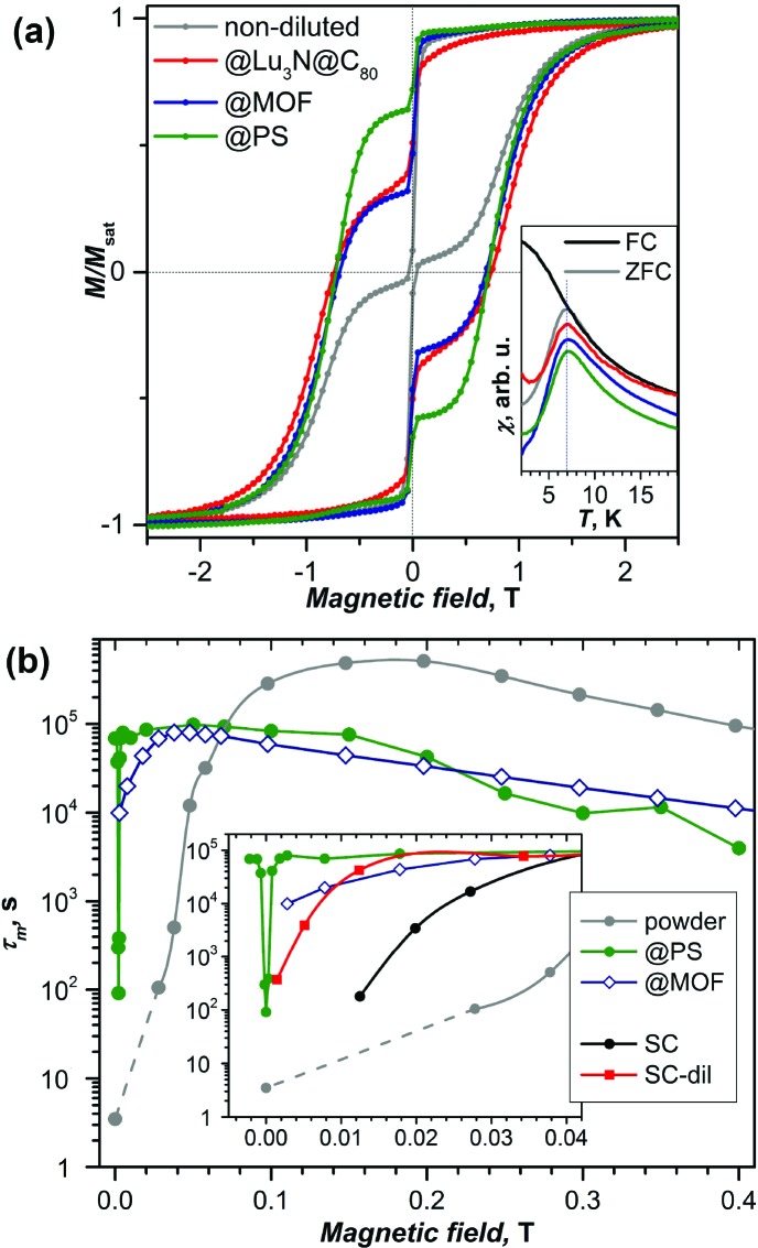
This article outlines the magnetic properties of single molecule magnets based on Dy-encapsulating endohedral metallofullerenes. The factors that govern these properties, such as the influence of different non-metal species in clusterfullerenes, the cage size, and cage isomerism are discussed, as well as the recent successful isolation of dimetallofullerenes with unprecedented magnetic properties. Finally, recent advances towards the organization of endohedral metallofullerenes in 1D, 2D, and 3D ordered structures with potential for devices are reviewed.


Figures

Fig. 1
Fig. 1. Molecular structures of selected Dy metallofullerenes showing single molecule magnetism. Dy is shown in green, Sc – magenta, Ti – cyan, Y – violet, N – blue, C – gray, and S – yellow. Only a part of the benzyl group of Dy2@C80(CH2Ph) can be seen.
Fig. 2
Fig. 2. (a) Magnetic hysteresis of non-diluted DySc2N@C80 powder measured at 1.8 K compared to the sample diluted with the diamagnetic fullerene Lu3N@C80, absorbed in the metal–organic framework DUT-51(Zr) (@MOF), and dispersed in polymer polystyrene (@PS). Strong variation of the QTM-induced drop of magnetization near zero-field with dilution can be seen. The inset shows that all samples have the same blocking temperature of 7 K. (b) Relaxation times of magnetization measured at 1.8 K in different magnetic fields for non-diluted powder and for diluted samples in MOF, polystyrene (PS) and in a single-crystal (non-diluted, SC, and diluted with Lu3N@C80, SC-dil). The inset zooms into the small field range. Reproduced from ref. 13.
Fig. 3
Fig. 3. (a) Hysteresis curves for DyxSc3–xN@C80-Ih (from left to right: x = 1, 2, and 3) recorded using SQUID magnetometry at 2 K with a field sweep rate of 0.8 mT s–1. Reprinted with permission from Westerström et al., Phys. Rev. B: Condens. Matter Mater. Phys., 2014, 89, 060406. Copyright 2014 by the American Physical Society. (b) Relaxation times of magnetization of DySc2N@C80 at temperatures of 2–87 K. Zero-field values are shown as full dots, and in-field (0.2 T) values are denoted as open dots. Relaxation times for non-diluted DySc2N@C80 are shown in black, and the values for diluted samples are shown in blue (dilution with MOF) and green (diluted with polystyrene, PS). The times longer or shorter than 10 s were determined by DC and AC magnetometry, respectively. The blue line is the fit of the points in the 2–5 K range with the Orbach relaxation mechanism, and the black line represents the fit of the QTM-like zero-field relaxation with the power function of temperature. Reproduced from ref. 13. (c) Relaxation times of the magnetization of Dy2ScN@C80. Green dots denote the values from DC measurements in zero field; two in-field points (red crosses) are also shown. AC values are measured with MPMS XL (7–50 K; open, magenta, and blue dots) and with PPMS (brown dots, 52–76 K). Magenta and blue dots denote long and short times from double-τ fits of the AC data, respectively, and open dots denote single-τ fits. Reproduced from ref. 14b.
Fig. 4
Fig. 4. Magnetization curves for (a) Dy2S@C82-Cs and (b) Dy2S@C82-C3v measured at T = 1.8–5 K with a magnetic field sweep rate of 8.33 mT s–1. The inset in each panel zooms into the region near zero-field. The inset in (b) shows the determination of TB of Dy2S@C82-C3v from the peak in the susceptibility of the zero-field cooled sample (magnetic field: 0.2 T, temperature sweep rate: 5 K min–1). (c) Magnetization relaxation times of Dy2S@C82-C3v; dots are experimental points, red lines are results of a global fit with three Orbach processes; and green, magenta, and brown lines represent contributions of individual Orbach processes. The inset shows an enhancement of the high-temperature range. Reproduced from ref. 15.
Fig. 5
Fig. 5. (a) Magnetic hysteresis in Dy2@C80(CH2Ph) between 2 and 22 K, field sweep rate: 2.9 mT s–1. (b) Magnetization relaxation times of Dy2@C80(CH2Ph) in zero-field and in a field of 0.4 T. The inset shows the out-of-phase dynamic susceptibility χ′′ measured at different temperatures between 23 and 33 K. (c) Alignment of magnetic moments in the ground state of Dy2@C80(CH2Ph) and respective spin Hamiltonian (CF denotes the crystal field). Reproduced from ref. 18.
Fig. 6
Fig. 6. (a) X-ray absorption spectra of Dy2ScN@C80 encapsulated in SWCNTs recorded using right (I+) and left (I) circularly polarized X-rays. (b) A comparison of the normalized total absorption and XMCD spectra from bulk Dy2ScN@C80 and Dy2ScN@C80 encapsulated in SWCNTs. The temperature is 2 K, and an external magnetic field of 6.5 T is applied parallel to the X-ray beam and the surface normal to the samples. (c) TEM image and structural model of the [DySc2N@C80]@SWCNT peapod. (d) Magnetization curves of [DySc2N@C80]@SWCNT measured at different temperatures by SQUID magnetometry; (e) comparison of magnetic hysteresis curves for bulk DySc2N@C80 and [DySc2N@C80]@SWCNT peapod (T = 1.8 K). (a) and (b) reproduced from ref. 33. Reprinted with permission from Nakanishi et al., J. Am. Chem. Soc., 2018, 140, 10955. Copyright 2018 by the American Chemical Society.
Fig. 7
Fig. 7. (a) Sub-monolayer (ML) of Dy2ScN@C80/Rh(111), T = 4 K, μ0H = 6.5 T; measurement geometry is shown in the inset. The polarization dependent X-ray absorption spectra (left panel), and the corresponding XMCD spectra (right panel) measured at incidence angles of θ = 0° and θ = 60°. Strong angular dependence points to the preferential alignment of Dy spins parallel to the surface. (b, c) Hysteresis curves measured by XMCD from a multilayer (b) and a sub-ML (c) of Dy2ScN@C80/Rh(111) at a magnetic field sweep rate of 2 T min–1 and a sample temperature of ∼4 K. The drop in magnetization at zero field is a consequence of the time of 30 s it takes the magnet to switch polarity. Reprinted with permission from Westerström et al., Phys. Rev. Lett., 2015, 114, 087201. Copyright 2015 by the American Physical Society.
Fig. 8
Fig. 8. (a) Scheme of a Prato reaction to obtain EMF-R derivatives (EMF = DySc2N@C80 (1), and Dy2ScN@C80 (2), R denotes the functional group with a thioether linker). (b, c) Magnetization curves of (b) 1-R and 1, and (c) 2-R and 2 measured by SQUID magnetometry at T = 2 K (field sweep rate: 2.9 mT s–1); the insets show determination of the blocking temperatures of magnetization TB (temperature sweep rate: 5 K min–1). (d, e) magnetization curves of sub-monolayers of 1-R (d) and 2-R (e) on Au(111) measured by XMCD at 2 K with a sweep rate of 2 T min–1 (averaging over five measured curves, and error bars are standard deviations). Reproduced from ref. 38.

References

    1. Sessoli R., Gatteschi D., Caneschi A., Novak M. A. Nature. 1993;365:141.
    2. Gatteschi D., Sessoli R. and Villain J., Molecular Nanomagnets, Oxford University Press, New York, 2006.
    3. Introduction to Molecular Magnetism: From Transition Metals to Lanthanides, ed. Benelli C. and Gatteschi D., Wiley-VCH Verlag GmbH & Co. KGaA, Weinheim, 2015.
    1. Sorace L., Benelli C., Gatteschi D. Chem. Soc. Rev. 2011;40:3092. - PubMed
    2. Zhang P., Guo Y.-N., Tang J. Coord. Chem. Rev. 2013;257:1728.
    3. Liddle S. T., van Slageren J. Chem. Soc. Rev. 2015;44:6655. - PubMed
    4. Zhu Z., Guo M., Li X.-L., Tang J. Coord. Chem. Rev. 2019;378:350.
    5. Liu J.-L., Chen Y.-C., Tong M.-L. Chem. Soc. Rev. 2018;47:2431. - PubMed
    6. Ishikawa N., Sugita M., Ishikawa T., Koshihara S., Kaizu Y. J. Am. Chem. Soc. 2003;125:8694. - PubMed
    7. Giraud R., Wernsdorfer W., Tkachuk A. M., Mailly D., Barbara B. Phys. Rev. Lett. 2001;87:057203. - PubMed
    8. Woodruff D. N., Winpenny R. E. P., Layfield R. A. Chem. Rev. 2013;113:5110. - PubMed
    9. Rinehart J. D., Long J. R. Chem. Sci. 2011;2:2078.
    1. Goodwin C. A. P., Ortu F., Reta D., Chilton N. F., Mills D. P. Nature. 2017;548:439. - PubMed
    2. Guo F.-S., Day B. M., Chen Y.-C., Tong M.-L., Mansikkamäki A., Layfield R. A. Angew. Chem., Int. Ed. 2017;56:11445. - PubMed
    3. Guo F.-S., Day B. M., Chen Y.-C., Tong M.-L., Mansikkamäki A., Layfield R. A. Science. 2018;362:1400. - PubMed
    4. McClain K. R., Gould C. A., Chakarawet K., Teat S., Groshens T. J., Long J. R., Harvey B. G. Chem. Sci. 2018;9:8492. - PMC - PubMed
    1. Westerström R., Dreiser J., Piamonteze C., Muntwiler M., Weyeneth S., Brune H., Rusponi S., Nolting F., Popov A., Yang S., Dunsch L., Greber T. J. Am. Chem. Soc. 2012;134:9840. - PubMed
    1. Lu X., Bao L., Akasaka T., Nagase S. Chem. Commun. 2014;50:14701. - PubMed
    2. Lu X., Akasaka T., Nagase S. Chem. Commun. 2011;47:5942. - PubMed