. 2019 Apr;23(4):2995-3009.
doi: 10.1111/jcmm.14206. Epub 2019 Feb 12.

Role of mitochondrial calcium uniporter-mediated Ca2+ and iron accumulation in traumatic brain injury

Affiliations

Role of mitochondrial calcium uniporter-mediated Ca2+ and iron accumulation in traumatic brain injury

Li Zhang et al. J Cell Mol Med. 2019 Apr.

Abstract

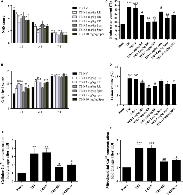
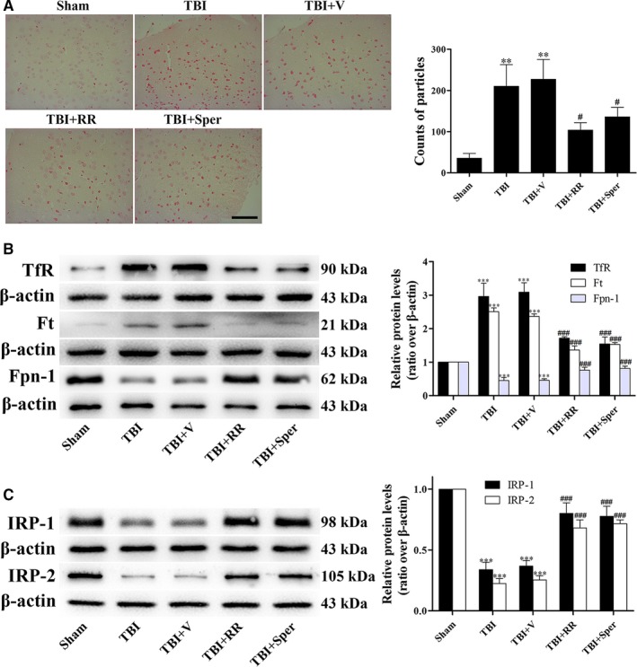
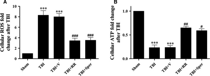
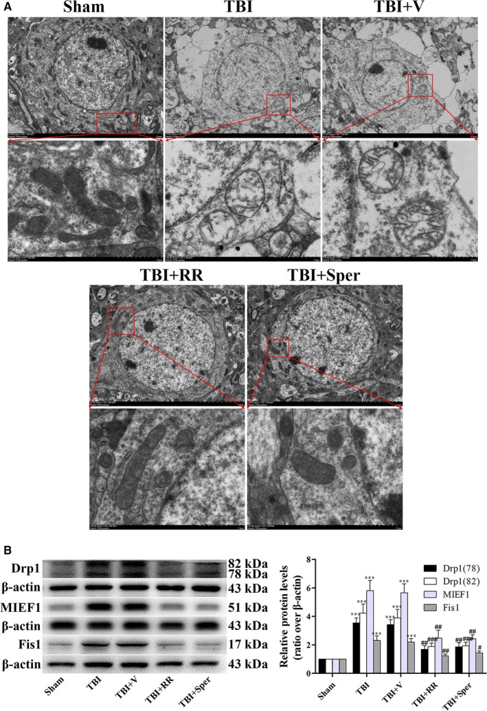
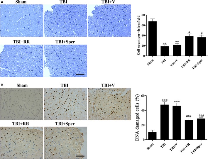
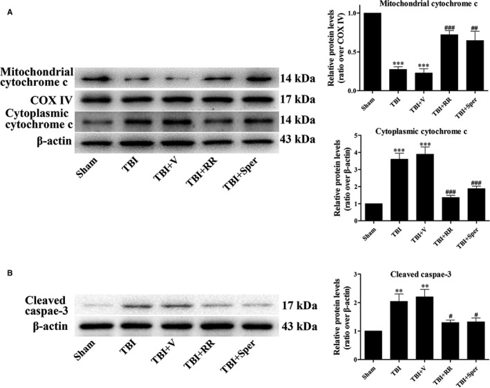
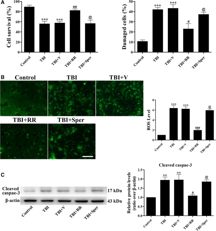
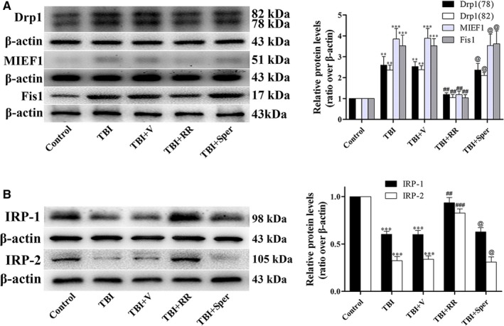
Previous studies have suggested that the cellular Ca2+ and iron homeostasis, which can be regulated by mitochondrial calcium uniporter (MCU), is associated with oxidative stress, apoptosis and many neurological diseases. However, little is known about the role of MCU-mediated Ca2+ and iron accumulation in traumatic brain injury (TBI). Under physiological conditions, MCU can be inhibited by ruthenium red (RR) and activated by spermine (Sper). In the present study, we used RR and Sper to reveal the role of MCU in mouse and neuron TBI models. Our results suggested that the Ca2+ and iron concentrations were obviously increased after TBI. In addition, TBI models showed a significant generation of reactive oxygen species (ROS), decrease in adenosine triphosphate (ATP), deformation of mitochondria, up-regulation of deoxyribonucleic acid (DNA) damage and increase in apoptosis. Blockage of MCU by RR prevented Ca2+ and iron accumulation, abated the level of oxidative stress, improved the energy supply, stabilized mitochondria, reduced DNA damage and decreased apoptosis both in vivo and in vitro. Interestingly, Sper did not increase cellular Ca2+ and iron concentrations, but suppressed the Ca2+ and iron accumulation to benefit the mice in vivo. However, Sper had no significant impact on TBI in vitro. Taken together, our data demonstrated for the first time that blockage of MCU-mediated Ca2+ and iron accumulation was essential for TBI. These findings indicated that MCU could be a novel therapeutic target for treating TBI.

Keywords: Ca2+; iron; mitochondrial calcium uniporter; neuroprotection; traumatic brain injury.


Conflict of interest statement

The authors declare no potential conflicts of interest.

Figures

Figure 1
Figure 1
Administration of ruthenium red (RR) or Sper protected mice against secondary brain injury and decreased Ca2+ concentrations after traumatic brain injury (TBI). (A, B, C) Mice were subjected to TBI and then received 1 mg/kg, 3 mg/kg, 5 mg/kg of RR or 2 mg/kg, 5 mg/kg, 10 mg/kg of Sper ip injection or vehicle 30 min after TBI. NSS and Grip test score were evaluated at 1, 3 and 7 days after TBI while brain water content was examined at 1 day after TBI. (A, B) All doses of RR or Sper had an improved motor performance within 3 days; however, larger doses such as 5 mg/kg of RR and 10 mg/kg of Sper did not exhibit a better neuroprotection. This effect was no longer significant at 7 days after TBI, n = 6 per group. (C) Mice subjected to TBI or treated with vehicle had an increased brain water content as compared with the sham group. Brain water content was significantly lower in the groups treated with RR or Sper than the vehicle‐treated group. Moreover, doses of 3 mg/kg of RR and 5 mg/kg of Sper had the best effect in relieving brain oedema, n = 6 each group. (D) TBI‐induced profound tissue loss of the brain was reversed by RR or Sper, and doses of 3 mg/kg of RR and 5 mg/kg of Sper had the best effect. (E, F) RR or Sper treatment decreased Ca2+ concentration following TBI. Restored cellular (E) and mitochondrial (F) concentrations of Ca2+ by RR (3 mg/kg) or Sper (5 mg/kg) treatment after TBI, n = 6 each group. Data are presented as mean ± SEM; **P < 0.01, ***P < 0.001 vs sham group; # P < 0.05, ## P < 0.01, ### P < 0.001 vs TBI + vehicle group. Scale bar: 50 μm
Figure 2
Figure 2
TBI induced cellular iron accumulation while ruthenium red (RR) or Sper administration partially reversed the effects. (A) Mice were subjected to traumatic brain injury (TBI) and then administrated RR (3 mg/kg), Sper (5 mg/kg) or vehicle 30 min after TBI. Ipsilateral cortex was collected 1 day after TBI and then subjected to iron staining. The left panel shows representative brain sections of cortex with iron stained (red) cells. The right panel is quantification of the digitized images showing the count of particles and fraction of total iron staining in each group. (B, C) TBI‐induced disruption of cellular iron homeostasis was improved by RR or Sper. Mice were subjected to TBI and treated with RR (3 mg/kg), Sper (5 mg/kg) or vehicle 30 min after TBI. Ipsilateral brain tissues were collected 1 day after TBI. The protein levels of TfR, Ft, Fpn‐1, IRP‐1 and IRP‐2 were measured by Western blot. TfR and Ft proteins were up‐regulated while Fpn‐1, IRP‐1 and IRP‐2 proteins were down‐regulated after TBI, however RR or Sper treatment reversed these effects. Data are presented as mean ± SEM, n = 6 per group; **P < 0.01, ***P < 0.001 vs sham group; # P < 0.05, ### P < 0.001 vs TBI + vehicle group. β‐actin was used as a loading control
Figure 3
Figure 3
Treatment of ruthenium red (RR) or Sper stabilized ROS and ATP homeostasis following traumatic brain injury (TBI). (A) Cellular ROS levels after TBI were repressed by RR (3 mg/kg) or Sper (5 mg/kg) administration. (B) Cellular ATP was resupplied after TBI by RR (3 mg/kg) or Sper (5 mg/kg) administration. Data are presented as mean ± SEM, n = 6 per group; ***P < 0.001 vs sham group; # P < 0.05, ## P < 0.01, ### P < 0.001 vs TBI + vehicle group
Figure 4
Figure 4
Treatment of ruthenium red (RR) or Sper ameliorated traumatic brain injury (TBI)‐induced deformation of mitochondria. (A) Electron photomicrographs of mitochondria after TBI. A representative of a mitochondrion with normal shape from the cortex of the sham group, a swelling mitochondrion with collapsed cristae from the cortex of the TBI group, a mitochondrion with quite normal shape and clear cristae of the TBI + RR (3 mg/kg) and TBI + Sper (5 mg/kg) groups. (B) Comparison of the expression of Drp1, MIEF1 and Fis1 using Western blot. Mice were subjected to TBI and treated with RR (3 mg/kg), Sper (5 mg/kg) or vehicle 30 min after TBI. Ipsilateral brain tissues were collected 1 day after TBI and the protein levels of Drp1, MIEF1 and Fis1 were measured by Western blot. TBI increased the protein levels of Drp1, MIEF1 and Fis1, while RR or Sper treatment significantly decreased them. Data are presented as mean ± SEM, n = 6 per group; ***P < 0.001 vs sham group; # P < 0.05, ## P < 0.01, ### P < 0.001 vs TBI + vehicle group. β‐actin was used as a loading control for cytoplasmic and whole‐cell extracts
Figure 5
Figure 5
Ruthenium red (RR) or Sper treatment suppressed traumatic brain injury (TBI)‐induced neuronal cell death and DNA damage. (A) Nissl staining to visualize the neuronal cell outline and structure. TBI reduced the number of the neurons and treatment of RR (3 mg/kg) or Sper (5 mg/kg) preserved neurons from damage. (B) DNA damage was determined using TUNEL assays 1 day after TBI. The DNA damaged cells were significantly increased after TBI compared to the sham group. RR (3 mg/kg) or Sper (5 mg/kg) treatment significantly decreased the percentage of DNA damaged cells after TBI. Data are presented as mean ± SEM, n = 6 each group; **P < 0.01, ***P < 0.001 vs sham group; # P < 0.05, ### P < 0.001 vs TBI + vehicle group. Scale bar: 50 μm
Figure 6
Figure 6
Ruthenium red (RR) or Sper treatment inhibited traumatic brain injury (TBI)‐induced up‐regulation of apoptosis‐related proteins. (A, B) Mice were subjected to TBI and treated with RR (3 mg/kg), Sper (5 mg/kg) or vehicle 30 min after TBI. Ipsilateral brain tissues were collected 1 day after TBI and the protein levels of cytochrome c and cleaved caspase‐3 were measured by Western blot. RR or Sper treatment significantly decreased apoptosis induced by TBI. Data are presented as mean ± SEM, n = 6 per group; **P < 0.01, ***P < 0.001 vs sham group; # P < 0.05, ## P < 0.01, ### P < 0.001 vs TBI + vehicle group. COX IV was used as a loading control for mitochondria extracts. β‐actin was used as a loading control for cytoplasmic and whole‐cell extracts
Figure 7
Figure 7
Ruthenium red (RR) instead of Sper treatment protected primary cultured neurons from traumatic brain injury (TBI). (A) Primary cortical neurons were subjected to scratch injury and then treated with RR (10 μM), Sper (10 μM) or saline for 1 day. The LDH release assay and TB staining were used to evaluate cell viability. The percentage of damaged cells significantly increased after TBI compared to the control group. RR treatment significantly decreased damaged cells after TBI, however, Sper treatment had no significant impact compared to the TBI + vehicle group. (B) Cells were subjected to scratch injury and subsequently treated with RR (10 μM), Sper (10 μM) or saline for 1 day. Then cells were incubated with DCFH‐DA and subjected to fluorescent microscopy analysis. The intracellular ROS was significantly increased after TBI compared to the sham group, and administration of RR instead of Sper significantly repressed ROS production as compared to the TBI + vehicle group. (C) RR (10 μM) treatment significantly decreased the expression of cleaved caspase‐3 after TBI, however, Sper treatment had no obvious effect. Data are presented as mean ± SEM, n = 6 per group; **P < 0.01, ***P < 0.001 vs control group; # P < 0.05, ## P < 0.01, ### P < 0.001 vs TBI + vehicle group; @ P > 0.05 vs TBI + vehicle group. β‐actin was used as a loading control. Scale bar: 50 μm
Figure 8
Figure 8
Traumatic brain injury (TBI)‐induced damage of mitochondria and disruption of iron homeostasis were improved by ruthenium red (RR) in primary cultured neurons. (A, B) RR treatment significantly decreased the expression of Drp1, MIEF1 and Fis1 (A) and increased the expression of IRP1/2 (B) after TBI, however, Sper treatment had no obvious effect on these proteins. Data are presented as mean ± SEM, n = 6 per group; **P < 0.01, ***P < 0.001 vs control group; ## P < 0.01, ### P < 0.001 vs TBI + vehicle group; @ P > 0.05 vs TBI + vehicle group. β‐actin was used as a loading control

Similar articles

Cited by

References

    1. Brooks JC, Strauss DJ, Shavelle RM, Paculdo DR, Hammond FM, Harrison‐Felix CL. Long‐term disability and survival in traumatic brain injury: results from the National Institute on Disability and Rehabilitation Research Model Systems. Arch Phys Med Rehabil. 2013;94:2203‐2209. - PubMed
    1. Ding K, Wang H, Xu J, et al. Melatonin stimulates antioxidant enzymes and reduces oxidative stress in experimental traumatic brain injury: the Nrf2‐ARE signaling pathway as a potential mechanism. Free Radic Biol Med. 2014;73:1‐11. - PubMed
    1. Zhang L, Wang H. Targeting the NF‐E2‐related factor 2 pathway: a novel strategy for traumatic brain injury. Mol Neurobiol. 2017. - PubMed
    1. Sun M, Zhao Y, Gu Y, Zhang Y. Protective effects of taurine against closed head injury in rats. J Neurotrauma. 2015;32:66‐74. - PubMed
    1. Baughman JM, Perocchi F, Girgis HS, et al. Integrative genomics identifies MCU as an essential component of the mitochondrial calcium uniporter. Nature. 2011;476:341‐345. - PMC - PubMed

Publication types

MeSH terms