. 2019 Apr 12;5(4):570-581.
doi: 10.1021/acsinfecdis.8b00315. Epub 2019 Feb 28.

Mechanistic and Structural Basis for the Actions of the Antibacterial Gepotidacin against Staphylococcus aureus Gyrase

Affiliations

Mechanistic and Structural Basis for the Actions of the Antibacterial Gepotidacin against Staphylococcus aureus Gyrase

Elizabeth G Gibson et al. ACS Infect Dis. .

Abstract

Gepotidacin is a first-in-class triazaacenaphthylene novel bacterial topoisomerase inhibitor (NBTI). The compound has successfully completed phase II trials for the treatment of acute bacterial skin/skin structure infections and for the treatment of uncomplicated urogenital gonorrhea. It also displays robust in vitro activity against a range of wild-type and fluoroquinolone-resistant bacteria. Due to the clinical promise of gepotidacin, a detailed understanding of its interactions with its antibacterial targets is essential. Thus, we characterized the mechanism of action of gepotidacin against Staphylococcus aureus gyrase. Gepotidacin was a potent inhibitor of gyrase-catalyzed DNA supercoiling (IC50 ≈ 0.047 μM) and relaxation of positively supercoiled substrates (IC50 ≈ 0.6 μM). Unlike fluoroquinolones, which induce primarily double-stranded DNA breaks, gepotidacin induced high levels of gyrase-mediated single-stranded breaks. No double-stranded breaks were observed even at high gepotidacin concentration, long cleavage times, or in the presence of ATP. Moreover, gepotidacin suppressed the formation of double-stranded breaks. Gepotidacin formed gyrase-DNA cleavage complexes that were stable for >4 h. In vitro competition suggests that gyrase binding by gepotidacin and fluoroquinolones are mutually exclusive. Finally, we determined crystal structures of gepotidacin with the S. aureus gyrase core fusion truncate with nicked (2.31 Å resolution) or intact (uncleaved) DNA (2.37 Å resolution). In both cases, a single gepotidacin molecule was bound midway between the two scissile DNA bonds and in a pocket between the two GyrA subunits. A comparison of the two structures demonstrates conformational flexibility within the central linker of gepotidacin, which may contribute to the activity of the compound.

Keywords: Staphylococcus aureus; gepotidacin; gyrase; novel bacterial topoisomerase inhibitors; single-stranded DNA cleavage.


Conflict of interest statement

Potential conflicts of interest: BB and PFC have shares in GlaxoSmithKline and PFC is an employee of GlaxoSmithKline. The authors declare no other competing financial interests.

Figures

Figure 1:
Figure 1:
Structure of the novel bacterial topoisomerase inhibitor (NBTI) gepotidacin (GSK2140944). Gepotidacin is composed of a triazaacenaphthylene on the left-hand side (LHS), a central linker region, a basic nitrogen, and a pyranopyridine on the right-hand side (RHS).
Figure 2:
Figure 2:
S. aureus gyrase removes positive supercoils more rapidly than it introduces negative supercoils into relaxed DNA. Time courses are shown for the relaxation of positively supercoiled plasmid followed by the introduction of negative supercoils (top) and the negative supercoiling of relaxed plasmid (bottom). The positions of positively supercoiled [(+)SC], relaxed, and negatively supercoiled [(−)SC] DNA are indicated on the gels. The gel images are representative of at least three independent experiments.
Figure 3:
Figure 3:
Gepotidacin and moxifloxacin inhibit DNA supercoiling and relaxation reactions catalyzed by S. aureus gyrase. The effects of gepotidacin (blue, top panels) and moxifloxacin (red, bottom panels) on the supercoiling of relaxed DNA (left panels) and the relaxation of positively supercoiled DNA (right panels) are shown. Error bars represent the standard deviation (SD) of at least three independent experiments.
Figure 4:
Figure 4:
Gepotidacin induces single-stranded DNA breaks in the presence of gyrase. The gel shows DNA products following cleavage reactions containing 5 or 200 μM gepotidacin or moxifloxacin in the absence or presence of S. aureus gyrase. The positions of negatively supercoiled [(−)SC], nicked (Nick), and linear (Lin) DNA are indicated on the gels. The generation of single- and double-stranded DNA breaks were monitored by the conversion of negatively supercoiled substrates to nicked and linear DNA products, respectively. The gel images are representative of at least three independent experiments.
Figure 5:
Figure 5:
Gepotidacin is a potent enhancer of gyrase-mediated single-stranded DNA cleavage. The left panel shows the effects of gepotidacin on S. aureus gyrase-mediated single- (SS, closed circles) and double-stranded (DS, open circles) DNA cleavage of negatively (blue) and positively (red) supercoiled DNA. The right panel shows the effects of moxifloxacin on gyrase-mediated single- and double-stranded DNA cleavage of negatively (black) and positively (green) supercoiled DNA. Error bars represent the SD of at least three independent experiments. The gels shown at the top are representative cleavage assays with negatively supercoiled DNA. The mobilities of negatively supercoiled DNA [(−)SC], nicked circular DNA (Nick), and linear DNA (Lin) are indicated on the gels.
Figure 6:
Figure 6:
Gepotidacin stabilizes only single-stranded DNA breaks mediated by S. aureus gyrase. The enhancement of single-stranded (SS, closed circles) and double-stranded (DS, open circles) DNA cleavage over time in the presence of 5 μM (blue) and 200 μM (red) gepotidacin are shown. Error bars represent the SD of at least three independent experiments.
Figure 7:
Figure 7:
Gepotidacin enhances only single-stranded DNA breaks mediated by S. aureus gyrase in the presence of ATP. The enhancement of gyrase-mediated single-stranded (SS, closed circles) or double-stranded (DS, open circles) DNA breaks generated by gyrase in the presence of 1.5 mM ATP is shown. Error bars represent the SD of at least three independent experiments.
Figure 8:
Figure 8:
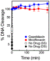
Gepotidacin induces stable DNA cleavage complexes formed by S. aureus gyrase. The persistence of ternary gyrase–DNA–drug cleavage complexes was monitored by the loss of single-stranded DNA breaks in the presence of 5 μM gepotidacin (blue) or double-stranded DNA cleavage in the presence of 25 μM moxifloxacin (red), or the loss of single- (open circle, white) or double-stranded DNA cleavage in the absence of drug (closed circle, black). Levels of DNA cleavage were set to 100% at time zero to allow for direct comparisons. Error bars represent the SD of at least three independent experiments.
Figure 9:
Figure 9:
Gepotidacin suppresses double-stranded DNA breaks generated by S. aureus gyrase. The effects of gepotidacin on S. aureus gyrase-mediated single-stranded (SS, closed circles) and double-stranded (DS, open circles) DNA cleavage are shown. Reactions were carried out in the presence of Ca2+ rather than Mg2+ to increase levels of baseline DNA cleavage. Error bars represent the SD of at least three independent experiments. The gel shown at the top is representative of at least three independent experiments. The mobilities of negatively supercoiled DNA [SC], nicked DNA (Nick), and linear DNA (Lin) are indicated on the gels.
Figure 10:
Figure 10:
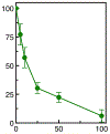
The actions of gepotidacin and moxifloxacin on S. aureus gyrase-mediated DNA cleavage are mutually exclusive. A DNA cleavage/ligation equilibrium was formed in the presence of a saturating concentration of moxifloxacin (25 μM) plus 0–100 μM gepotidacin. Competition was monitored by the loss of moxifloxacin-induced double-stranded DNA breaks. Error bars represent the SD of at least 3 independent experiments.
Figure 11:
Figure 11:
Views of a gepotidacin complex formed with S. aureus gyrase and doubly nicked DNA at a resolution of 2.31Å. The top left panel shows gepotidacin binding on the twofold axis of the complex midway between the two DNA cleavage sites; the top right panel is an approximately orthogonal (90°) view of the same structure. The bottom left and right panels show the same views as the corresponding top panels, but zoomed out to show the subunits of gyrase. In these panels, gepotidacin is shown as spheres, DNA with semi-transparent surface, and proteins as ribbons. In all panels, carbon atoms in the DNA are green, those in the first GyrBA core fusion truncate subunit are cyan/blue in GyrA and magenta in GyrB, and those in the second subunit are grey or black. Carbon atoms in gepotidacin are yellow, and oxygen, nitrogen, and sulfur atoms are red, blue, and yellow, respectively. Water molecules are shown as small red spheres.
Figure 12:
Figure 12:
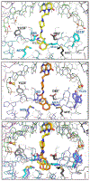
Comparison of two gepotidacin crystal structures with S. aureus DNA gyrase and DNA. (top panel) A 2.31Å crystal structure of gepotidacin with doubly nicked DNA (atoms not bonded are arrowed). (middle panel) A 2.37Å crystal structure of gepotidacin with intact (uncleaved) DNA. (bottom panel) Structures from top and middle are superimposed based on GyrA subunits shown with grey/black carbons. Note the ~1.2Å shift of atoms in the GyrA subunit with cyan/blue carbons and the similar shift in the right-hand side of gepotidacin. Colors are as shown in Figure 11, except that the carbon atoms in gepotidacin in the complex with intact DNA (middle and bottom panels) are shown in orange.

References

    1. Hooper DC (1999) Mechanisms of fluoroquinolone resistance. Drug Resist. Updat 2 (1), 38–55. DOI: 10.1054/drup.1998.0068. - DOI - PubMed
    1. Anderson VE, Osheroff N (2001) Type II topoisomerases as targets for quinolone antibacterials: turning Dr. Jekyll into Mr. Hyde. Curr. Pharm. Des 7 (5), 337–353. DOI: 10.2174/1381612013398013 - DOI - PubMed
    1. Drlica K, Hiasa H, Kerns R, Malik M, Mustaev A, Zhao X (2009) Quinolones: action and resistance updated. Curr. Top. Med. Chem 9 (11), 981–998. DOI: 10.2174/156802609789630947. - DOI - PMC - PubMed
    1. Chen SH, Chan NL, Hsieh TS (2013) New mechanistic and functional insights into DNA topoisomerases. Annu. Rev. Biochem 82, 139–170. DOI: 10.1146/annurev-biochem-061809-100002. - DOI - PubMed
    1. Aldred KJ, Kerns RJ, Osheroff N (2014) Mechanism of quinolone action and resistance. Biochemistry. 53 (10), 1565–1574. DOI: 10.1021/bi5000564. - DOI - PMC - PubMed

Publication types

MeSH terms