. 2019 Apr;28(4):374-382.
doi: 10.1111/exd.13901. Epub 2019 Mar 13.

Dissection of Merkel cell formation in hairy and glabrous skin reveals a common requirement for FGFR2-mediated signalling

Affiliations

Dissection of Merkel cell formation in hairy and glabrous skin reveals a common requirement for FGFR2-mediated signalling

Minh Binh Nguyen et al. Exp Dermatol. 2019 Apr.

Abstract

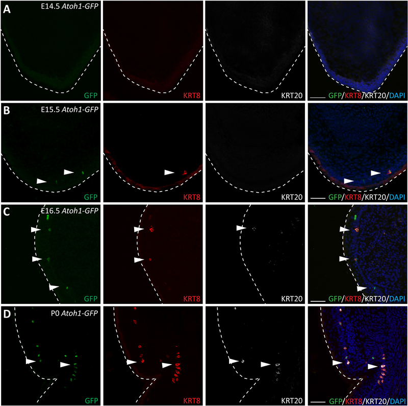
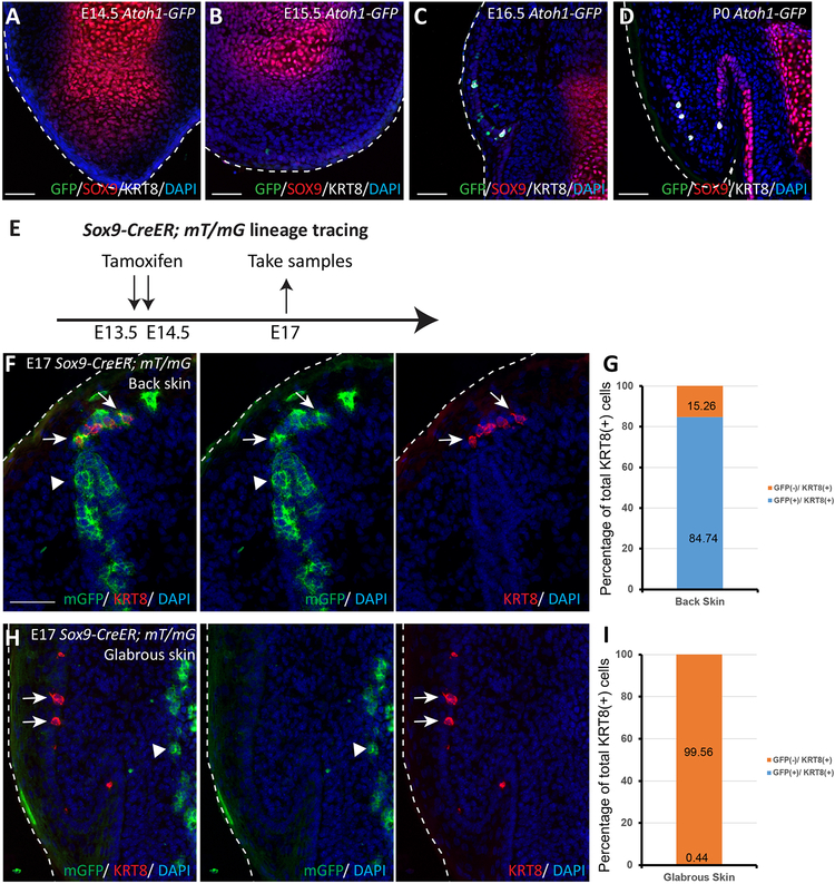
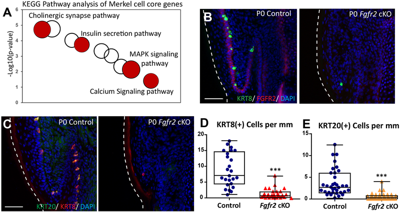
Merkel cells are mechanosensory cells involved in tactile discrimination. Merkel cells have been primarily studied in the murine back skin, where they are found in specialized structures called touch domes located around primary hair follicles. Yet, little is known about the morphogenesis of Merkel cells in areas of the skin devoid of hair, such as the glabrous paw skin. Here, we describe Merkel cell formation in the glabrous paw skin during embryogenesis. We first found in the glabrous paw skin that Merkel cells were specified at E15.5, 24 hours later, compared to in the back skin. Additionally, by performing lineage-tracing experiments, we found that unlike in the back skin, SOX9(+) cells do not give rise to Merkel cells in the glabrous paw skin. Finally, we compared the transcriptomes of Merkel cells in the back and the glabrous paw skin and showed that they are similar. Genetic and transcriptome studies showed that the formation of Merkel cells in both regions was controlled by similar regulators. Among them was FGFR2, an upstream factor of MAPK signalling that was reported to have a critical function in Merkel cell formation in the back skin. Here, we showed that FGFR2 is also required for Merkel cell development in the glabrous paw skin. Taken together, our results demonstrate that Merkel cells in the murine back skin and glabrous paw skin are similar, and even though their formation is controlled by a common genetic programme, their precursor cells might differ.

Keywords: FGFR2; Merkel cells; glabrous skin; hair follicle.


Conflict of interest statement

CONFLICT OF INTERESTS

The authors have declared no conflicting interests.

Figures

Figure 1.
Figure 1.. Merkel Cells are formed at Embryonic day 15.5 in the glabrous paw skin
(A–D). Immunofluorescence analysis of early, intermediate, and late Merkel cell differentiation markers ATOH1-GFP (green), KRT8 (red), and KRT20 (white), respectively, in the glabrous paw skin of Atoh1-GFP mice 14.5 (A), E15.5 (B), E16.5 (C) and P0 (D). White arrows indicate Merkel cells. White dotted lines indicate the edges of the tissue. Note that ATOH1-GFP(+) Merkel cells and KRT8(+) Merkel cells were first observed at E15.5, KRT20(+) Merkel cells were first observed at E16.5. Scale = 50μm in all panels.
Figure 2.
Figure 2.. During embryogenesis, SOX9(+) cells do not give rise to Merkel cells in the glabrous paw skin at E14.5
(A–D). Immunofluorescence analysis of SOX9 (red), Merkel cell markers ATOH1-GFP (green), and KRT8 (white) at E14.5 (A), E15.5 (B), E16.5 (C), and at P0 (D) in the glabrous paw skin of Atoh1-GFP mice. White dotted lines indicate the edges of the tissue. Note that ATOH1-GFP(+) and KRT8(+) Merkel cells do not overlap with SOX9(+) cells. (E). Experimental design for Sox9-CreER; mT/mG lineage tracing experiment. (F). Sox9-CreER; mT/mG lineage tracing in the back skin. White arrows point to Merkel cells; arrow heads point to SOX9-traced cells. SOX9-traced cells are GFP(+) (green). KRT8(+) Merkel cells are stained in red. Note the overlap between SOX9 progenies and KRT8 staining. (G). Quantification of Merkel cells positive for KRT8 and GFP in Sox9-CreER; mT/mG back skin sections at E17. n = 6. Data is presented as a percentage of the total 401 cells quantified. The bar graph shows the percentage of KRT8(+) cells, which are co-labelled with GFP (blue) or not co-labelled with GFP (orange). (H) Sox9-CreER; mT/mG lineage tracing in the glabrous paw skin. White arrows point to Merkel cells; white arrow heads point to SOX9-traced cells. SOX9-traced cells are GFP(+) (green). KRT8(+) Merkel cells are stained in red. Right side panels show separated color channels. (I). Quantification of Merkel cells positive for KRT8 and GFP in Sox9-CreER; mT/mG glabrous paw skin sections at E17. n = 6. Data is presented as percentages of the total 325 cells quantified. The bar graph shows the percentage of KRT8(+) cells that are co-labelled with GFP (blue) or not co-labelled with GFP (orange). White dotted lines indicate the edges of the tissue. Scale bar is 50μm in (A-D), (F) and (H).
Figure 3.
Figure 3.. RNA-seq transcriptional profiling of Merkel cells isolated from back and glabrous paw skins revealed common core Merkel cell genes.
(A). Principal component analysis based on the top 5000 most variable genes in the back skin and the glabrous paw skin. (B). Hierarchical clustering of the significantly differentially expressed genes between Merkel cells and IFE in either the back skin or glabrous paw skin (Fig. S3), indicating distinct clusters of genes with either shared or unique gene expression in all isolated skin cell types. (C). List of core Merkel cell genes. Selected signature genes from the 514 genes that were commonly upregulated in Merkel cells of back and the glabrous paw skin regions, compared to IFE of the corresponding skin region and selected by these criteria: FPKM > 2, fold-change > 5 and adjusted p-value < 0.01, are organized according to functional categories. (D). Differential expression analysis of genes expressed in Merkel cells in the glabrous paw skin versus the back skin. Genes with mean FPKM > 2, absolute fold-change > 2 and adjust p-value < 0.01 were considered as differentially expressed genes.
Figure 4.
Figure 4.. FGFR2 is critical for Merkel cell formation in the glabrous paw skin.
(A). KEGG pathway analysis of Merkel cell core genes from the back skin and glabrous paw skin. Selected KEGG terms are in red. The size of a cycle indicates the number of genes in a KEGG term. (B). Immunofluorescence analysis of Merkel cell markers KRT8 (green) and FGFR2 (red) in P0 control and Fgfr2 cKO glabrous paw skin. Note that FGFR2 (red) is completely gone in P0 Fgfr2 cKO. (C). Immunofluorescence analysis of Merkel cell markers KRT20 (green) and KRT8 (red) in P0 control and P0 Fgfr2 cKO glabrous paw skin. (D). Quantification of KRT8 (+) Merkel cells in P0 control and P0 Fgfr2 cKO, p < 0.0001, n = 4. (E). Quantification of KRT20(+) Merkel cells in P0 control and P0 Fgfr2 cKO, p < 0.0001, n = 4. White dotted lines indicate the edges of the tissue. Scale = 50μm in (B) and (C). The data presented in box plots (D, E) show the median with 25th and 75th percentile borders. Whiskers extend from minimum to maximum. ***p < 0.001 (per Mann-Whitney test).

Similar articles

Cited by

References

    1. Winkelmann RK, Breathnach AS. The Merkel cell. J Invest Dermatol. 1973;60(1):2–15. - PubMed
    1. Haeberle H, Lumpkin EA. Merkel Cells in Somatosensation. Chemosens Percept. 2008;1(2):110–118. - PMC - PubMed
    1. Lumpkin EA, Marshall KL, Nelson AM. The cell biology of touch. J Cell Biol. 2010;191(2):237–248. - PMC - PubMed
    1. Maricich SM, Morrison KM, Mathes EL, Brewer BM. Rodents rely on Merkel cells for texture discrimination tasks. J Neurosci. 2012;32(10):3296–3300. - PMC - PubMed
    1. Iggo A, Muir AR. The structure and function of a slowly adapting touch corpuscle in hairy skin. J Physiol. 1969;200(3):763–796. - PMC - PubMed

Publication types

Substances