Review
. 2019 May 8;119(9):5775-5848.
doi: 10.1021/acs.chemrev.8b00451. Epub 2019 Feb 13.

Emerging Diversity in Lipid-Protein Interactions

Affiliations
Review

Emerging Diversity in Lipid-Protein Interactions

Valentina Corradi et al. Chem Rev. .

Abstract

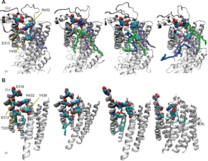
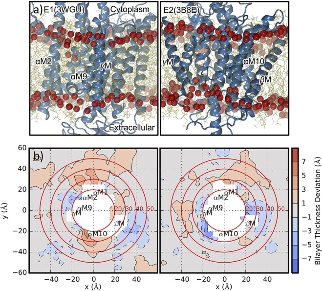
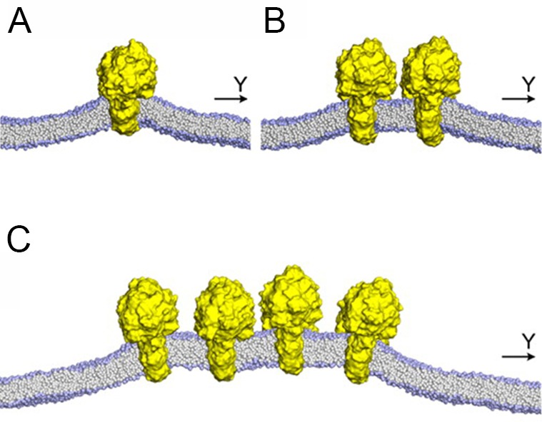
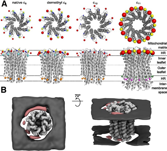
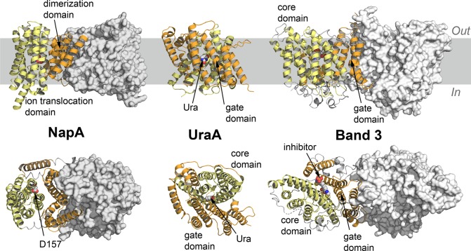
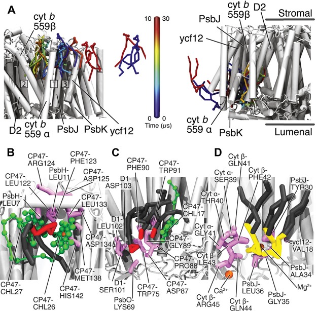
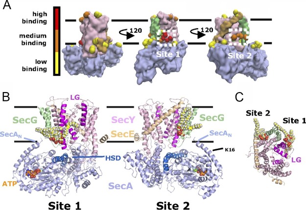
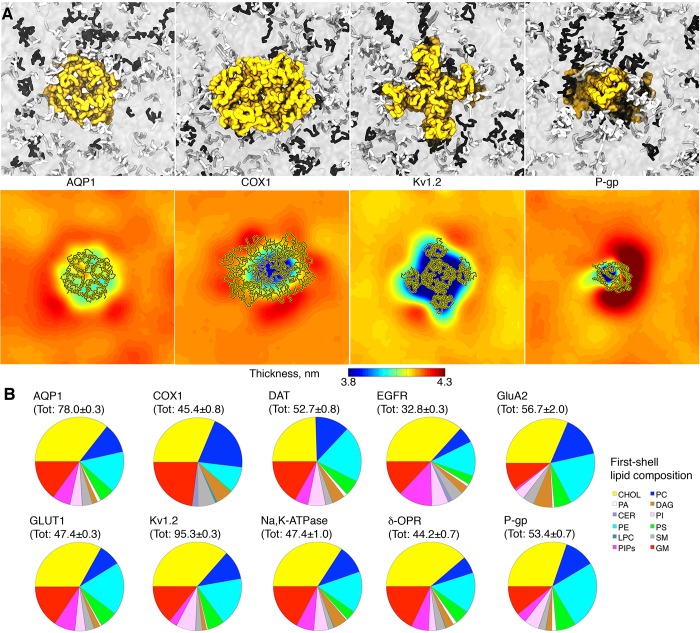
Membrane lipids interact with proteins in a variety of ways, ranging from providing a stable membrane environment for proteins to being embedded in to detailed roles in complicated and well-regulated protein functions. Experimental and computational advances are converging in a rapidly expanding research area of lipid-protein interactions. Experimentally, the database of high-resolution membrane protein structures is growing, as are capabilities to identify the complex lipid composition of different membranes, to probe the challenging time and length scales of lipid-protein interactions, and to link lipid-protein interactions to protein function in a variety of proteins. Computationally, more accurate membrane models and more powerful computers now enable a detailed look at lipid-protein interactions and increasing overlap with experimental observations for validation and joint interpretation of simulation and experiment. Here we review papers that use computational approaches to study detailed lipid-protein interactions, together with brief experimental and physiological contexts, aiming at comprehensive coverage of simulation papers in the last five years. Overall, a complex picture of lipid-protein interactions emerges, through a range of mechanisms including modulation of the physical properties of the lipid environment, detailed chemical interactions between lipids and proteins, and key functional roles of very specific lipids binding to well-defined binding sites on proteins. Computationally, despite important limitations, molecular dynamics simulations with current computer power and theoretical models are now in an excellent position to answer detailed questions about lipid-protein interactions.


Conflict of interest statement

The authors declare no competing financial interest.

Figures

Figure 1
Figure 1
Representative structures of GPCRs. From left to right, the structures of rhodopsin, the β2 adrenergic receptor (β2AR), and the smoothened receptor are shown as cartoons, with the 7 transmembrane (TM) helices highlighted in different colors. For β2AR, the three subunits of the G protein (Gα, Gβ, and Gγ) are shown in light gray and blue cartoons. For the smoothened receptor, the extracellular cysteine-rich domain (CRD) and the long extracellular loop 3 are shown in light blue and gray cartoons, respectively. The top panels show a side view of the receptors, while the bottom panels provide a view from the extracellular side. Substrates or agonists are labeled and shown in white spheres, and the hydrophobic region of the membrane is highlighted in gray. In, intracellular side; Out, extracellular side; ICL, intracellular loop; ECL, extracellular loop.
Figure 2
Figure 2
Effect of cholesterol on β2AR activation. A. Definition of two distance parameters used to measure the conformational changes as a function of time. The D3.32–S5.46 Cα atom distance (denoted LL) measures fluctuations in the ligand-binding site of the receptor, and the R3.50–E6.30 Cα atom distance (denoted LG) captures fluctuations in the G protein binding interface. B and C. The conformational space probed by the simulations in pure DOPC and DOPC–10% Chol concentration bilayer, respectively, plotted as a function of these two distance parameters. Cholesterol significantly decreases the conformational space sampled by the receptor. Adapted with permission from ref (177). Copyright 2016 Manna et al. Licensed under Creative Commons Attribution 4.0.
Figure 3
Figure 3
Experimental structures of GPCR dimers. Side view (upper panels) and the view from the extracellular side (bottom panels) of the (left) μ opioid receptor (μOR) dimer and (right) the chemokine receptor type 4 (CXCR4) dimer. The 7 transmembrane (TM) helices of the receptors are highlighted in different colors and shown as cartoons, while one of the monomers is also represented with a transparent gray surface. Substrates bound to these dimers are not shown for clarity.
Figure 4
Figure 4
Different structural topologies of selected ion channels. From left to right the structures of the Shaker Kv1.2, the two-pore domains K2P1, the inwardly rectifying Kir2.2 potassium channels, and the structure of the NavAb sodium channel are shown in cartoons. All structures, with one notable exception of K2P1 channels, are functional tetramers comprised of four identical monomers. Monomers are colored from yellow to orange to dark brown. The trans-membrane helices forming voltage-sensing domains (VSD) are labeled S1 to S4, while two transmembrane helices forming the pore domain (PD) in voltage-gated channels are labeled as S5 to S6; Kir2.2 pore is comprised of two transmembrane helices, outer and inner corresponding to S5 and S6 helices in Kv1.2 or NaVab channels. The location of the pore helix present in many structures of ion channels is shown on the top-view of Kir 2.2. The functional dimer of the K2P1 channel is comprised of two pore domains (P1 and P2) with each domain formed by TM helices M1 to M4, where helices M1 and M3 are outer helices of P1 and P2 protomers. For the K2P1 and the Kir2.2 channels the positions of potassium ions in the filter region are shown by purple spheres. In, intracellular side; out, extracellular side. The membrane region is highlighted in gray.
Figure 5
Figure 5
State-dependent PIP2 lipid interactions in the Kv7.2 channel. Zhang et al. used atomistic MD simulations to model the interactions of PIP2 lipids in the open (A) and closed (B) state of the channel. Shown are the trajectories of PIP2 lipid molecules as a function of time near the channel (viewed from the intracellular side). PIP2 migrates toward the S4–S5 linker in the open state (A) and toward the S2–S3 linker in the closed state (B). Adapted with permission from ref (292). Copyright 2013 Zhang et al.
Figure 6
Figure 6
Rosenhouse-Dantsker et al. identified specific cholesterol-protein interactions in the Kir2.1 channel by combining molecular docking, MD simulations, mutagenesis, and electrophysiology experiments. A. Cholesterol binding sites identified via molecular docking and MD simulations. In blue are residues lining the cholesterol binding sites, while in red are residues whose mutation alters cholesterol sensitivity. B. Schematic representation of the identified cholesterol interaction sites with respect to the architecture of the channel. Reproduced with permission from ref (299). Copyright 2013 The American Society for Biochemistry and Molecular Biology, Inc.
Figure 7
Figure 7
Kir2.2 cholesterol binding sites identified with CG MD simulations. Simulation snapshots of Kir2.2 with cholesterol molecules (in yellow) bound to sites I and II in the open state and to sites III, IV and Ib in the closed state. Reproduced with permission from ref (302). Copyright 2018 Biophysical Society.
Figure 8
Figure 8
Lipid distribution around Kv1.2 described by CG MD simulations. MD simulations of Kv1.2 in a plasma membrane mixture revealed an asymmetric distribution of polyunsaturated (PU), fully saturated (FS) lipids and cholesterol (CHOL) between upper and lower. The simulation system consisted of 4 copies of Kv1.2 and ca. 6000 lipids. Shown are 2D distribution maps for PU, FS lipids, CHOL, and the class Others (which groups all the remaining lipid classes of the plasma membrane mixtures), highlighting enrichment (red) and depletion (blue) with respect to the average value of the corresponding class in each leaflet. Figure generated as described in Corradi et al. for Aquaporin 1.
Figure 9
Figure 9
Ligand-gated channels. From left to right are the structures of the nicotinic acetylcholine receptor (nAchR), the P2X3 channel and the TRPV1 channel. For each structure, the individual monomers are colored from light yellow to dark brown. ATP molecules bound to P2X3 and resiniferatoxin (RTX) bound to TRPV1 are shown in white spheres. ECD, extracellular domain; ICD, intracellular domain. The top panels show a side view of the proteins, with the membrane region highlighted in gray. The lower panels are the view from the extracellular side. The functional assembly of nAchR includes five protomers labeled α to γ, with each monomer comprised of trans-membrane sections (helices M1 to M4) and the extra-cellular domain (ECD). P2X3 channels are assembled as functional trimers with each monomer containing a trans-membrane section comprised of 2 TM helices, TM1 and TM2. The nomenclature of the TRPV1 channel is similar to that of voltage-gated ion channels shown in Figure 4.
Figure 10
Figure 10
Open and closed structures of the MscS channel. Side view (upper panels) and view from the periplasmic side (bottom panels) of an open and closed MscS channel., Each monomer is shown in cartoons and colored on a scale from light yellow to dark brown. Per, periplasm; In, intracellular side. The hydrophobic region of the membrane is highlighted in gray.
Figure 11
Figure 11
CLs stabilize supercomplexes in large scale CG MD simulations. Top: Snapshot of final configuration of the system containing CIII and CIV, indicating the presence of many supercomplexes. Bottom: Close-up view on a particular supercomplex with CLs present at the protein–protein interface. Figure courtesy of Clement Arnarez.
Figure 12
Figure 12
Structural features of an ABC exporter. The two halves of P-glycoprotein are shown in orange and light yellow cartoons for the (left) inward-facing and (right) outward-facing state. ATP molecules bound to the nucleotide binding domains (NBD1 and NBD2) of the outward-facing state are shown in white spheres. TMD1 and TMD2 indicate the two transmembrane domains, embedded in the hydrophobic region of the membrane here highlighted in gray.
Figure 13
Figure 13
Lipid organization around the ABC transporter McjD. CG simulations of the McjD transporter were carried out in different mixtures (A–D) of POPE, POPG, and cardiolipin. Preferential interactions in the binary and tertiary mixtures with the headgroup of anionic lipids were driven by electrostatic interactions with several positively charged residues. Shown are the headgroup number density maps for the four simulated systems, with the densities of POPE, POPG, and cardiolipin in cyan, magenta, and yellow, respectively, for upper and lower leaflet. Adapted with permission from ref (415). Copyright 2016 The American Society for Biochemistry and Molecular Biology, Inc.
Figure 14
Figure 14
Lipid access to the central cavity of P-glycoprotein. CG simulations of P-glycoprotein in different mixtures of POPE and POPC lipids were used to identify the residues involved in interactions with lipids at the portals and inside the cavity. A. Displayed are examples of pathways sampled by different lipids to gain access to (L1 to L4) and to leave the central cavity ((L1′ to L4′) during the simulation time. P-glycoprotein is viewed from the extracellular side. Reprinted with permission from ref (432). Copyright 2018 Barreto-Ojeda et al. B. Snapshot of P-glycoprotein with a POPC lipid (PC1) located at portal 1 (yellow helices), a second lipid (PC2) located at portal 2 (orange helices), and a third lipid (PC3) inside the cavity. Highlighted are residues identified in the simulations as hot-spots for lipid interactions at the portals (green spheres) and inside the cavity (cyan spheres).
Figure 15
Figure 15
Examples of OMPs discussed in this review. From left to right: OmpA, the OmpF trimer, Hia, LptD and LptE, and BamA. The five POTRA domains of BamA are labeled P1–P5. Individual subunits are shown as cartoons and colored in light yellow, orange, and brown. Upper and lower panels show a side view and a view from the extracellular side of the proteins, respectively. Out, extracellular side; Per, periplasm. The membrane region is highlighted in gray.
Figure 16
Figure 16
Simulation snapshots of the OmpF trimer embedded in atomistic OM models of increasing complexity. PE, PG and cardiolipin types of lipids of the inner leaflet are shown in blue, orange and magenta spheres; Lipid A is shown as pink spheres; a model of the LPS core is shown in gray stick, and polysaccharides of the O-antigen are shown in orange sticks. Ca2+, K+ and Cl ions are shown as cyan, green and magenta spheres. Reprinted with permission from ref (484). Copyright 2016 Biophysical Society, by Elsevier Inc.
Figure 17
Figure 17
Lipid scrambling and structural rearrangements in N. haematococca TMEM16 as described by MD simulations. A. Snapshots showing, from left to right, the structural rearrangements needed for lipid molecules to access the groove from the extracellular side. R432 initially interacts with E313 but as lipids approach it switches to the interactions with E318. B. Snapshots showing, from left to right, lipid translocation from the intracellular side to the extracellular side. The transmembrane region of TMEM16 is shown as white cartoons, while relevant residues involved in key interactions and lipids molecules are shown as spheres and sticks, respectively. Adapted with permission from ref (507). Copyright 2018 Lee et al. Licensed under Creative Commons Attribution 4.0. Panels in B are only a subset of those in the original figure. We thank G. Khelashvili and H. Weinstein for providing the original figures.
Figure 18
Figure 18
Membrane deformation around the Na+,K+-ATPase pump. MD simulations revealed distinct local perturbations of membrane thickness for two conformational states of the pump, linked to specific transmembrane helices. A. Snapshots of the pump in the E1 (left) and E2 (right) state. B. Thickness deviations profiles obtained from the last 50 ns of the simulations for the E1 (left) and E2 (right) state of the pump. Transmembrane helices linked to thickness changes are labeled. Reprinted with permission from ref (540). Copyright 2017 Elsevier B.V.
Figure 19
Figure 19
Mitochondrial membrane shaping induced by ATP synthase dimers. CG MD simulations showed how ATP synthase dimers (yellow surface) contribute to the formation of mitochondrial cristae via curvature effects. A, one dimer; B, two dimers; C, four dimers. Adapted with permission from ref (557). Copyright 2012 Davies et al.
Figure 20
Figure 20
Selective binding of cardiolipin around c-rings of different size, as detected from MD simulation studies. A. Location of cardiolipin headgroup densities, determined based on radial distribution function analyses, around the c-rings are shown as spheres, color-coded accordingly to specific residues involved in the interactions. The size of the spheres reflects the intensity of the density signal. B. Time-averaged lipid density. Density for POPC and POPE headgroups is shown in gray, and density for cardiolipin headgroup is shown in pink. Reprinted with permission from ref (386). Copyright 2016 Duncan et al.
Figure 21
Figure 21
Organization of lipids around AQP0. To study the effect of temperature, lipid phase, and protein mobility on the reorganization of lipids around AQP0, Briones et al. performed MD simulations using different simulation setups and found that protein mobility has the strongest effect on lipid packing in close proximity of the protein. Displayed are lipid densities around AQP0 monomers as a function of simulation conditions. Reprinted with permission from ref (570). Copyright 2017 Briones et al. Licensed under Creative Commons Attribution 4.0.
Figure 22
Figure 22
Lipid–protein interactions in RTKs and cytokine receptors as detected in MD simulations. A. General structure of an RTK. B. The transmembrane helix and juxtamembrane region of EphA2 receptor, colored according to the extent of the interactions with PIP lipids. A and B are reprinted with permission from ref (585). Licensed under Creative Commons Attribution 4.0. C. Snapshots after 5 μs of simulation time (left, upper leaflet; right, lower leaflet) of a plasma membrane model with multiple transmembrane segments of a cytokine receptor. GM3, PIP lipids and sodium ions are shown in magenta, yellow and blue spheres, respectively. D. Extent of the interactions from blue to red for PIP lipids and cholesterol with the transmembrane helix of the receptor. C and D are reprinted with permission from ref (591). Copyright 2014 Koldso et al. Licensed under Creative Commons Attribution 4.0.
Figure 23
Figure 23
MFS fold and the LeuT fold in the SLC superfamily. Left. LacY structure in the inward-facing state with the two repeats (Rep1 and Rep2) shown in light yellow and orange cartoons. The side chains of residues known to interact with the sugar substrate are represented in white spheres. Right. Structure of a LeuT dimer, with the Na+ ions represented as blue spheres and the Leu substrate as white spheres. For one monomer, the two repeats are shown in light yellow and orange cartoons, while the second monomer is shown as a white surface. The top panels represent a side view of the proteins, with the membrane region highlighted in gray. The bottom panels represent the view from the periplasmic side. Per, periplasm; In, intracellular side.
Figure 24
Figure 24
Cholesterol interaction sites in NSSs obtained from CG simulations. Shown are different views of the selected NSSs with the cholesterol sites identified based on occupancy maps obtained in the presence of 20% cholesterol. The maps are drawn at occupancy values at least three times higher than bulk values. Adapted with permission from ref (663). Copyright 2018 Zeppelin et al. Licensed under Creative Commons Attribution 4.0.
Figure 25
Figure 25
Representative structures of other inverted repeat folds in the SLC superfamily. Left. NapA dimer, with the ion translocation domain in light yellow cartoons and the dimerization domain in orange cartoons. The second monomer is shown as a white surface. Aspartate 157 required for ion binding is highlighted in white spheres. Center. The UraA structure, with the gate and the core domains in orange and light yellow cartoons, respectively, and the Ura substrate highlighted in white spheres. Right. The structure of a Band 3 dimer, with the gate and core domain of one monomer in orange and yellow cartoons, respectively. The second monomer is shown as a white surface and the bound inhibitor is shown as white spheres.
Figure 26
Figure 26
Ammonium channel AmtB. The three monomers of AmtB are shown in light yellow, orange, and brown cartoons, with the sites for NH3 and NH4 binding highlighted by blue and red spheres, respectively. The upper panel is the side view of the protein, with the membrane region highlighted in gray. The bottom panel is the top view from the periplasmic side. Per, periplasm; In, intracellular side.
Figure 27
Figure 27
Interactions between protein and lipids. (A) Initial (top) and final (bottom) back-mapped coordinates of a representative E/M dimer (DENVimpl) complexed with lipids. The E protein ectodomains (red, yellow, and blue corresponding to domains I, II, and III, respectively) and E protein TM/stem regions (purple) as well as entire M protein (green) are shown in cartoon representation, with lipids shown in CPK format. (B) The percentage contacts, averaged over 200 ns, between lipid phosphate groups and residues mapped onto the E/M protein heterodimer for the three complete dengue envelope systems, together with a list of the residues in the E/M proteins that contacted lipid phosphates in >80% of frames analyzed across all systems. A contact was recorded if a given amino acid was within 0.45 nm of a phosphate particle. Arginine/lysine residues are excluded from this analysis and are instead shown in C. (C) The basic residues that made a contact with a lipid phosphate in >80% of the simulation frames across all systems. Basic residues in the E and M proteins are shown in orange and pink, respectively, while the protein is represented in white molecular surface format. Reproduced with permission from ref (724). Copyright 2018 Elsevier.
Figure 28
Figure 28
Cholesterol-regulated mechanism of CD81. Cholesterol regulates CD81 equilibrium between the capped state (left) and the open state (right). The conformational transition between these states was studied with MD simulations, and it is believed to be an important regulatory step for the interactions between CD81 and other protein, such as CD19 (modeled in the picture). Reprinted with permission from ref (819). Copyright 2016 Elsevier Inc.
Figure 29
Figure 29
Lipid–protein interactions in the photosystem II. A. Examples of two lipid diffusion events from the stromal (left) and luminal (right) leaflet through the plastoquinone exchange cavity. The two lipids are colored relatively to the time the diffusion events took place. B–D. Binding sites for (B–C) MGDG (headgroup in red) and (D) SQDG (headgroup in yellow) lipids identified in the simulations. Chlorophyll molecules are shown in green. Adapted with permission from ref (822). Copyright 2017 Biophysical Society. Licensed under Creative Commons Attribution 4.0.
Figure 30
Figure 30
Cardiolipin interaction sites on the SecYEG complex. CG simulations of the SecYEG-SecA complex embedded in lipid bilayers with different concentrations of cardiolipins identified specific protein residues responsible for stable interactions with cardiolipins. A. CG representation of the complex, with SecY in pink, SecE in tan, SecG in green, and SecA in light blue. Residues involved in the interactions with cardiolipins are highlighted as spheres and colored from yellow to red according to occupancy values determined from the simulations. The newly identified sites 1 and 2 are marked. B. Selected frames from the CG simulations were back-mapped to an atomistic representation to highlight site 1 and 2 with cardiolipin molecules bound. C. Cytoplasmic view of the SecYEG complex in B. SecA is not shown for clarity. Adapted with permission from ref (826). Copyright 2018 Corey et al.
Figure 31
Figure 31
Corradi et al. used CG MD simulations to investigate lipid reorganization around different membrane proteins embedded in a plasma membrane model. Each system consisted of 4 copies of a given membrane protein, and ca. 6000 lipid molecules of more than 60 different types. A. Snapshots at 30 μs of four selected systems, namely AQP1, COX1, Kv1.2, and P-gp, viewed from the extracellular side. Fully saturated and polyunsaturated lipids are shown in white and black licorice, respectively, while all other lipid types are shown as a transparent surface. Different lipid distribution around the proteins is reflected in different thickness profiles, averaged between 25 and 30 μs of simulation time and over the 4 protein copies of a given system. B. Unique lipid composition is found around ten different proteins in the plasma mixture. The total number of lipids found within 0.7 nm cutoff is reported in parentheses as average number of lipids obtained from the four protein copies of each system, between 25 and 30 μs of simulation time.

References

    1. Pereto J.; Lopez-Garcia P.; Moreira D. Ancestral Lipid Biosynthesis and Early Membrane Evolution. Trends Biochem. Sci. 2004, 29, 469–477. 10.1016/j.tibs.2004.07.002. - DOI - PubMed
    1. Lombard J.; Lopez-Garcia P.; Moreira D. The Early Evolution of Lipid Membranes and the Three Domains of Life. Nat. Rev. Microbiol. 2012, 10, 507–515. 10.1038/nrmicro2815. - DOI - PubMed
    1. Rask-Andersen M.; Masuram S.; Schioth H. B. The Druggable Genome: Evaluation of Drug Targets in Clinical Trials Suggests Major Shifts in Molecular Class and Indication. Annu. Rev. Pharmacol. Toxicol. 2014, 54, 9–26. 10.1146/annurev-pharmtox-011613-135943. - DOI - PubMed
    1. Escriba P. V.; Gonzalez-Ros J. M.; Goni F. M.; Kinnunen P. K. J.; Vigh L.; Sanchez-Magraner L.; Fernandez A. M.; Busquets X.; Horvath I.; Barcelo-Coblijn G. Membranes: A Meeting Point for Lipids, Proteins and Therapies. J. Cell. Mol. Med. 2008, 12, 829–875. 10.1111/j.1582-4934.2008.00281.x. - DOI - PMC - PubMed
    1. Escriba P. V. Membrane-Lipid Therapy: A New Approach in Molecular Medicine. Trends Mol. Med. 2006, 12, 34–43. 10.1016/j.molmed.2005.11.004. - DOI - PubMed

Publication types