

Establishment and maintenance of blood-lymph separation
- PMID: 30758642
- PMCID: PMC6482084
- DOI: 10.1007/s00018-019-03042-3
Establishment and maintenance of blood-lymph separation
Abstract
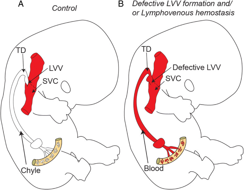
Hippocratic Corpus, a collection of Greek medical literature, described the functional anatomy of the lymphatic system in the fifth century B.C. Subsequent studies in cadavers and surgical patients firmly established that lymphatic vessels drain extravasated interstitial fluid, also known as lymph, into the venous system at the bilateral lymphovenous junctions. Recent advances revealed that lymphovenous valves and platelet-mediated hemostasis at the lymphovenous junctions maintain life-long separation of the blood and lymphatic vascular systems. Here, we review murine models that exhibit failure of blood-lymph separation to highlight the novel mechanisms and molecular targets for the modulation of lymphatic disorders. Specifically, we focus on the transcription factors, cofactors, and signaling pathways that regulate lymphovenous valve development and platelet-mediated lymphovenous hemostasis, which cooperate to maintain blood-lymph separation.
Keywords: Blood; Hemostasis; Lymph; Lymphatic development; Lymphovenous valve; Platelet.
Conflict of interest statement
The authors have declared that no conflict of interest exists.
Figures

References
-
- Drake CJ, Fleming PA. Vasculogenesis in the day 6.5 to 9.5 mouse embryo. Blood. 2000;95:1671–1679. - PubMed
Publication types
MeSH terms
Grants and funding
LinkOut - more resources
Full Text Sources
