

Role of MRI in staging and follow-up of endometrial and cervical cancer: pitfalls and mimickers
- PMID: 30758678
- PMCID: PMC6375059
- DOI: 10.1186/s13244-019-0696-8
Role of MRI in staging and follow-up of endometrial and cervical cancer: pitfalls and mimickers
Abstract
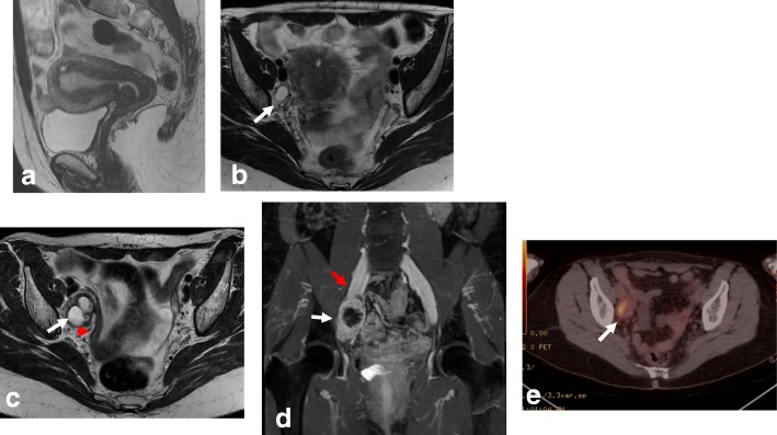
MRI plays important roles in endometrial and cervical cancer assessment, from detection to recurrent disease evaluation. Endometrial cancer (EC) is the most common malignant tumor of the female genital tract in Western countries. EC patients are divided into risk categories based on histopathological tumor type, grade, and myometrial invasion depth. EC is surgically staged using the International Federation of Gynecology and Obstetrics (FIGO) system. Since FIGO (2009) stage correlates with prognosis, preoperative staging is essential for tailored treatment. MRI reveals myometrial invasion depth, which correlates with tumor grade and lymph node metastases, and thus correlates with prognosis. Cervical cancer (CC) is the second most common cancer, and the third leading cause of cancer-related death among females in developing countries. The FIGO Gynecologic Oncology Committee recently revised its CC staging guidelines, allowing staging based on imaging and pathological findings when available. The revised FIGO (2018) staging includes node involvement and thus enables both therapy selection and evaluation, prognosis estimation, and calculation of end results. MRI can accurately assess prognostic indicators, e.g., tumor size, parametrial invasion, pelvic sidewall, and lymph node invasion. Despite these important roles of MRI, radiologists still face challenges due to the technical and interpretation pitfalls of MRI during all phases of endometrial and cervical cancer evaluation. Awareness of mimics that can simulate both cancers is critical. With careful application, functional MRI with DWI and DCE sequences can help establish a correct diagnosis, although it is sometimes necessary to perform biopsy and histopathological analysis.
Keywords: Cervical cancer; Diffusion; Endometrial cancer; Lymph nodes; Magnetic resonance.
Conflict of interest statement
Competing interests
The authors declare that they have no competing interests.
Publisher’s Note
Springer Nature remains neutral with regard to jurisdictional claims in published maps and institutional affiliations.
Figures
References
-
- Mahajan A, Sable NP, Popat PB et al (2016) Magnetic resonance imaging of gynecological malignancies: role in personalized management. Semin Ultrasound CT MRI 10.1053/j.sult.2016.11.005 - PubMed
-
- Sala E, Rockall AG, Freeman SJ, Mitchell DG, Reinhold C. The added role of MR imaging in treatment stratification of patients with gynecologic malignancies: what the radiologist needs to know. Radiology. 2013;266:717–740. - PubMed
-
- Miccò M, Sala E, Lakhman Y, Hricak H, Vargas HA (2014) Role of imaging in the pretreatment evaluation of common gynecological cancers. Womens Health (Lond) 10(3):299–321 - PubMed
-
- Nougaret S, Lakhman Y, Vargas HA, et al. From staging to prognostication: achievements and challenges of MR imaging in the assessment of endometrial cancer. Magn Reson Imaging Clin N Am. 2017;25:611–633. - PubMed
-
- Colombo N, Creutzberg C, Amant F, et al. ESMO-ESGO-ESTRO consensus conference on endometrial cancer: diagnosis, treatment and follow-up. Radiother Oncol. 2015;117(3):559–581. - PubMed
Publication types
LinkOut - more resources
Full Text Sources
