

Plexiform Fibrohistiocytic Tumor Presenting as a Central Neck Mass Clinically Mimicking a Thyroglossal Duct Cyst: An Unusual Case Reported with Histo-cytopathologic Correlation and a Review of the Cytopathology Literature
- PMID: 30758757
- PMCID: PMC7021857
- DOI: 10.1007/s12105-019-01022-4
Plexiform Fibrohistiocytic Tumor Presenting as a Central Neck Mass Clinically Mimicking a Thyroglossal Duct Cyst: An Unusual Case Reported with Histo-cytopathologic Correlation and a Review of the Cytopathology Literature
Abstract
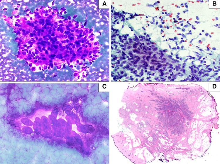
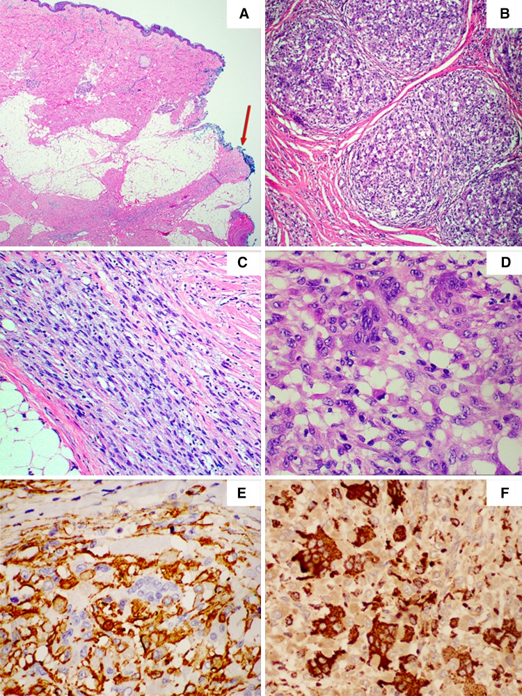
We present the case of an uncommon example of a plexiform fibrohistiocytic tumor (PFHT) occurring in the anterior central neck region of a 40 year-old female with previous subtotal thyroidectomy. The tumor clinically mimics a complicated thyroglossal duct cyst. On fine needle aspiration cytology, the tumor was composed of sheets of bland spindle cells and nests of plump histiocytoid cells in vaguely whorled arrangements. Occasional multinucleated giant cells were also identified. The excised specimen showed an irregular, highly infiltrative subcutaneous tumor arranged in a nodular/plexiform pattern concentrated to the center of the tumor mass. In addition, the tumor contained numerous tongue-like extensions composed of variably cellular, fibroblastic/fibromatosis-like areas. These fibroblastic/fibromatosis-like extensions reached far from the epicenter of the tumor and were associated with scattered small plexiform nodules of histiocytic cells. These tongue-like extensions multifocally involved the surgical margins. The fibroblastic and histiocytoid cells showed diffuse smooth muscle actin (SMA) expression. The multinucleated giant cells and also the histiocytoid proliferation were positive for CD68. This case illustrates an uncommon both anatomical and demographic manifestation of PFHT and also characterize the fine needle aspiration cytologic features in this tumor, which previously have been reported in a few cases.
Keywords: Fine needle aspiration cytology; Neck; Plexiform fibrohistiocytic tumor; Thyroglossal duct cyst.
Conflict of interest statement
The authors declare that they have no conflict of interest.
Figures
Similar articles
-
Papillary carcinoma in thyroglossal duct cyst: Diagnosis by fine-needle aspiration cytology and immunocytochemistry.Diagn Cytopathol. 2018 Sep;46(9):797-800. doi: 10.1002/dc.23968. Epub 2018 May 8. Diagn Cytopathol. 2018. PMID: 29737627
-
Expression of MiTF may be helpful in differentiating cellular neurothekeoma from plexiform fibrohistiocytic tumor (histiocytoid predominant) in a partial biopsy specimen.Am J Dermatopathol. 2012 Apr;34(2):157-60. doi: 10.1097/DAD.0b013e3182286a03. Am J Dermatopathol. 2012. PMID: 22441367
-
Thyroglossal Duct Cyst Associated with Xanthogranulomatous Inflammation.Head Neck Pathol. 2015 Dec;9(4):530-3. doi: 10.1007/s12105-015-0628-y. Epub 2015 Apr 21. Head Neck Pathol. 2015. PMID: 25896144 Free PMC article.
-
Invasive Thyroglossal Duct Cyst Papillary Carcinoma: A Case Report and Review of the Literature.Am J Case Rep. 2018 Jun 28;19:757-762. doi: 10.12659/AJCR.907313. Am J Case Rep. 2018. PMID: 29950556 Free PMC article. Review.
-
Squamous cell carcinoma arising in a thyroglossal duct cyst.Arch Otolaryngol Head Neck Surg. 1992 May;118(5):538-41. doi: 10.1001/archotol.1992.01880050092022. Arch Otolaryngol Head Neck Surg. 1992. PMID: 1571131 Review.
Cited by
-
Study on the value of multi-dimensional conformal radiotherapy and functional imaging in tumor bioimaging.Transl Cancer Res. 2022 Oct;11(10):3780-3789. doi: 10.21037/tcr-22-2005. Transl Cancer Res. 2022. PMID: 36388020 Free PMC article.
-
A rare case of congenital plexiform fibrohistiocytic tumor of the foot in a 4-year-old boy: case report and literature review.Case Reports Plast Surg Hand Surg. 2021 Oct 1;8(1):164-168. doi: 10.1080/23320885.2021.1986049. eCollection 2021. Case Reports Plast Surg Hand Surg. 2021. PMID: 34621916 Free PMC article.
-
Morphological, genetic and clinical correlations in infantile hemangiomas and their mimics.Rom J Morphol Embryol. 2020 Jul-Sep;61(3):687-695. doi: 10.47162/RJME.61.3.07. Rom J Morphol Embryol. 2020. PMID: 33817710 Free PMC article.
References
Publication types
MeSH terms
LinkOut - more resources
Full Text Sources
Medical
